Correction: Epitalon increases telomere length in human cell lines through telomerase upregulation or ALT activity
Sarah Al-dulaimi, Ross Thomas, Sheila Matta, Terry Roberts

Abstract
Genes, proteins, chemicals, diseases, species, mutations and cell lines named across the full text — each resolved to its canonical identifier and authoritative record.
Click any figure to enlarge with its caption.
 Figure 1
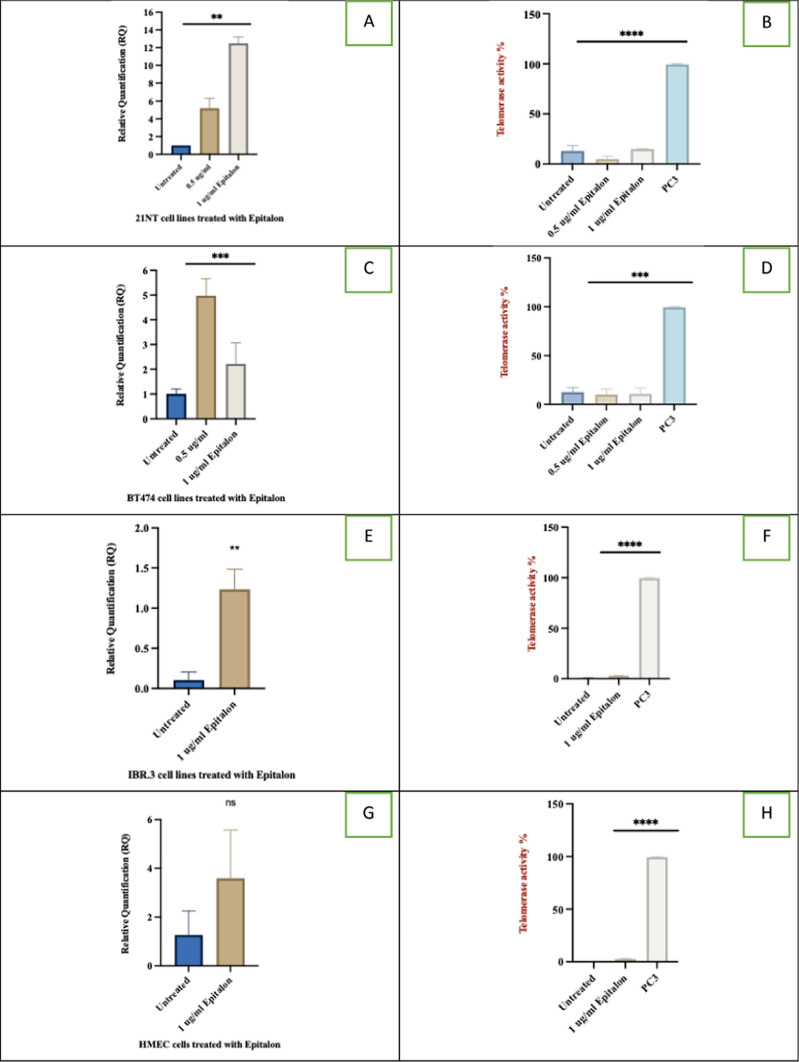 Figure 2
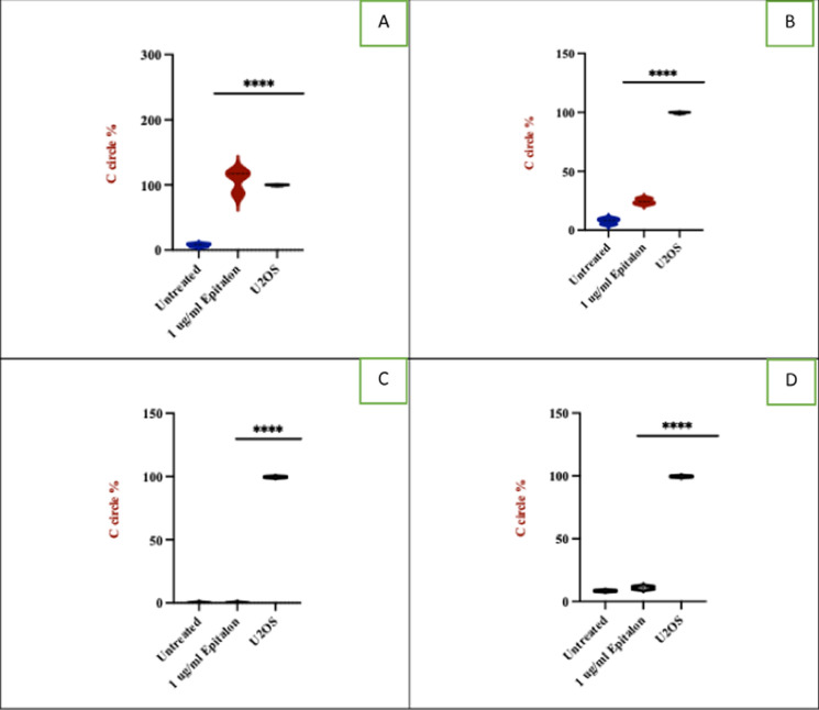 Figure 3
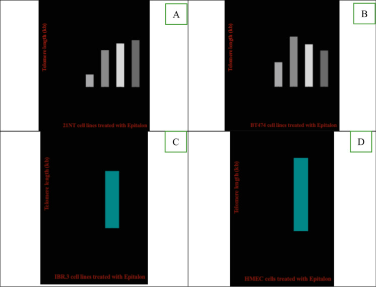 Figure 4
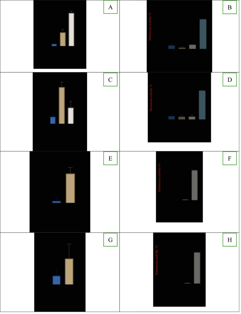 Figure 5
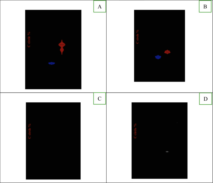 Figure 6
Peer Reviews
No public reviews on file for this paper yet. If you reviewed it on a platform where reviews are public (OpenReview, ICLR, NeurIPS, ICML), you can paste yours below so the community can read it here.
Videos
No videos yet. Explain this paper in a talk, walkthrough, or lecture? Add one.
Taxonomy
TopicsTelomeres, Telomerase, and Senescence · Nuclear Structure and Function · Muscle Physiology and Disorders
Correction to: Biogerontology 10.1007/s10522-025-10315-x
In this article, the wrong figures appeared in Figs. 1, 2 and 3. The incorrect and correct versions of Figs. 1, 2 and 3 are provided below:
Incorrect version of Fig. 1:Telomere length of breast cancer cells and normal cells treated with epitalon. A 21NT cells B BT474 cells. Both cells were treated for 4 days with varying concentrations of epitalon. (0.1, 0.2, 0.5 and 1 ug/ml). Untreated cells were used as a control. C Telomere length of IBR.3 cells (P.14) treated with 1 μg /ml epitalon for 3 weeks. D telomere length of HMEC (P.14) treated with 1 μg /ml epitalon for 3 weeks. Untreated cells were included as a control
Correct Version of Fig. 1:
Fig. 1. Telomere length of breast cancer cells and normal cells treated with epitalon. A 21NT cells B BT474 cells. Both cells were treated for 4 days with varying concentrations of epitalon. (0.1, 0.2, 0.5 and 1 ug/ml). Untreated cells were used as a control. C Telomere length of IBR.3 cells (P.14) treated with 1 μg /ml epitalon for 3 weeks. D telomere length of HMEC (P.14) treated with 1 μg /ml epitalon for 3 weeks. Untreated cells were included as a control
Incorrect Version of Fig. 2:hTERT expression (RQ) and telomerase enzyme activity in breast cancer cells and normal fibroblast cells treated with epitalon. A and B hTERT expression and telomerase activity for 21NT treated with 0.5 and 1 μg/ml of epitalon for 4 days. C and D hTERT expression and telomerase activity of BT474 treated with 0.5 and 1 μg/ml of epitalon for 4 days. PC3 was included as a positive control for telomerase activity. E and F hTERT expression and telomerase activity for IBR.3 were treated with 1 μg/ml of epitalon for three weeks. G and H hTERT expression and telomerase activity for HMEC treated with 1 ug/ml of epitalon for three weeks
Correct Version of Fig. 2:
Fig. 2hTERT expression (RQ) and telomerase enzyme activity in breast cancer cells and normal fibroblast cells treated with epitalon. A and B hTERT expression and telomerase activity for 21NT treated with 0.5 and 1 μg/ml of epitalon for 4 days. C and D hTERT expression and telomerase activity of BT474 treated with 0.5 and 1 μg/ml of epitalon for 4 days. PC3 was included as a positive control for telomerase activity. E and F hTERT expression and telomerase activity for IBR.3 were treated with 1 μg/ml of epitalon for three weeks. G and H hTERT expression and telomerase activity for HMEC treated with 1 ug/ml of epitalon for three weeks
Incorrect Version of Fig. 3:ALT activity in breast cancer cells and normal cells treated with epitalon. A and B ALT activity in 21NT and BT474 treated with 1 μg/ml of epitalon for 4 days. Untreated cells and the ALT positive U2OS were included as controls. C IBR.3 and D HMEC treated with 1 μg/ml of epitalon for three weeks. E Immunofluorescence to detect PML bodies in 21NT and BT474 cells treated with epitalon for 4 days. The colocalization PML bodies (green staining) within the nucleus (blue staining) for 21NT and BT474 treated with epitalon indicated the presence of ALT. PML bodies were detected using Lecia microscope with X 100 objective
Correct Version of Fig. 3:
Fig. 3ALT activity in breast cancer cells and normal cells treated with epitalon. A and B ALT activity in 21NT and BT474 treated with 1 μg/ml of epitalon for 4 days. Untreated cells and the ALT positive U2OS were included as controls. C IBR.3 and D HMEC treated with 1 μg/ml of epitalon for three weeks. E Immunofluorescence to detect PML bodies in 21NT and BT474 cells treated with epitalon for 4 days. The colocalization PML bodies (green staining) within the nucleus (blue staining) for 21NT and BT474 treated with epitalon indicated the presence of ALT. PML bodies were detected using Lecia microscope with X 100 objective
The original article has been corrected.