Distance‐based assessment of spatial artifact extension in the prostate from fiducial markers in diffusion‐weighted magnetic resonance imaging
Mizgin Coskun, Patrik Brynolfsson, Christian Jamtheim Gustafsson, Adalsteinn Gunnlaugsson, Lars E. Olsson

TL;DR
This study measures how fiducial markers in prostate MRI scans create image artifacts, finding that artifacts are smaller in patient images than in phantoms and that motion increases their size.
Contribution
The study quantifies artifact extension distances in prostate MRI caused by fiducial markers and evaluates the impact of motion and SNR on artifact size.
Findings
Artifacts were smaller in ADC maps compared to DWI images.
Motion increased artifact extension distances by up to 2–3 mm in both phantom and patient images.
Lower SNR resulted in smaller visible artifact extensions.
Abstract
Fiducial markers in image‐guided prostate cancer radiotherapy reduce geometric uncertainty during daily patient setup and enable assessment of target position changes. Diffusion‐weighted magnetic resonance imaging (MRI) for target delineation improves prostate cancer localization, beneficial for intraprostatic focal boost. Artifacts from fiducial markers on prostate diffusion‐weighted MRI (DWI) need to be investigated, as they could be detrimental for target delineation. This study aims to determine the distances of artifact extensions caused by fiducial markers in DWI and in the apparent diffusion coefficient (ADC) maps and to assess how motion and signal‐to‐noise ratio (SNR) influence the artifact size in ADC maps. Three phantoms were used: two homogeneous gel phantoms—one containing three cylindrical gold fiducial markers (GFM) and the other containing three spherical gold anchor…
Genes, proteins, chemicals, diseases, species, mutations and cell lines named across the full text — each resolved to its canonical identifier and authoritative record.
Click any figure to enlarge with its caption.
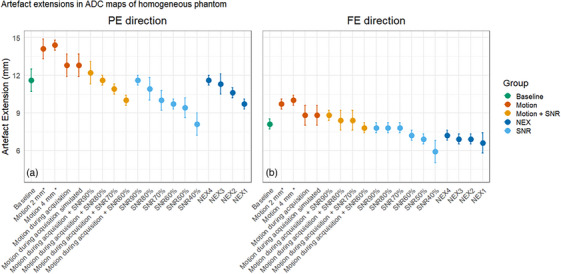 Figure 1
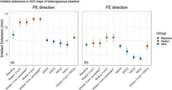 Figure 2
 Figure 3
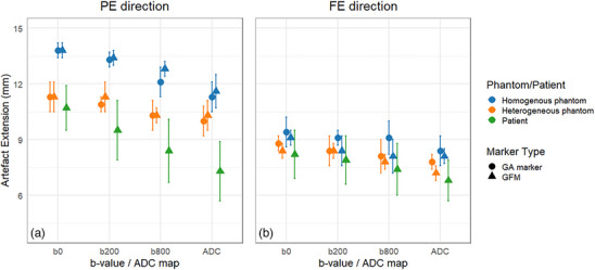 Figure 4
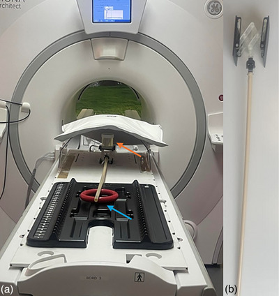 Figure 5
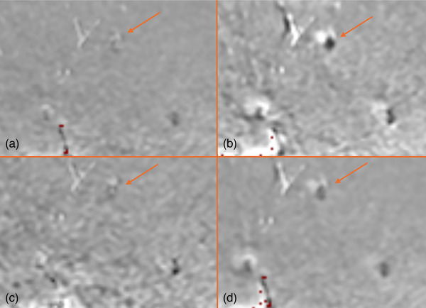 Figure 6
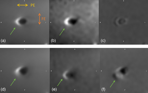 Figure 7
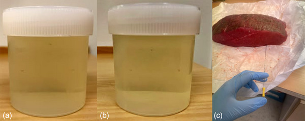 Figure 8
 Figure 9
Peer Reviews
No public reviews on file for this paper yet. If you reviewed it on a platform where reviews are public (OpenReview, ICLR, NeurIPS, ICML), you can paste yours below so the community can read it here.
Videos
No videos yet. Explain this paper in a talk, walkthrough, or lecture? Add one.
Taxonomy
TopicsMRI in cancer diagnosis · Prostate Cancer Diagnosis and Treatment · Advanced Neuroimaging Techniques and Applications