Improved brain tumor diagnostics and follow-up with novel magnetic resonance imaging methods: A single center study protocol
Jesse Lohela, Kaisa Lehtiö, Kalle Inget, Sakari S. Karhula, Susanna Piironen, Angélica Suutari, Antti Knuutinen, Miro Jänkälä, Eveliina Lammentausta, Michaela K. Bode, Juha Nikkinen, Niina Salokorpi, Tuija Keinänen

TL;DR
This study aims to improve brain tumor diagnosis and monitoring using advanced MRI techniques to provide more detailed physiological and molecular insights.
Contribution
The study introduces a multiparametric MRI framework to enhance diagnostic accuracy and personalize treatment decisions for brain tumors.
Findings
Advanced MRI techniques provide physiological and molecular insights beyond conventional anatomical imaging.
The study hypothesizes that combining advanced MRI sequences improves diagnostic accuracy compared to conventional MRI.
The protocol aims to model spatial recurrence risk and distinguish true progression from pseudoprogression using quantitative imaging biomarkers.
Abstract
This protocol outlines a prospective study aimed at enhancing the diagnosis and monitoring of brain tumors through advanced non-invasive imaging techniques. While magnetic resonance imaging (MRI) is a cornerstone of brain tumor diagnostics, it often lacks the specificity required for definitive diagnosis, which typically relies on invasive tissue sampling. To address this, the study will evaluate advanced MRI techniques—such as perfusion, diffusion, blood-oxygen-level-dependent imaging, magnetic resonance spectroscopy, and amide proton transfer-weighted imaging— that offer valuable physiological and molecular insights, beyond conventional anatomical imaging. Despite their potential, clinical adoption of these methods remains limited. MRI also plays a central role in treatment response assessment and follow-up, yet conventional anatomical sequences may not detect early physiological…
Genes, proteins, chemicals, diseases, species, mutations and cell lines named across the full text — each resolved to its canonical identifier and authoritative record.
Click any figure to enlarge with its caption.
 Figure 1
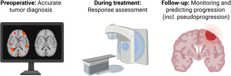 Figure 2
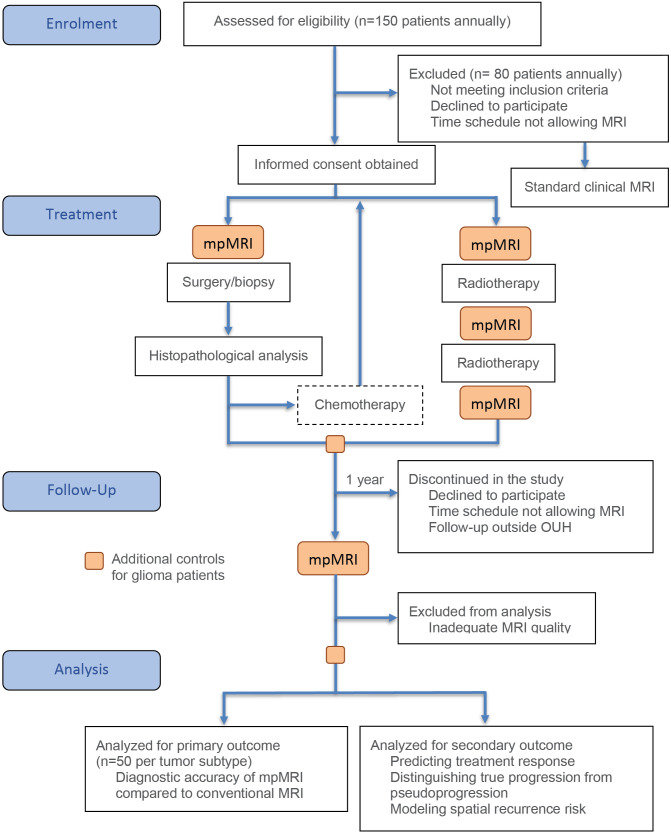 Figure 3
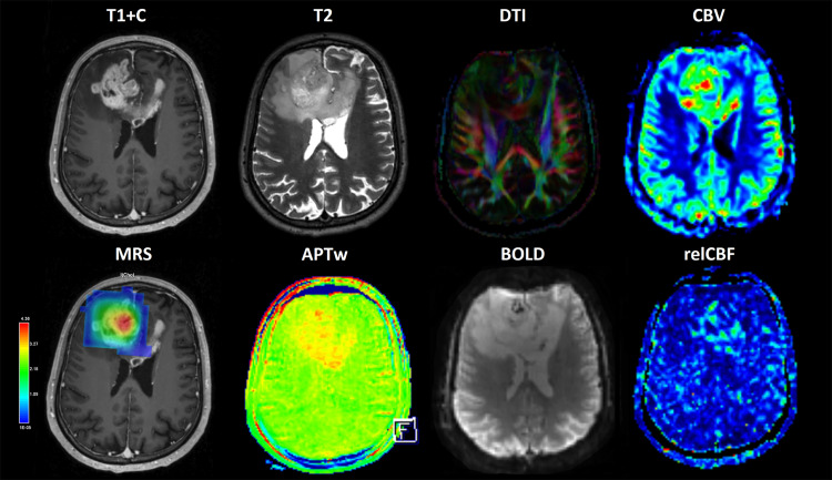 Figure 4
Peer Reviews
No public reviews on file for this paper yet. If you reviewed it on a platform where reviews are public (OpenReview, ICLR, NeurIPS, ICML), you can paste yours below so the community can read it here.
Videos
No videos yet. Explain this paper in a talk, walkthrough, or lecture? Add one.
Taxonomy
TopicsAdvanced MRI Techniques and Applications · Glioma Diagnosis and Treatment · MRI in cancer diagnosis