T2 Dixon Imaging in the Evaluation of Hibernoma: Reliable Identification of Macroscopic Fat
Sander Gurdeep Singh, Wouter Huysse, Frederiek Laloo

TL;DR
T2 Dixon imaging helps identify fat in soft tissue lesions, distinguishing benign from suspicious areas using water-only and fat-only images.
Contribution
The paper highlights the reliability of T2 Dixon imaging in identifying macroscopic fat within hibernoma lesions.
Findings
Water-only T2 Dixon images highlight fluid or edema in soft tissue lesions.
Fat-only T2 Dixon images effectively identify macroscopic fat and delineate fat-devoid regions.
The technique helps distinguish benign from suspicious areas in soft tissue evaluation.
Abstract
Teaching point: T2 Dixon imaging is valuable in assessing soft tissue lesions. Water-only images highlight fluid or edema, while fat-only images identify macroscopic fat and help delineate fat-devoid regions, which often indicate suspicious areas—or confirm their absence in benign lesions.
Genes, proteins, chemicals, diseases, species, mutations and cell lines named across the full text — each resolved to its canonical identifier and authoritative record.
Click any figure to enlarge with its caption.
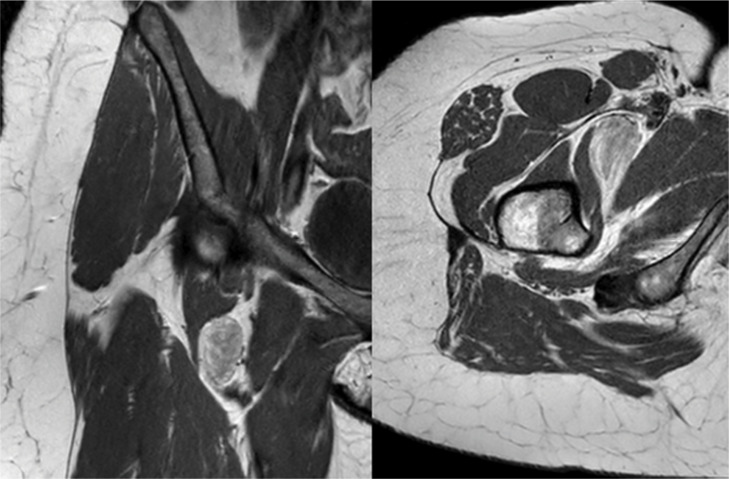 Figure 1
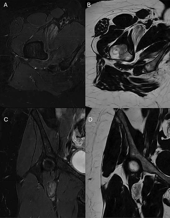 Figure 2
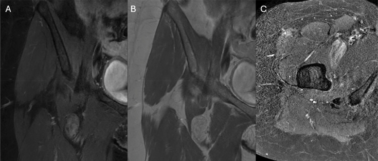 Figure 3
Peer Reviews
No public reviews on file for this paper yet. If you reviewed it on a platform where reviews are public (OpenReview, ICLR, NeurIPS, ICML), you can paste yours below so the community can read it here.
Videos
No videos yet. Explain this paper in a talk, walkthrough, or lecture? Add one.
Taxonomy
TopicsSarcoma Diagnosis and Treatment · Soft tissue tumor case studies · Musculoskeletal synovial abnormalities and treatments