Actin arginylation alters myosin engagement and F-actin patterning despite structural conservation
Clyde Savio Pinto, Saskia E. Bakker, Andrejus Suchenko, Isabella M. Kolodny, Hamdi Hussain, Tomoyuki Hatano, Karuna Sampath, Krishna Chinthalapudi, Sarah M. Heissler, Masanori Mishima, Mohan Balasubramanian

TL;DR
Arginylation of actin changes how it interacts with myosin and organizes into structures in cells, even though its structure remains mostly the same.
Contribution
The study reveals that arginylated actin alters myosin-II interactions and F-actin organization despite structural conservation.
Findings
Arginylated β-actin filaments have nearly identical structures to non-arginylated actin.
Arginylation disrupts myosin-II interactions and affects cytokinesis in cells.
R-actin alters actin filament organization in vivo, impacting subcellular structures.
Abstract
Actin is vital for cell functions and is regulated by posttranslational modifications like arginylation. The structure of arginylated β-actin (R-β-actin) is nearly identical to non-modified actin. However, arginylation alters myosin-II interactions and impacts actin filament organization and cytokinesis in cells. Actin is a conserved protein with crucial roles in cell polarity, division, and muscle contraction. Its function is regulated in part by posttranslational modifications, one of which is N-terminal arginylation. What is the structure of arginylated-β-actin (R-β-actin), and how does it regulate F-actin function? Here we report the 3.6 Å structures of ADP-R-β-actin filaments, which are nearly identical to that of non-arginylated F-actin. In vitro assays reveal that the interaction between myosin-II and actin is altered upon actin arginylation, characterized by frequent detachment…
Genes, proteins, chemicals, diseases, species, mutations and cell lines named across the full text — each resolved to its canonical identifier and authoritative record.
Click any figure to enlarge with its caption.
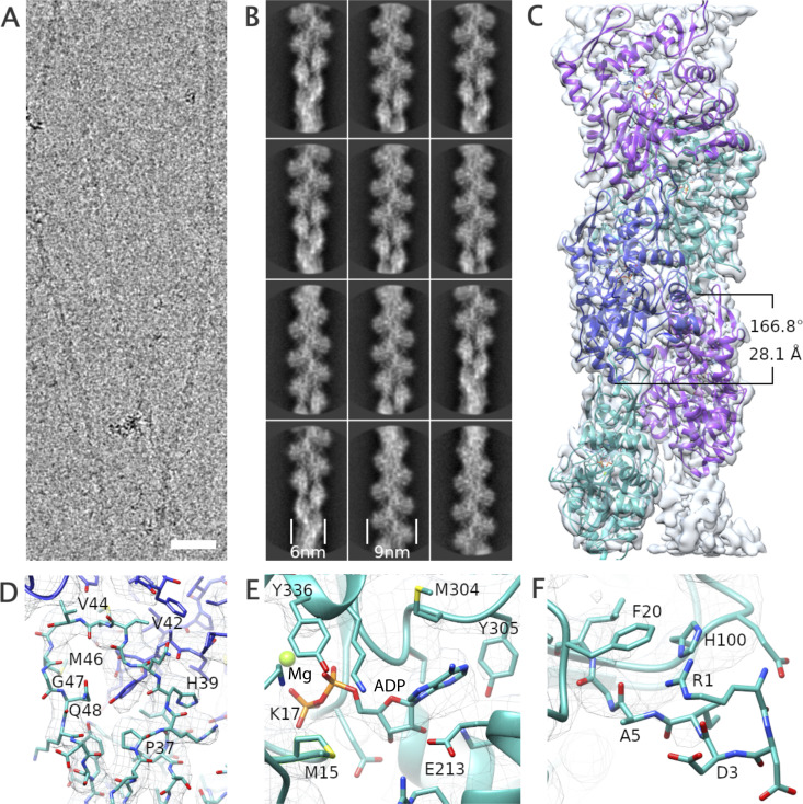 Figure 1
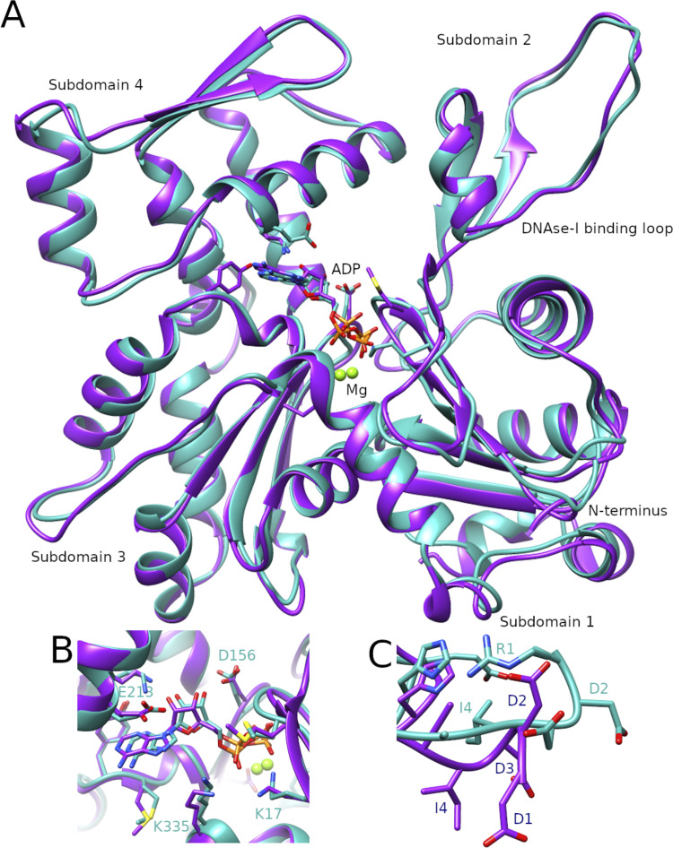 Figure 2
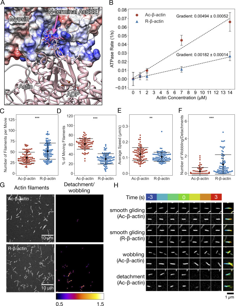 Figure 3
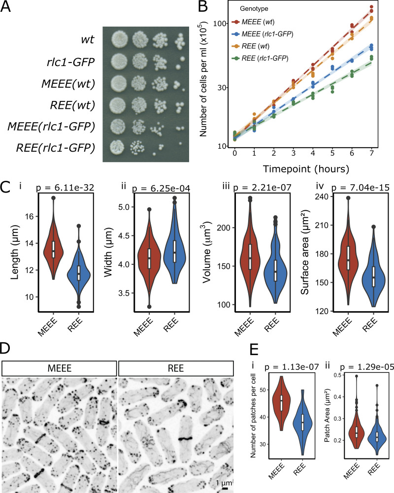 Figure 4
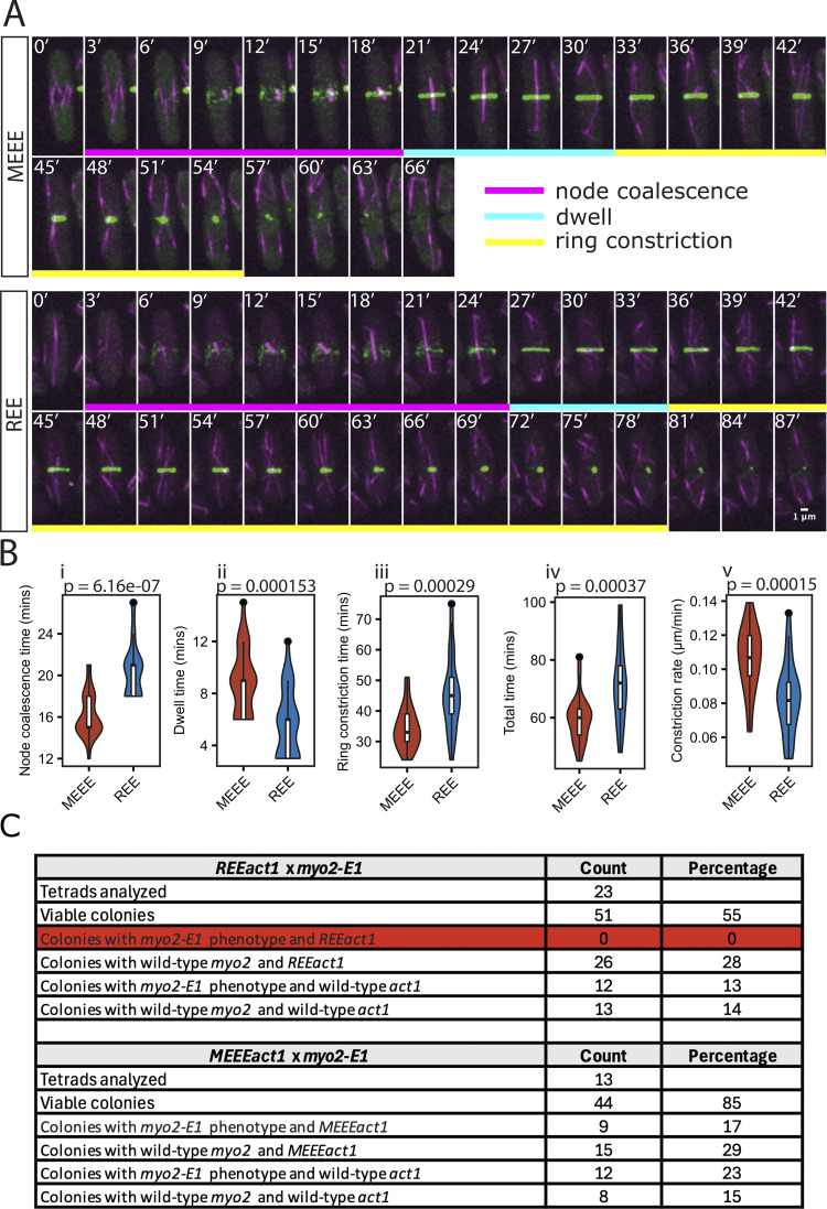 Figure 5
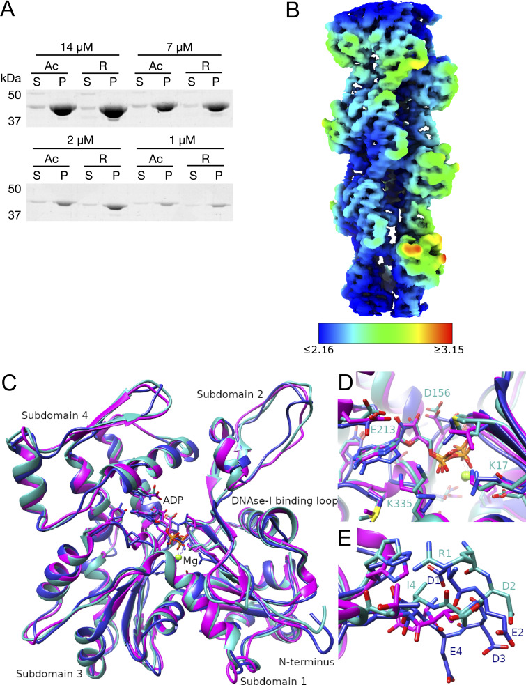 Figure 6
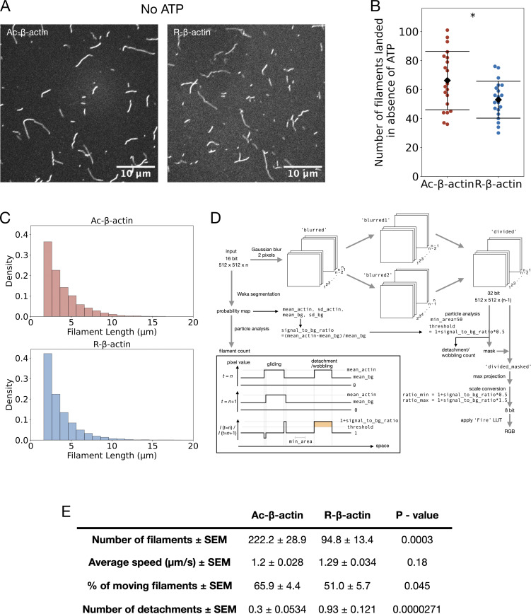 Figure 7
 Figure 8
 Figure 9
Peer Reviews
No public reviews on file for this paper yet. If you reviewed it on a platform where reviews are public (OpenReview, ICLR, NeurIPS, ICML), you can paste yours below so the community can read it here.
Videos
No videos yet. Explain this paper in a talk, walkthrough, or lecture? Add one.
Taxonomy
TopicsUbiquitin and proteasome pathways · Peptidase Inhibition and Analysis · Connective tissue disorders research