Quantitative comparison of a novel wide-field OCT-angiography device with ultrawide-field fluorescein angiography in detecting retinal nonperfusion in vascular retinopathies
Michael Hafner, Tina R. Herold, Viktoria Deiters, Bettina von Livonius, Siegfried G. Priglinger, Maximilian J. Gerhardt

TL;DR
This study compares a new wide-field OCT-angiography device with an existing standard for detecting retinal nonperfusion, showing it works well for mild to moderate cases but has limitations in severe cases.
Contribution
First quantitative comparison of DREAM WF-OCTA with UWF-FA for retinal nonperfusion assessment.
Findings
WF-OCTA showed strong correlation with UWF-FA for retinal ischemia assessment.
Montage WF-OCTA had good agreement with UWF-FA in mild to moderate ischemia.
Bland-Altman analysis revealed underestimation at higher nonperfusion levels.
Abstract
Reliable assessment of retinal nonperfusion is critical in managing vascular retinopathies. While ultrawide-field fluorescein angiography (UWF-FA) is the clinical standard, it is invasive and dye-dependent. Previous wide-field optical coherence tomography angiography (WF-OCTA) systems have been limited by insufficient peripheral coverage. To the best of our knowledge, this is the first quantitative comparison of DREAM WF-OCTA with UWF-FA. The study leverages the device’s increased field of view (≈130° single scan, > 200° montage) and demonstrates that the previously published, semi-automated VMseg approach can also be applied to DREAM data. 24 eyes from 13 patients with diabetic retinopathy or retinal vein occlusion underwent both UWF-FA (Optos Silverstone, 200°) and WF-OCTA and were analyzed. The ischemic index (ISI) was calculated for each modality using previously developed…
Genes, proteins, chemicals, diseases, species, mutations and cell lines named across the full text — each resolved to its canonical identifier and authoritative record.
Click any figure to enlarge with its caption.
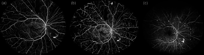 Figure 1
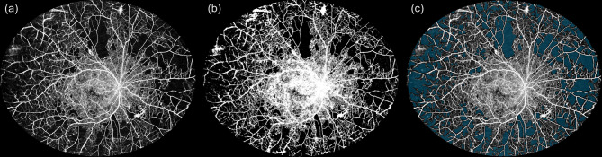 Figure 2
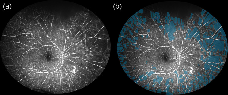 Figure 3
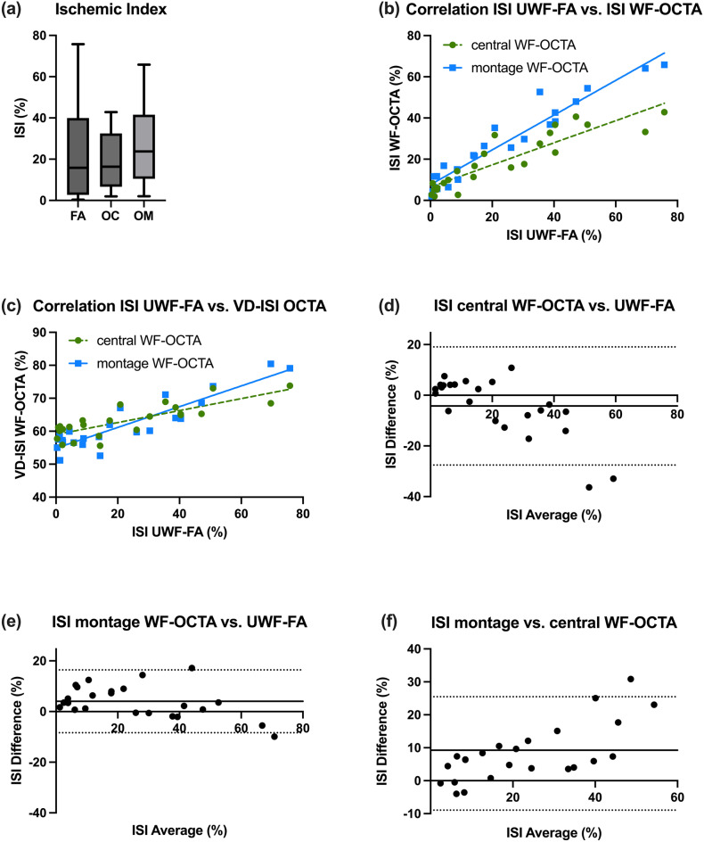 Figure 4
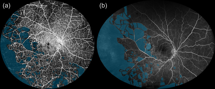 Figure 5
Peer Reviews
No public reviews on file for this paper yet. If you reviewed it on a platform where reviews are public (OpenReview, ICLR, NeurIPS, ICML), you can paste yours below so the community can read it here.
Videos
No videos yet. Explain this paper in a talk, walkthrough, or lecture? Add one.
Taxonomy
TopicsRetinal Imaging and Analysis · Retinal Diseases and Treatments · Retinal and Optic Conditions