Multiphoton microscopy imaging of fibrous meningiomas based on the combination of multichannel mode and lambda mode
Linghan You, Linjing Shi, Yuqing Huang, Lingxin Pan, Shiying Zheng, Yingyuan Wang, Zanyi Wu, Xingfu Wang, Jianxin Chen, Na Fang

TL;DR
This study uses multiphoton microscopy to image fibrous meningiomas, revealing collagen and other components with high resolution and contrast.
Contribution
The novel use of combined multichannel and lambda modes in MPM to analyze fibrous meningioma microstructures and quantify collagen content.
Findings
Collagen is the most abundant component in fibrous meningiomas, with a relative ratio of 0.952.
Combined MPM imaging provides high-resolution visualization of tumor microenvironment features.
SHG and 32-channel spectral imaging both show similar average collagen content (around 0.537 and 0.503 respectively).
Abstract
Fibrous meningiomas, known for their dense and tough texture, present unique challenges in diagnosis and surgical treatment. This study explores the potential of multiphoton microscopy (MPM) for visualizing the microstructures of fibrous meningiomas by combining multichannel and lambda modes. Using MPM, we imaged 14 fibrous meningioma samples collected from neurosurgical procedures. The multichannel mode captured second harmonic generation (SHG) and two-photon excitation fluorescence (TPEF) signals, while the lambda mode provided detailed spectral imaging across 32 channels. Image analysis algorithms were developed to quantify collagen content and assess morphological features. Spectroscopic analysis revealed the intrinsic components of fibrous meningiomas, with collagen being the most abundant component (relative ratio: 0.952), followed by structural proteins (0.502), free-form NADH…
Genes, proteins, chemicals, diseases, species, mutations and cell lines named across the full text — each resolved to its canonical identifier and authoritative record.
Click any figure to enlarge with its caption.
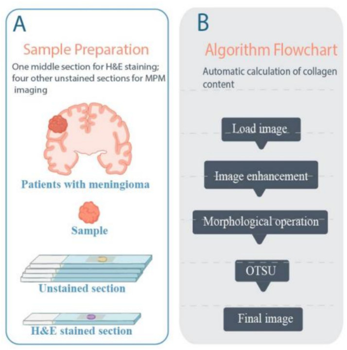 Figure 1
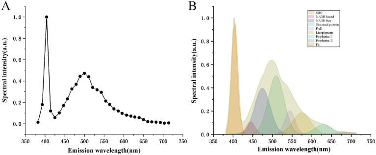 Figure 2
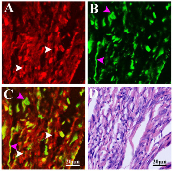 Figure 3
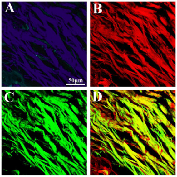 Figure 4
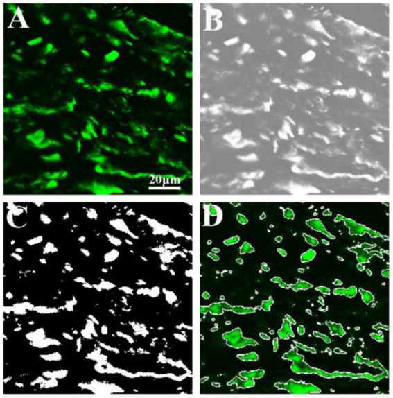 Figure 5
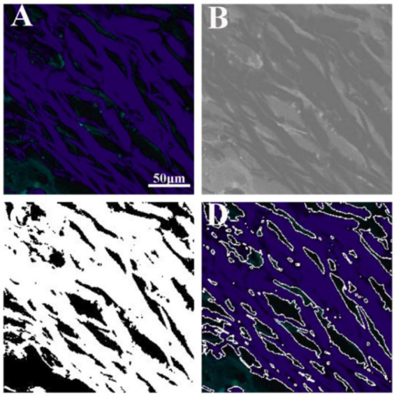 Figure 6
Peer Reviews
No public reviews on file for this paper yet. If you reviewed it on a platform where reviews are public (OpenReview, ICLR, NeurIPS, ICML), you can paste yours below so the community can read it here.
Videos
No videos yet. Explain this paper in a talk, walkthrough, or lecture? Add one.
Taxonomy
TopicsAdvanced Fluorescence Microscopy Techniques · Advanced X-ray Imaging Techniques · Optical Coherence Tomography Applications