Imaging Findings of Paratesticular Adenomatoid Tumor
Wenqin Liu, Zhifan Yuan, Limei Liang, Xiaoling Leng

TL;DR
This paper discusses the imaging characteristics of a rare benign tumor near the epididymis and its effective treatment through surgery.
Contribution
The paper provides insights into the diagnostic imaging features of paratesticular adenomatoid tumors.
Findings
These tumors are typically asymptomatic and appear as hyperechoic masses on ultrasound.
MRI is useful for difficult diagnostic cases.
Surgical excision is curative with low recurrence risk.
Abstract
Paratesticular adenomatoid tumors are benign growths usually found near the epididymis. They often show no symptoms and appear as hyperechoic masses on ultrasound. Magnetic resonance imaging helps in difficult cases. Surgical removal is curative with minimal recurrence risk.
Genes, proteins, chemicals, diseases, species, mutations and cell lines named across the full text — each resolved to its canonical identifier and authoritative record.
Click any figure to enlarge with its caption.
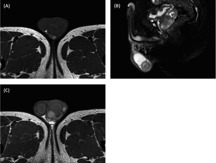 Figure 1
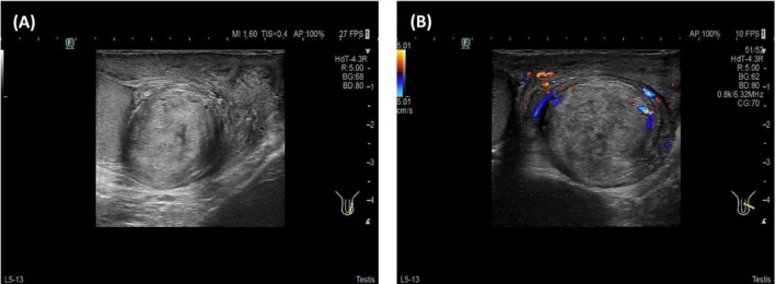 Figure 2
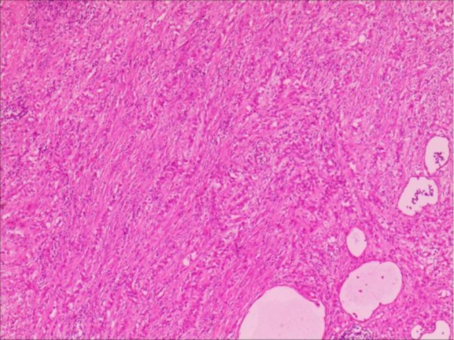 Figure 3
Peer Reviews
No public reviews on file for this paper yet. If you reviewed it on a platform where reviews are public (OpenReview, ICLR, NeurIPS, ICML), you can paste yours below so the community can read it here.
Videos
No videos yet. Explain this paper in a talk, walkthrough, or lecture? Add one.
Taxonomy
TopicsTesticular diseases and treatments · Adrenal and Paraganglionic Tumors · Intraperitoneal and Appendiceal Malignancies
Paratesticular adenomatoid tumor, an uncommon benign neoplasm derived from mesothelial cells, predominantly arises in the epididymal vicinity of the testes. This entity comprises roughly 30% of all paratesticular tumors and approximately 60% of benign paratesticular lesions [1]. It can manifest across all age groups in males, with a predilection for sexually active young adults aged 20 to 50 years. Asymptomatic in nature, the detection of these tumors often occurs fortuitously, though a minority of patients may experience scrotal discomfort or pain secondary to mass enlargement. Here, we present a case of paratesticular adenomatoid tumor, its ultrasound, magnetic resonance imaging, and subsequent pathological confirmation.
A 47‐year‐old male patient presented to our hospital with a progressively enlarging left scrotal mass spanning 7 years. Physical examination revealed a drooping and swollen left scrotum harboring a palpable, round, smooth‐surfaced, painless mass that remained unchanged in size with positional manipulation. The transillumination test yielded negative results. High‐frequency ultrasound disclosed a heterogeneous, hyperechoic mass located at the caudal aspect of the left epididymis, demarcated by a clear boundary and interspersed with irregular internal echoes (Figure 1A). Upon probe compression, relative mobility was observed between the mass and the scrotal wall, accompanied by the discernment of short, rod‐like vascular signals both within and adjacent to the mass (Figure 1B). Magnetic resonance imaging provided further insights, revealing an enlarged left epididymis with irregular contours compared to the contralateral side. An abnormal signal intensity lesion was identified in the lower internal quadrant, exhibiting mildly prolonged T1 and T2 relaxation times. Moreover, focal, patchy areas with contrasting T1 shortening (Figure 2A) and T2 prolongation (Figure 2B) were noted. Enhanced magnetic resonance imaging scans demonstrated pronounced yet heterogeneous enhancement of the abnormal signal nodules within the lower inner aspect of the left epididymis and scrotum (Figure 2C). The patient underwent left epididymal mass resection, and pathological assessment confirmed the diagnosis of adenomatoid tumor with localized necrosis (Figure 3). Immunohistochemical profiling revealed positive staining for CK, Vim, WT‐1, EMA, CK5/6, CK7, CR, MC, and CD31/CD34, whereas CEA, CK20, Prohibitin a, Oct‐4, and SALL4 were negative. The Ki‐67 index was approximately 3%.
Ultrasound serves as the initial modality of choice for diagnosing testicular adenomatoid tumors. Tumors in the epididymal tail commonly manifest as hyperechoic nodules, whereas those in the head may exhibit varied echogenicity. Tumors are typically located between the epididymal head and the testicular upper pole [2]. In cases where ultrasound diagnosis is challenging or local infiltration is suspected, magnetic resonance imaging offers a more exhaustive assessment, enabling precise localization, sizing, morphological delineation, and assessment of lesion‐tissue relationships, thereby facilitating differential diagnosis from other scrotal pathologies [3]. However, magnetic resonance imaging is usually unnecessary and may be cost‐prohibitive in most cases. The mainstay of paratesticular adenomatoid tumor management involves local surgical excision without the necessity for testicular resection, ensuring complete tumor removal to prevent recurrence. Surgical intervention typically results in a complete cure, with low rates of postoperative recurrence and malignancy.
Author Contributions
Wenqin Liu: data curation, writing – original draft. Zhifan Yuan: data curation, formal analysis. Limei Liang: data curation, formal analysis. Xiaoling Leng: supervision, writing – review and editing.
Disclosure
Transparency Statement: We can confirm that this manuscript is an honest, accurate, and transparent account of the case being reported and that no important aspects have been omitted.
Consent
Written informed consent was obtained from the patient to publish this manuscript by the journal's patient consent policy.
Conflicts of Interest
The authors declare no conflicts of interest.
The reference list from the paper itself. Each links out to its DOI / PubMed record.
- 1F. Pagliuca , S. Lucà , M. D. Sio , et al., “Testicular/Paratesticular Mesothelial Tumours: Uncommon Histopathologic Entities in a Very Complex Anatomical Site,” Pathology, Research and Practice 253 (2024): 155069.38181581 10.1016/j.prp.2023.155069 · doi ↗ · pubmed ↗
- 2R. Pichler , G. Tulchiner , F. Steinkohl , et al., “Adenomatoid Tumor of the Testis Mimicking Malignant Testicular Cancer on Multiparametric Ultrasound,” European Journal of Medical Research 23, no. 1 (2018): 3.29325584 10.1186/s 40001-018-0301-5PMC 5765709 · doi ↗ · pubmed ↗
- 3V. Rafailidis , D. Y. Huang , and P. S. Sidhu , “Paratesticular Lesions: Aetiology and Appearances on Ultrasound,” Andrology 9, no. 5 (2021): 1383–1394.33864338 10.1111/andr.13021 · doi ↗ · pubmed ↗