Computer Vision-Assisted Spatial Analysis of Mitoses and Vasculature in Lung Cancer
Anna Timakova, Alexey Fayzullin, Vladislav Ananev, Egor Zemnuhov, Vadim Alfimov, Alexey Baranov, Yulia Smirnova, Vitaly Shatalov, Natalia Konukhova, Evgeny Karpulevich, Peter Timashev, Vladimir Makarov

TL;DR
This paper uses computer vision to analyze lung cancer tissue patterns, helping identify potential therapy targets based on vascular and proliferative features.
Contribution
The study introduces two AI frameworks for analyzing vascular and mitotic features in lung cancer, revealing distinct patterns linked to tumor aggressiveness.
Findings
SegFormer achieved high accuracy in vessel segmentation with IoU = 0.96 and AUC-ROC = 0.98.
RetinaNet + CNN ensemble showed high specificity (0.96) and sensitivity (0.97) for mitotic detection.
Distinct trophic patterns were identified, which could guide targeted therapies like antiangiogenic treatment.
Abstract
Background/Objectives: Lung cancer is characterized by a significant microstructural heterogenicity among different histological types. Artificial intelligence and digital pathology instruments can facilitate morphological analysis by introducing calculated metrics allowing for the distinguishment of different tissue patterns. Methods: We used computer vision models to calculate a number of morphometric features of tumor vascularization and proliferation. We used two frameworks to process whole-slide images: (1) LVI-PathNet framework for vascular detection, based on the SegFormer architecture; and (2) Mito-PathNet framework for mitotic figure detection, based on the RetinaNet detector and an ensemble classification model. The results were visualized in the segmented and gradient heatmaps. Results: SegFormer for vessel segmentation achieved the following quality metrics: IoU = 0.96,…
Genes, proteins, chemicals, diseases, species, mutations and cell lines named across the full text — each resolved to its canonical identifier and authoritative record.
Click any figure to enlarge with its caption.
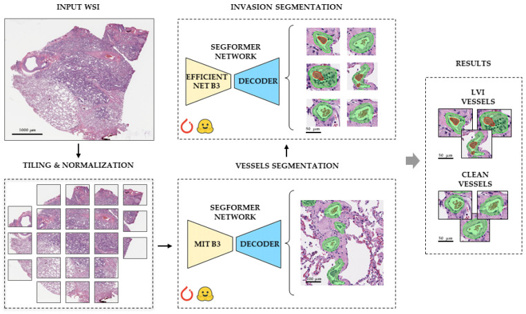 Figure 1
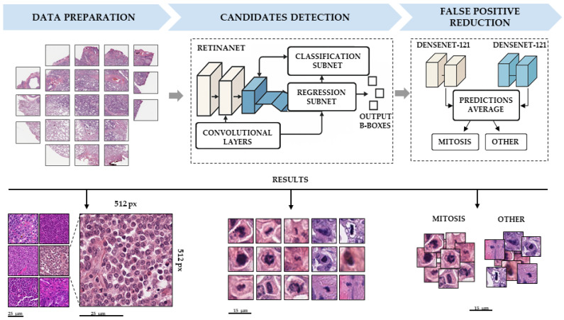 Figure 2
 Figure 3
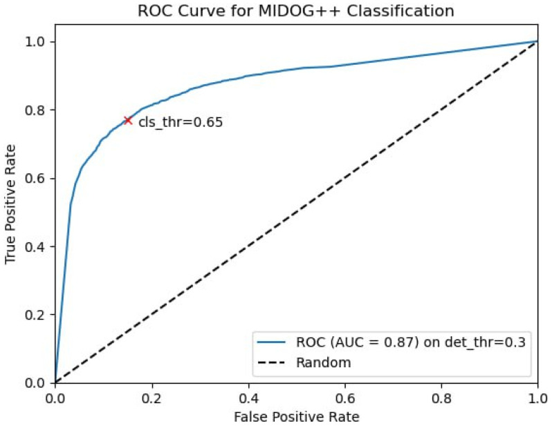 Figure 4
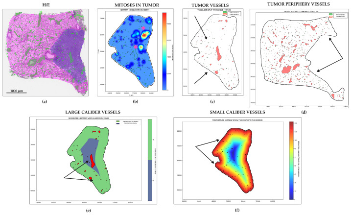 Figure 5
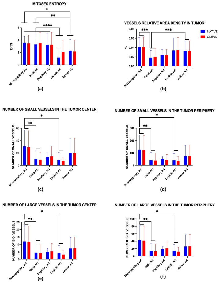 Figure 6
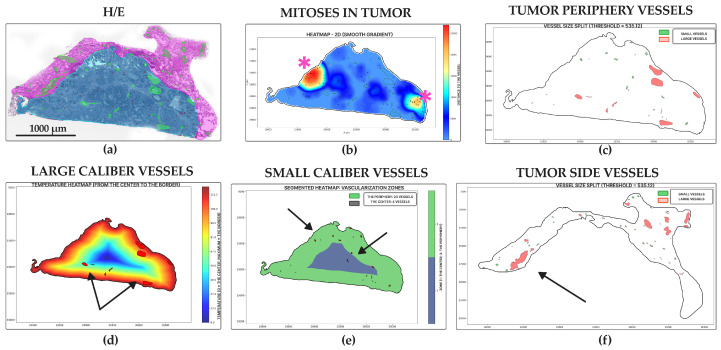 Figure 7
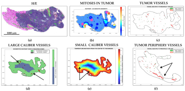 Figure 8
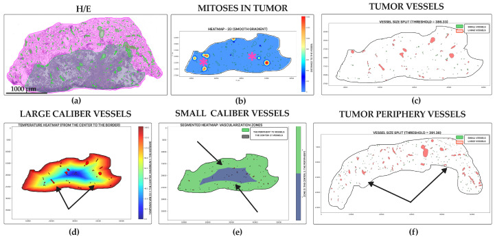 Figure 9
 Figure 10
 Figure 11
 Figure 12
 Figure 13
 Figure 14
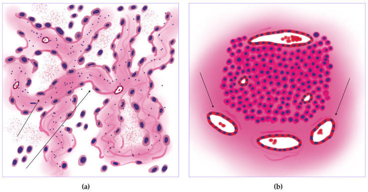 Figure 15
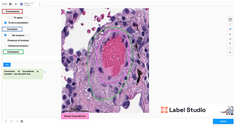 Figure 16
Peer Reviews
No public reviews on file for this paper yet. If you reviewed it on a platform where reviews are public (OpenReview, ICLR, NeurIPS, ICML), you can paste yours below so the community can read it here.
Videos
No videos yet. Explain this paper in a talk, walkthrough, or lecture? Add one.
Taxonomy
TopicsAI in cancer detection · Radiomics and Machine Learning in Medical Imaging · Digital Imaging for Blood Diseases