Magnetisation Transfer 3D-Radial Zero Echo Time MR Imaging at 7T
Mark Symms, Paulina Kozioł, Catarina Rua, Douglas Kelley, Natalia Pietroń, Katarzyna Wiśniewska, Anna Niedziałek, Anna Jamroz-Wiśniewska, Andrzej Stepniewski, Radosław Pietura

TL;DR
A new MRI technique called SilentMT shows promise in detecting brain lesions in multiple sclerosis patients more effectively than traditional methods.
Contribution
The study introduces a novel 3D-radial Zero Echo Time MT-weighted MRI sequence optimized for 7T scanners to detect short-T2 signals in neurodegenerative disorders.
Findings
SilentMT detected MS lesions missed by 7T FLAIR imaging, including in the temporal lobe and brain stem.
Increased Magnetisation Transfer Ratio (MTR) was observed in specific brain regions of an MS patient.
The sequence mitigates RF field inhomogeneity issues at 7T, enabling uniform MTR maps.
Abstract
Background/Objectives: Magnetisation Transfer (MT) MRI is used for neuro-degenerative disorders, including Multiple Sclerosis (MS), providing an indirect measure of large biomolecular MR signal sources which cannot be observed directly because their typical T2 is usually much shorter than the echo time (TE) of conventional MR sequences. We investigated a 3D-radial Zero Time of Echo (ZTE) MT-weighted sequence with potentially enhanced sensitivity to short-T2 MR signals indirectly (via MT weighting) and directly (due to the short TE). Methods: The sequence runs on a human 7T MR scanner, producing whole-brain MT-weighted images with isotropic 0.8 mm resolution in 6.5 minutes. One RF pulse is used to suppress the fat signal and generate MT weighting, reducing RF power deposition to moderate levels. The small excitation pulses and the “quasi-adiabatic” MT pulse mitigate the negative effects…
Genes, proteins, chemicals, diseases, species, mutations and cell lines named across the full text — each resolved to its canonical identifier and authoritative record.
Click any figure to enlarge with its caption.
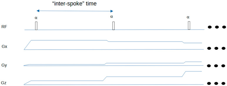 Figure 1
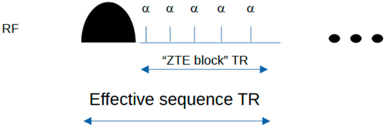 Figure 2
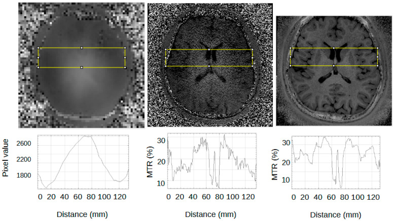 Figure 3
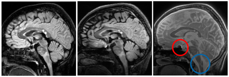 Figure 4
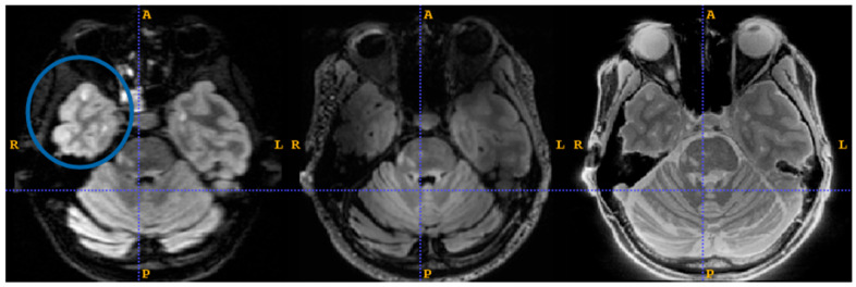 Figure 5
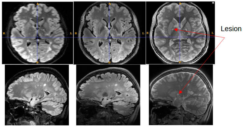 Figure 6
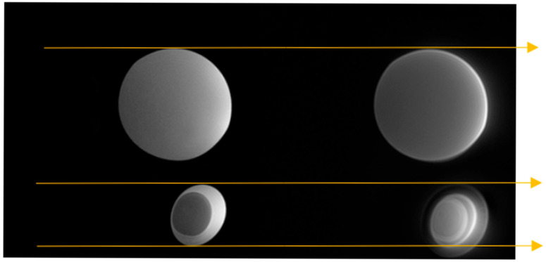 Figure 7
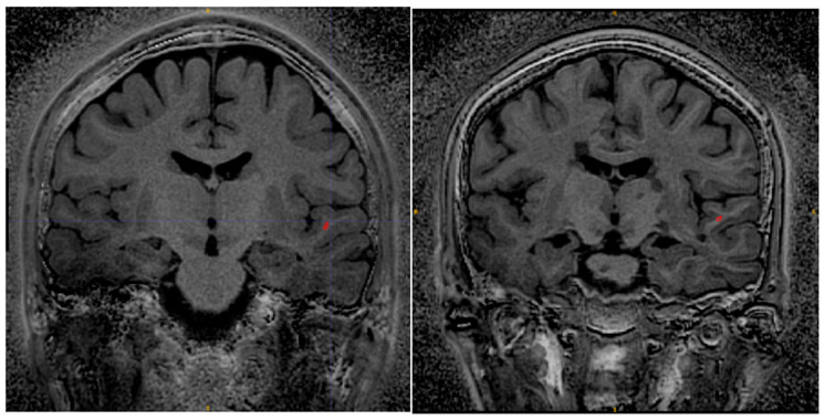 Figure 8
Peer Reviews
No public reviews on file for this paper yet. If you reviewed it on a platform where reviews are public (OpenReview, ICLR, NeurIPS, ICML), you can paste yours below so the community can read it here.
Videos
No videos yet. Explain this paper in a talk, walkthrough, or lecture? Add one.
Taxonomy
TopicsAdvanced MRI Techniques and Applications · Multiple Sclerosis Research Studies · Advanced Neuroimaging Techniques and Applications