Dynamic Distance Mapping Enhances Hallux Valgus Progression Visualization
Dror Robinson, Hamza Murad, Muhammad Khatib, Muhamad Kiwan Mahamid, Eitan Lavon, Mustafa Yassin

TL;DR
A new technique called dynamic distance mapping helps visualize how a foot deformity called hallux valgus progresses, offering insights that could improve surgical planning.
Contribution
The study introduces dynamic distance mapping as a novel method to visualize hallux valgus progression and identify spatial features linked to severity.
Findings
Seven distances correlated negatively and seven positively with hallux valgus angles (HVA), involving the distal phalanx, sesamoids, and second metatarsal.
Eleven distances showed strong positive correlation with intermetatarsal angle (IMA), reflecting displacement patterns.
Dynamic distance mapping visualizations highlighted progressive spatial changes in hallux valgus deformity.
Abstract
Background/Objectives: Hallux valgus (HV), a common foot deformity, is difficult to quantify beyond traditional angular measurements. This study introduces a novel dynamic distance mapping technique to visualize HV progression and identify spatial features linked to severity. Methods: A retrospective analysis of 335 feet from 178 patients undergoing HV surgery at Hasharon Hospital, Israel (2014–2024), utilized custom Python software to annotate 24 landmarks on preoperative standing anteroposterior radiographs. This generated 276 normalized Euclidean distances, analyzed via Pearson correlation against HV angles (HVA, IMA, DMAA, HIA). Results: Seven distances correlated negatively (r > 0.4, p < 0.05) and seven positively with HVA, involving the distal phalanx, sesamoids, and second metatarsal. Eleven distances showed strong positive correlation (r > 0.4, p < 0.05) with IMA, reflecting…
Genes, proteins, chemicals, diseases, species, mutations and cell lines named across the full text — each resolved to its canonical identifier and authoritative record.
Click any figure to enlarge with its caption.
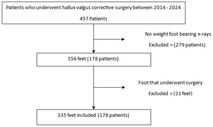 Figure 1
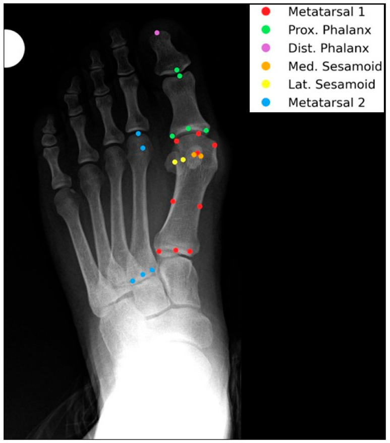 Figure 2
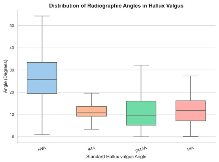 Figure 3
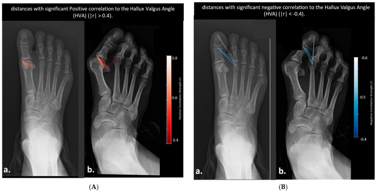 Figure 4
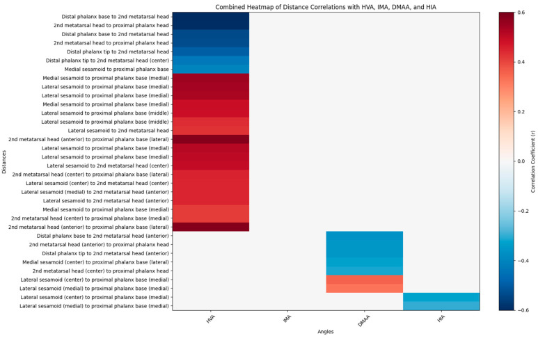 Figure 5
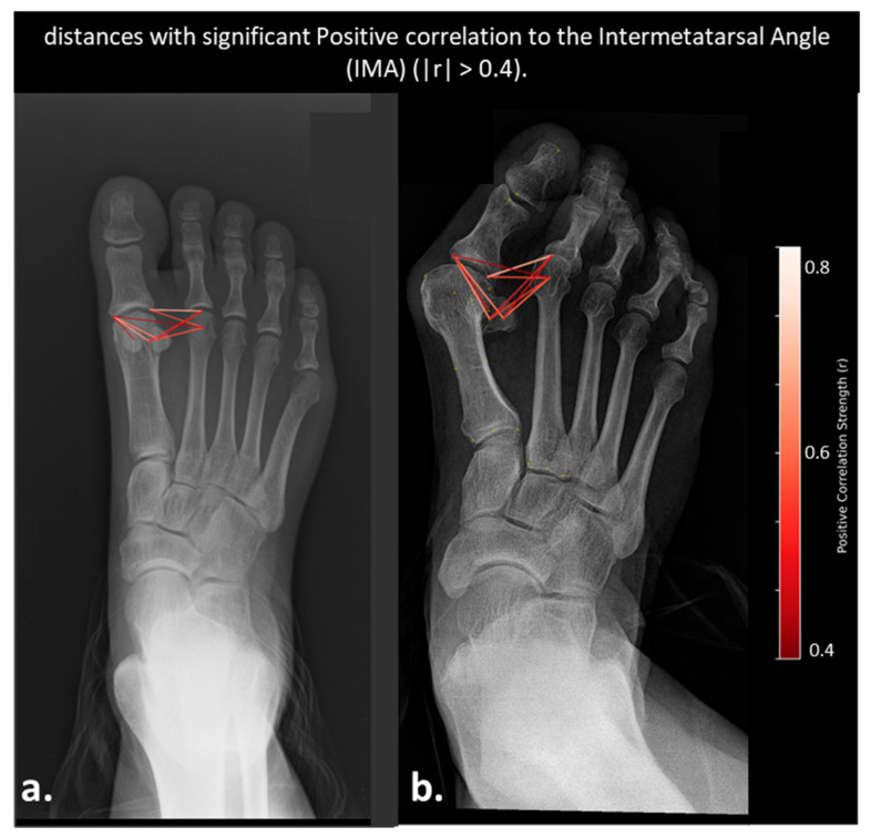 Figure 6
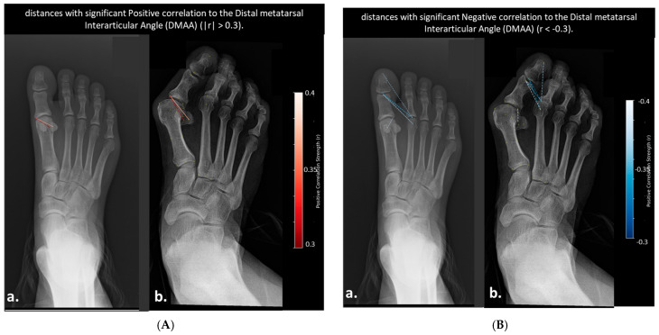 Figure 7
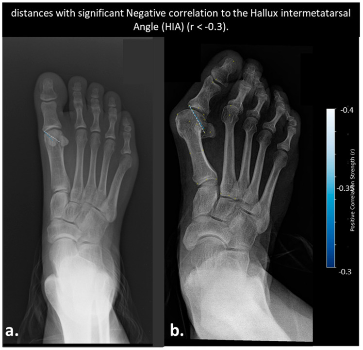 Figure 8
Peer Reviews
No public reviews on file for this paper yet. If you reviewed it on a platform where reviews are public (OpenReview, ICLR, NeurIPS, ICML), you can paste yours below so the community can read it here.
Videos
No videos yet. Explain this paper in a talk, walkthrough, or lecture? Add one.
Taxonomy
TopicsFoot and Ankle Surgery · Lower Extremity Biomechanics and Pathologies · Diabetic Foot Ulcer Assessment and Management