Automated Analysis of the Foveal Avascular Zone in Optical Coherence Tomography Angiography Before and After Phacoemulsification
María S. Pighin, Evangelos Tsiroukis, Agniezska Dyrda, Ignasi Jürgens

TL;DR
This study compares two methods for measuring the foveal avascular zone in eye scans before and after cataract surgery, finding that a machine learning approach is reliable and shows changes after surgery.
Contribution
The study introduces a machine learning-based method for measuring the foveal avascular zone that is comparable to traditional semiautomated algorithms.
Findings
The ML-based and script-based methods showed strong agreement in measuring the foveal avascular zone across three plexuses.
The foveal avascular zone in the superficial vascular plexus decreased significantly after phacoemulsification at 1 and 2 months.
The ML-based method is a practical alternative to the script-based algorithm for measuring the foveal avascular zone.
Abstract
Objective: This study aimed to compare two methods for measuring the foveal avascular zone (FAZ) before and after phacoemulsification: a script-based semiautomated algorithm and a machine learning (ML)-based semiautomated algorithm. Methods: Optical coherence tomography angiography (OCTA) images were obtained with a Spectralis OCTA system (Heidelberg Engineering, Germany) preoperatively and in three postoperative visits. The FAZ was measured using both methods. Results: The study analyzed 708 OCTA scans from 59 eyes. Correlation analyses showed strong agreement between the semiautomated script-based and ML-based methods in the three plexuses, with Pearson correlation coefficients of r = 0.836 (95% CI: 0.74–0.89), r = 0.646 (95% CI: 0.45–0.78), and r = 0.861 (95% CI: 0.78–0.92), respectively (p < 0.0001 for all). In longitudinal analysis, the FAZ in the SVP decreased significantly after…
Genes, proteins, chemicals, diseases, species, mutations and cell lines named across the full text — each resolved to its canonical identifier and authoritative record.
Click any figure to enlarge with its caption.
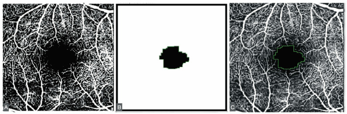 Figure 1
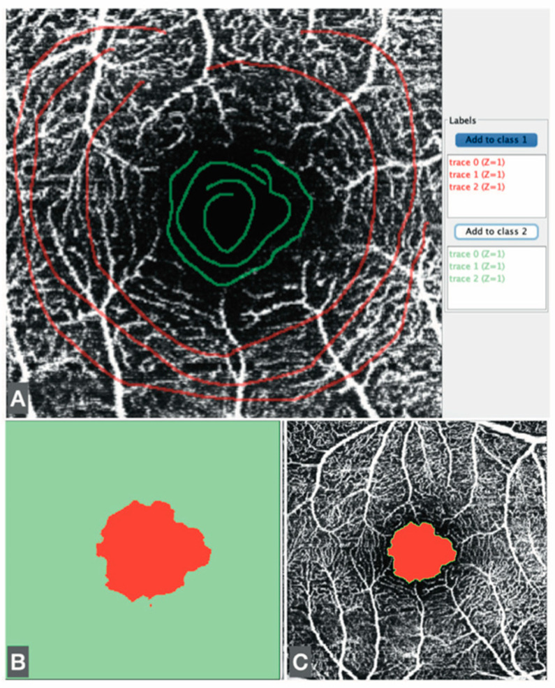 Figure 2
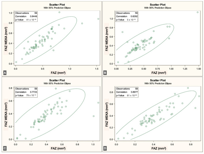 Figure 3
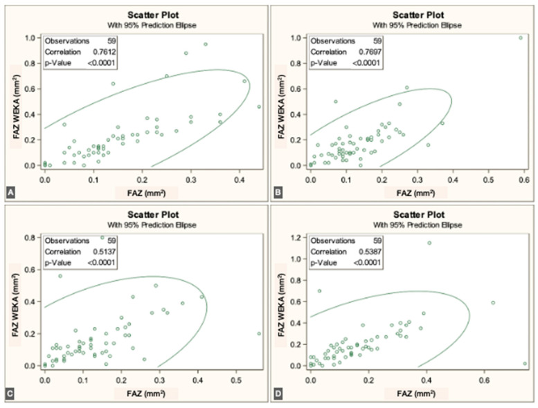 Figure 4
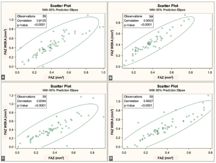 Figure 5
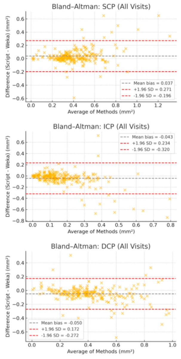 Figure 6
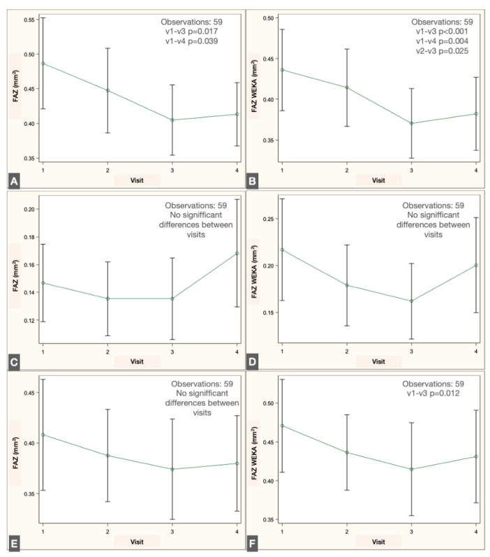 Figure 7
Peer Reviews
No public reviews on file for this paper yet. If you reviewed it on a platform where reviews are public (OpenReview, ICLR, NeurIPS, ICML), you can paste yours below so the community can read it here.
Videos
No videos yet. Explain this paper in a talk, walkthrough, or lecture? Add one.
Taxonomy
TopicsRetinal Diseases and Treatments · Retinal and Macular Surgery · Glaucoma and retinal disorders