Multiple, Segmental, Non-Syndromic Basal Cell Carcinomas—Clinical, Dermoscopic and Histopathological Features
Martyna Sławińska, Beata Zagórska, Wojciech Biernat, Michał Sobjanek

TL;DR
A 72-year-old woman had four non-melanotic skin tumors confirmed as basal cell carcinomas with unique dermoscopic features.
Contribution
This is the first report describing dermoscopic features of segmental/agminated basal cell carcinoma.
Findings
Four amelanotic tumors on the left arm were confirmed as basal cell carcinomas via histopathology.
Dermoscopy revealed polymorphic and arborizing vessels with non-specific malignant features.
No recurrence was observed during a 3-year follow-up period.
Abstract
We present a case of a 72-year-old woman with four amelanotic tumors on the left arm, without a history of skin cancer or sun exposure. Dermoscopy showed polymorphic and arborizing vessels, with some lesions displaying non-specific malignant features. Histopathology confirmed basal cell carcinoma (BCC) in all lesions. No signs of recurrence were observed during 3-year follow-up. Segmental/agminated basal cell carcinoma is a rare differential diagnosis of multiple clustered, painless pink tumors. To the best of our knowledge, this is the first report describing their dermoscopic features.
Genes, proteins, chemicals, diseases, species, mutations and cell lines named across the full text — each resolved to its canonical identifier and authoritative record.
Click any figure to enlarge with its caption.
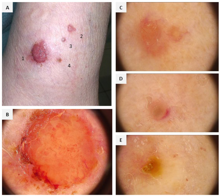 Figure 1
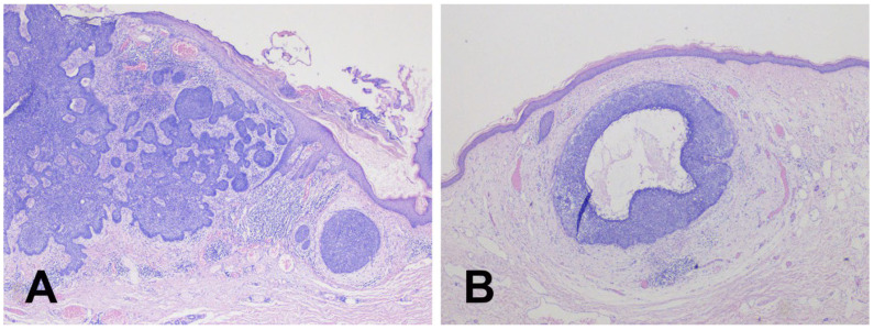 Figure 2
Peer Reviews
No public reviews on file for this paper yet. If you reviewed it on a platform where reviews are public (OpenReview, ICLR, NeurIPS, ICML), you can paste yours below so the community can read it here.
Videos
No videos yet. Explain this paper in a talk, walkthrough, or lecture? Add one.
Taxonomy
TopicsNonmelanoma Skin Cancer Studies · Cancer and Skin Lesions · Cutaneous Melanoma Detection and Management