WDR81 represses IKK-mediated expression of pro-survival genes to regulate apoptosis
Sannoong Hu, Pranav Danthi

TL;DR
WDR81 helps control cell death during virus infection by regulating survival signals linked to the NFκB pathway.
Contribution
WDR81 is newly identified as a regulator of apoptosis via its role in suppressing pro-survival gene expression.
Findings
WDR81 is required for apoptosis induction after reovirus infection.
WDR81-deficient cells show upregulated pro-survival gene expression.
Blocking IKK signaling in WDR81-deficient cells restores normal pro-survival gene expression and susceptibility to cell death.
Abstract
Apoptosis is a common host response to virus infection. The extent and timing of apoptosis following infection is controlled by the balance between the strength of signals that activate death-inducing and survival-promoting pathways in cells. In many cell types, infection with mammalian orthoreovirus (reovirus) results in induction of cell death by apoptosis late in infection. In this study, we uncovered that WD repeat-containing protein 81 (WDR81) is required for apoptosis induction after reovirus infection. The requirement for WDR81 for apoptosis induction is not unique to reovirus because cells lacking WDR81 are also resistant to apoptosis induced by other agonists. We find that in cells deficient in WDR81, expression of several pro-survival genes is upregulated. The expression of these genes is controlled by the inhibitor of κB kinase (IKK) complex-nuclear factor of kB (NFκB)…
Genes, proteins, chemicals, diseases, species, mutations and cell lines named across the full text — each resolved to its canonical identifier and authoritative record.
Click any figure to enlarge with its caption.
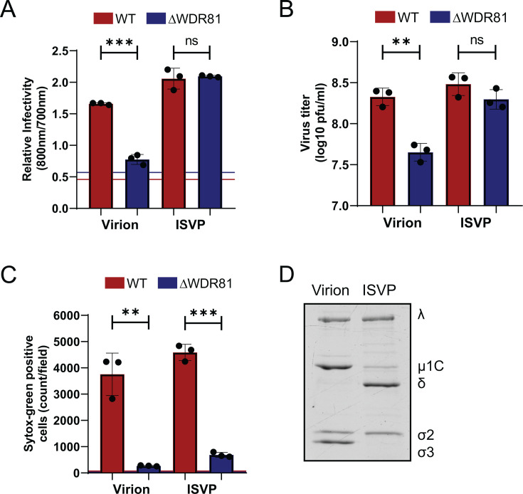 Fig 1
Fig 1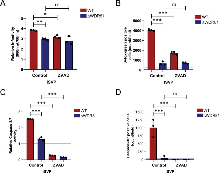 Fig 2
Fig 2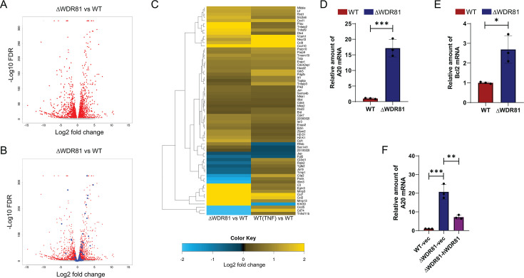 Fig 3
Fig 3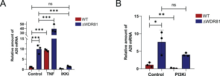 Fig 4
Fig 4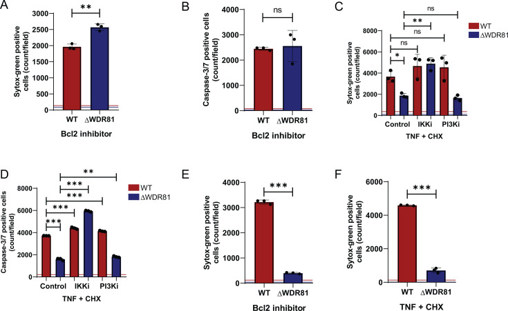 Fig 5
Fig 5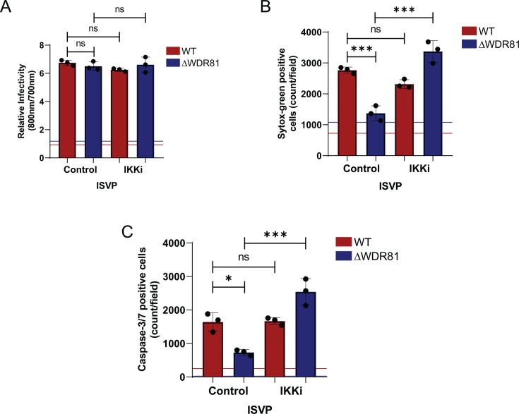 Fig 6
Fig 6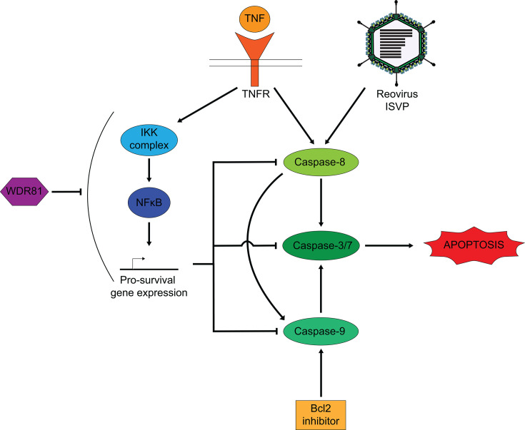 Fig 7
Fig 7- —National Institute of Allergy and Infectious Diseaseshttp://dx.doi.org/10.13039/100000060
Peer Reviews
No public reviews on file for this paper yet. If you reviewed it on a platform where reviews are public (OpenReview, ICLR, NeurIPS, ICML), you can paste yours below so the community can read it here.
Videos
No videos yet. Explain this paper in a talk, walkthrough, or lecture? Add one.
Taxonomy
TopicsViral gastroenteritis research and epidemiology · Virus-based gene therapy research · Animal Virus Infections Studies
INTRODUCTION
Apoptosis is a well-studied form of programmed cell death. Apoptosis is characterized by activation of a cascade of proteases called caspases (1, 2). The activation of these caspases ultimately leads to the death of the cell in a non-inflammatory manner (2). Apoptosis can be triggered by extrinsic or intrinsic pathways. Extrinsic signals such as tumor necrosis factor (TNF) can bind to their receptors and trigger the activation of an initiator caspase, caspase-8. In its activated form, caspase-8 cleaves effector caspases, caspase-3 and caspase-7. The intrinsic pathway involves the mitochondria and results in the activation of caspase-9, which, in turn, also activates effector caspases. Activated effector caspases cleave numerous cellular components, ultimately leading to cell death (2).
Cells express proteins that prevent the spontaneous activation of caspases (2–4). The expression of these pro-survival gene products is usually mediated by the inhibitor of κB kinase (IKK) complex via the nuclear factor of kB (NFκB) transcription factor (3, 4). Thus, in the absence of a cell death trigger, the cell continues to grow and divide without activating caspases. Apoptosis can be triggered to overcome survival signals by extrinsic ligands such as TNF or TNF-related apoptosis-inducing ligand (TRAIL) or by intrinsic imbalances such as stress or intracellular pathogens like viruses (5–7). Virus infection and replication within a cell is often detected by pathogen-associated molecular patterns, which trigger a variety of cellular immune responses, including cell death (5, 8). Mammalian orthoreovirus (reovirus) infection triggers apoptosis (9–11). While reovirus-activated apoptosis is not associated with limiting viral replication within the infected cell, it has been associated with disease pathogenesis in mouse models (12–14). Reovirus-induced apoptosis requires activation of both the extrinsic and the intrinsic apoptotic pathways (11, 15–17). While a few viral and cellular factors are implicated in this process (10, 18–22), the exact manner in which apoptotic pathways are activated in reovirus-infected cells is not understood.
We previously identified a function for a host protein, WD repeat-containing protein 81 (WDR81), in reovirus infection (23). WDR81 regulates endosomal maturation (24). It is present in the phosphoinositide-3-kinase (PI3K) complex on the outer leaflet of early endosomes and inhibits the function of class III PI3Ks, thus allowing the maturation of the early endosome to late endosome (24). In the absence of WDR81, reovirus virions attach to cell surface receptors, reach the endosome, and are disassembled to generate entry intermediates called infectious subvirion particles (ISVPs) by the action of endosomal proteases (23). However, the ISVPs are trapped in the endosomes and fail to enter the cytoplasm and launch infection. When infection is launched with in vitro generated ISVPs, which do not require endosomal uptake, infection proceeds normally (23). These data implicate WDR81 in regulating productive trafficking of reovirus particles through the endosomal pathway during the early stages of reovirus infection.
In this study, we analyzed the role of WDR81 in reovirus-induced programmed cell death. We demonstrate that even though WDR81 is dispensable for infection by reovirus ISVPs, it is required for ISVP-induced apoptosis. We show that in the absence of WDR81, other death agonists also fail to trigger apoptosis. Our data show that the absence of WDR81 results in increased expression of pro-survival genes. We demonstrate that blocking the IKK-NFκB pathway normalizes the expression of these pro-survival genes and restores the capacity of cells to succumb to apoptosis inducers. Together, our study describes a novel function of WDR81 in apoptosis. Furthermore, it reveals a previously unknown connection between WDR81 and the IKK-NFκB signaling pathway.
RESULTS
WDR81 is required for reovirus-induced programmed cell death
WDR81, a host protein critical for early-to-late endosome maturation, plays a key role in cellular trafficking (24). Previous work demonstrated that WDR81 is essential for infection by reovirus virions but dispensable for infection by ISVPs, an entry intermediate generated via in vitro chymotrypsin digestion of virions (23). To confirm this phenotype, control cells (WT mouse embryo fibroblast [MEFs]) and cells deficient in WDR81 (ΔWDR81 MEFs) were infected with reovirus virions or ISVPs. Virus infectivity was measured 24 h post-infection by indirect immunofluorescence. Consistent with previous work (23), reovirus virions showed lower infectivity in ΔWDR81 MEFs compared to WT MEFs, while infection with reovirus ISVPs showed no difference in infectivity between WT and ΔWDR81 MEFs (Fig. 1A). Virus production was also quantified by measuring viral titer 24 h post-infection by plaque assay. Consistent with previous results and indirect immunofluorescence, in comparison to WT MEFs, reovirus virions produced ~0.7 log_10_ lower infectious virus in the ΔWDR81 MEFs, while infection with reovirus ISVPs showed no difference in the amount of infectious titer produced in both cell types (Fig. 1B). To study the role of WDR81 in reovirus-induced programmed cell death, WT MEFs and ΔWDR81 MEFs were infected with reovirus virions or ISVPs and incubated for 48 h. Permeability of host cell membranes to Sytox Green nucleic acid dye, which only stains nuclei of dead or dying cells (25), was evaluated as a measure of cell death. This later time point was chosen as MEFs infected with reovirus do not show detectable cell death at 24 h post-infection (data not shown). When infected with virions, in comparison to WT MEFs, ΔWDR81 MEFs showed significantly fewer dead cells (Fig. 1C). This result was expected as reovirus virions are unable to establish infection or replicate in ΔWDR81 MEFs (Fig. 1A and B). Interestingly, upon infection with reovirus ISVPs, ΔWDR81 MEFs still showed a significantly lower number of Sytox Green-positive cells in comparison to the WT MEFs (Fig. 1C). This result was unexpected since reovirus ISVPs are capable of successful replication in ΔWDR81 MEFs, as shown in Fig. 1A and B. To confirm the in vitro generation of ISVPs from purified virions, virions and ISVPs were resolved on an SDS-PAGE gel (Fig. 1D). The cleavage of µ1C protein to δ and the loss of σ3 protein indicate the successful generation of ISVPs. Together, these results suggest that WDR81 plays a role in reovirus-induced programmed cell death. Because ISVPs efficiently infected ΔWDR81 MEFs, for the remainder of this study, we used ISVPs as a tool to dissect the role of WDR81 in cell death induction.
*WDR81 is required for ISVP-induced cell death. (A) MEFs were adsorbed with virions or ISVPs of reovirus T3DCD at a multiplicity of infection (MOI) of 10 PFU/cell.. Cells were then incubated for 24 h at 37°C. Infectivity was measured in fixed cells by indirect immunofluorescence using LI-COR Odyssey Scanner. Relative infectivity was calculated using the ratio of signal at 800 nm (reovirus) to signal at 700 nm (cells). Horizontal lines represent the ratio for mock-infected cells for each cell type. Error bars represent SD. ***, P < 0.0005 by Student’s t-test. (B) MEFs were adsorbed with virions or ISVPs of reovirus T3DCD at MOI 10 PFU/cell. Cells were incubated at 37°C for 24 h. Virus titer was measured by plaque assay on L929 cells. Error bars represent SD. **, P < 0.005. (C) MEFs were adsorbed with virions or ISVPs of reovirus T3DCD at MOI 10 PFU/cell. Cells were incubated for 48 h in media supplemented with Sytox Green (Sytox G) (50 nM). Cell death was quantified 48 h post-infection by analyzing the number of Sytox G-positive cells per field. Horizontal lines represent signals for mock-infected cells for each cell type. Error bars represent SD. ***, P < 0.0005 and *, P < 0.005 by Student's t-test. (D) Purified T3DCD virions (2 × 10¹¹ particles) were incubated with 200 µg/mL Na-p-tosyl-L-lysine chloromethyl ketone-treated chymotrypsin at 32°C for 30 min. The reaction was terminated by the addition of 1 mM phenylmethylsulfonyl fluoride. Purified virions and ISVPs were analyzed by stain-free 10% SDS-PAGE. Reovirus protein bands are labeled.
WDR81 is required for reovirus-induced caspase-3/7 activity
Reovirus induces cell death either through apoptosis, involving both the intrinsic and the extrinsic pathways, or necroptosis, mediated by receptor-interacting protein kinase 3 (RIPK3) and mixed lineage kinase domain like pseudokinase (MLKL) activation (9, 26). Reovirus infection of MEFs produces morphological and biochemical changes in cells that resemble apoptosis (22). To determine whether reovirus infection induces apoptosis in MEFs, we assessed the effect of ZVAD-fmk (27), a broad-spectrum caspase inhibitor, on cell death 48 h post-infection. Consistent with data shown in Fig. 1, a minimal difference was observed between infection of WT and ΔWDR81 cells with ISVPs. ZVAD-fmk treatment did not reduce ISVP infectivity in either cell type to a level that affects infectious virus output (Fig. 2A and not shown). We then assessed the ability of ZVAD-fmk to inhibit cell death induced by ISVPs and found that treatment with ZVAD-fmk significantly reduced the activation of cell death in WT MEFs (Fig. 2B). These data suggest that MEFs undergo cell death by apoptosis, following infection with reovirus ISVPs. Because ΔWDR81 cells fail to undergo cell death following infection by ISVPs, our results suggest that WDR81 is required for efficient induction of apoptosis. A hallmark of apoptosis is the activation of effector caspases—caspase-3 and caspase-7. To determine whether effector caspases are activated, WT and ΔWDR81 MEFs were infected with ISVPs. The activity of effector caspases in cell lysates at 48 h post-infection was measured using a caspase-3/7 substrate which shows enhanced luminescence upon cleavage. ISVP infection of WT MEFs resulted in a ~2.5-fold increase in caspase-3/7 activity, compared to only a ~1.5-fold increase in ΔWDR81 MEFs (Fig. 2C). Caspase-3/7 activation was also quantified using a cell-permeable substrate that fluoresces upon cleavage by active caspases. Upon infection of WT MEFs with reovirus ISVPs, we detected a large number of cells positive for caspase-3/7 activity. In contrast, ΔWDR81 MEFs exhibited markedly fewer cells with active caspase-3/7 (Fig. 2D). These data suggested that WDR81 is required for ISVP-induced caspase-3/7 activation. To confirm the effectiveness of ZVAD-fmk, we also assessed caspase activation by ISVPs in the presence of ZVAD-fmk. ZVAD-fmk was efficiently able to inhibit caspase-3/7 activation (Fig. 2C and D). Together, these data suggest that ISVPs trigger caspase-mediated apoptosis in MEFs and that WDR81 is required for this process.
*WDR81 regulates ISVP-induced caspase-3/7 activation. (A) MEFs were adsorbed with reovirus T3DCD ISVPs at an MOI of 10 PFU/cell. Cells were then incubated with either dimethyl sulfoxide (DMSO) control or ZVAD-fmk (20 µM) for 24 h at 37°C. Infectivity was measured in fixed cells by indirect immunofluorescence using LI-COR Odyssey Scanner. Relative infectivity was calculated using the ratio of signal at 800 nm (reovirus) to signal at 700 nm (cell control). Horizontal lines represent signals for mock-infected cells. Error bars represent SD. **, P < 0.005; *, P < 0.05, by Student’s t-test. (B) MEFs were adsorbed with reovirus T3DCD ISVPs at an MOI of 10 PFU/cell. Cells were incubated for 48 h in media with either DMSO control or ZVAD-fmk (20 µM) and with Sytox G (50 nM). Cell death was estimated 48 h post-infection by analyzing the number of Sytox G-positive cells per field. Horizontal lines represent signals for mock-infected cells. Error bars represent SD. ***, P < 0.0005 by Student's t-test. (C) MEFs were adsorbed with reovirus T3DCD ISVPs at a n MOI of 10 PFU/cell. Cells were incubated for 48 h in media with either DMSO control or ZVAD-fmk (20 µM). Caspase-3/7 activity was measured using Promega Caspase-3/7 Glo kit as per manufacturer’s instructions. Relative caspase-3/7 activity was calculated as the ratio of luminescence signal compared to background signal in mock-infected cells. A horizontal line represents a mock signal set to 1. Error bars represent SD. ***, P < 0.0005 by Student’s t-test. (D) MEFs were adsorbed with reovirus T3DCD ISVPs at at an MOI of 10 PFU/cell. Cells were incubated for 48 h in media with either DMSO control or ZVAD-fmk (20 µM) and with IncuCyte Caspase-3/7 dye (1:2,000). The number of cells showing caspase-3/7 activation per field was assessed by fluorescence microscopy. Horizontal lines represent signals for mock-infected cells. Error bars represent SD. **, P < 0.0005 by Student’s t-test.
WDR81-deficient cells express genes that are also induced by TNF treatment
We hypothesized that the resistance of ΔWDR81 MEFs to ISVP-induced apoptosis might result from altered expression of genes regulating cell death and survival. Gene expression differences between WT and ΔWDR81 MEFs were compared by RNA-seq. RNA-seq analysis, using a false discovery rate (FDR) cutoff of <0.0001, revealed numerous genes differentially expressed between WT and ΔWDR81 MEFs (Fig. 3A). Ingenuity pathway analysis (28) of differential gene expression revealed that WDR81 deficiency alters gene expression across multiple cellular pathways, including—but not limited to—those governing pathogen responses and cell-based immunity (data not shown). The large-scale, bidirectional changes in gene expression between WT and ΔWDR81 cells made it challenging to use these analyses to identify specific pathways driving the resistance of ΔWDR81 cells to ISVP-induced apoptosis. Many constitutively expressed host genes promote cell survival by repressing the activation of pathways of apoptosis (29). One reason for the reduced ability of ΔWDR81 MEFs to undergo apoptosis following ISVP infection could be because such pro-survival genes are expressed at a higher level in these cells at a basal level. In most cells, including MEFs, pro-survival genes can be upregulated by treatment with TNF (29). We therefore identified those genes whose expression is differentially regulated by TNF treatment (Fig. 3B). We identified 107 TNF-responsive genes in WT cells (using the same stringent FDR cutoff). We then asked if these genes were also differentially expressed in ΔWDR81 MEFs. Notably, 66 of 107 TNF-induced genes were differentially expressed at a basal level in untreated ΔWDR81 MEFs compared to untreated WT MEFs (Fig. 3C). To validate the differential gene expression analysis, transcript levels of A20 and Bcl2—two TNF-induced genes—were assessed by RT-qPCR (30, 31). RT-qPCR analysis confirmed that ΔWDR81 MEFs exhibit ~10-fold higher A20 mRNA levels and ~2.5-fold higher Bcl2 mRNA levels compared to WT MEFs (Fig. 3D and E). These findings indicate that ΔWDR81 MEFs exhibit elevated basal expression of genes typically induced by TNF. We have previously reported that expression of human WDR81 in ΔWDR81 MEFs restores virus infection and virus-induced cell death (23). While these data indicated that the phenotype observed in these cells is related to the absence of WDR81 and not an off-target effect, we sought to confirm that the unexpected effect of WDR81 loss on the expression of pro-survival genes is also restored by re-expression of WDR81. Trans-complementation of human WDR81 in ΔWDR81 cells significantly reduced the levels of A20 mRNA compared to control ΔWDR81 cells (Fig. 3F). Together, these data suggest that in the absence of WDR81, cells have higher levels of pro-survival genes that are typically upregulated by TNF.
*In the absence of WDR81, pro-survival gene expression is upregulated. (A) Total RNA was harvested from MEFs, and gene expression was analyzed by RNA-seq. A volcano plot of genes differentially expressed in WT and ΔWDR81 is shown. Genes that are differentially expressed with FDR <0.0001 are shown in red. (B) Genes in the volcano plot shown in A that are also differentially expressed in WT and TNF-treated WT cells are marked in blue. (C) Heat map of 66 genes that are differentially expressed both in ΔWDR81 and WT MEFs as well as in TNF-treated and control WT cells is shown. A scale bar showing log2 fold change in gene expression is included. (D) MEFs were grown in a 24-well plate, and total mRNA was extracted using Bio-Rad Aurum Total RNA mini kit according to manufacturer’s protocol. cDNA was synthesized using Applied Biosystems High-Capacity cDNA Reverse Transcription kit. qPCR for A20 gene expression was done using Applied Biosystems StepOnePlus Real-Time PCR system. A20 gene expression was calculated using the 2-ΔΔCT method using glyceraldehyde-3-phosphate dehydrogenase (GAPDH) as internal control. Error bars represent SD. ***, P < 0.0005 by Student’s t-test. (E) MEFs were grown in a 24-well plate, and total mRNA was extracted using Bio-Rad Aurum Total RNA mini kit according to manufacturer’s protocol. cDNA was synthesized using Applied Biosystems High-Capacity cDNA Reverse Transcription kit. qPCR for Bcl2 gene expression was done using Applied Biosystems StepOnePlus Real-Time PCR system. Bcl2 gene expression was calculated using the 2-ΔΔCT method using GAPDH as internal control. Error bars represent SD. *, P < 0.05 by Student's t-test. (F) MEFs were grown in standard media as described in Materials and Methods. Total mRNA was extracted using Bio-Rad Aurum Total RNA mini kit according to manufacturer’s protocol. cDNA was synthesized using Applied Biosystems High-Capacity cDNA Reverse Transcription kit. qPCR for A20 gene expression was done using Applied Biosystems StepOnePlus Real-Time PCR system. A20 gene expression was calculated using the 2-ΔΔCT method using GAPDH as internal control. Error bars represent SD. ***, P < 0.0005, *, P < 0.005, by one-way analysis of variance.
Pro-survival gene expression in ΔWDR81 MEFs depends on IKK activity
After engaging its receptor on the cell surface, TNF signals via the IKK complex to activate NFκB and drive expression of its genomic targets (29). Given the similarity between TNF-induced genes and those upregulated in ΔWDR81 MEFs, we hypothesized that IKK signaling may regulate pro-survival gene expression observed in WDR81-deficient cells. Treatment with an IKK inhibitor significantly reduced A20 mRNA levels in ΔWDR81 MEFs (Fig. 4A). These data suggest that at least some of the genes that are differentially expressed in the absence of WDR81 are controlled by IKK signaling. WDR81 negatively regulates endosomal type III PI3K, which generates phosphatidylinositol 3-phosphates (PtdIns3P) involved in endosomal signaling (24). Consequently, ΔWDR81 cells are expected to exhibit elevated PI3K activity. To assess if higher PI3K activity plays a role in higher pro-survival gene expression, WT and ΔWDR81 MEFs were treated with broad-spectrum PI3K inhibitor LY294002 (32). Treatment of ΔWDR81 MEFs with LY294002 at functional concentrations (data not shown) did not reduce A20 mRNA levels (Fig. 4B), indicating that elevated PI3K activity does not contribute to increased pro-survival gene expression in the absence of WDR81. These findings together indicate that signaling via IKK but not PI3K drives the upregulation of certain pro-survival genes in WDR81-deficient cells.
*Higher A20 mRNA levels in WDR81-deficient cells are driven by IKK but not PI3K signaling. (A) MEFs were incubated in media with either DMSO control, TNF (50 ng/mL), or IKK inhibitor (5 µM) for 3 h. Total mRNA was extracted using Bio-Rad Aurum Total RNA mini kit according to manufacturer’s protocol. cDNA was synthesized using Applied Biosystems High-Capacity cDNA Reverse Transcription kit. qPCR for A20 gene expression was done using Applied Biosystems StepOnePlus Real-Time PCR system. A20 gene expression was calculated using the 2-ΔΔCT method using GAPDH as internal control. Error bars represent SD. ***, P < 0.0005 by one-way analysis of variance (ANOVA). (B) MEFs were incubated in media with DMSO control or PI3K inhibitor (25 µM) for 3 h. Total mRNA was extracted using Bio-Rad Aurum Total RNA mini kit according to manufacturer’s protocol. cDNA was synthesized using Applied Biosystems High-Capacity cDNA Reverse Transcription kit. qPCR for A20 gene expression was done using Applied Biosystems StepOnePlus Real-Time PCR system. A20 gene expression was calculated using the 2-ΔΔCT method using GAPDH as internal control. Error bars represent SD. **, P < 0.005, , P < 0.05 by one-way ANOVA.
WDR81 controls the activation of the multiple pathways of apoptosis
TNF signaling via IKKs drives the expression of NFκB-dependent genes that enhance cell survival (29). We hypothesized that the upregulated pro-survival genes in ΔWDR81 cells may confer resistance to apoptosis induced by other triggers. ABT-737 inhibits the Bcl2 family of antiapoptotic proteins (33). Since the Bcl2 family of proteins typically inhibits the activation of intrinsic apoptotic pathways, ABT-737 treatment results in mitochondrial cytochrome c release, caspase-9 activation, and cell death. To test if WDR81 is required for apoptosis via the intrinsic pathway, we treated WT and ΔWDR81 MEFs with ABT-737 for 24 h. Treatment with ABT-737 resulted in comparable levels of cell death (Fig. 5A) and caspase-3/7 activation (Fig. 5B) in both WT and ΔWDR81 MEFs. These results indicate that in MEFs, WDR81 is dispensable for apoptosis induced by the intrinsic pathway. We next tested if WDR81 is required for extrinsic trigger-induced apoptosis. To evaluate this, we used TNF and cycloheximide (CHX) to induce death in WT and ΔWDR81 MEFs. This combination is a well-characterized inducer of death receptor-mediated, extrinsic apoptosis (34). While neither TNF nor CHX alone altered cell survival (data not shown), upon treatment with the combination for 24 h, WT MEFs succumbed to cell death. In contrast, ΔWDR81 MEFs showed significantly lower numbers of dead cells (Fig. 5C). Similarly, TNF-CHX treatment induced caspase-3/7 activity in WT MEFs, whereas it failed to do so in ΔWDR81 MEFs (Fig. 5D). Thus, in MEFs, WDR81 is required for apoptosis induced by extrinsic triggers.
*WDR81 controls apoptosis induction via intrinsic and extrinsic pathways. (A) MEFs were incubated in media with DMSO control or Bcl2 inhibitor (5 µM) and Sytox Green (Sytox G) (50 nM) for 24 h. Cell death was quantified by analyzing the number of Sytox G-positive cells per field. Horizontal lines represent signals for control DMSO-treated cells. Error bars represent SD. **, P < 0.005. (B) MEFs were incubated in media with DMSO control or Bcl2 inhibitor (5 µM) and IncuCyte Caspase-3/7 dye (1:2,000) for 24 h. The number of cells showing caspase-3/7 activation per field was assessed by fluorescence microscopy. Horizontal lines represent signals for control DMSO-treated cells. Error bars represent SD. (C) MEFs were incubated in media supplemented with TNF (50 ng/mL) + CHX (10 µg/mL) and either DMSO control, IKK inhibitor (5 µM), or PI3K inhibitor (25 µM) for 24 h. Cell death was quantified by analyzing the number of Sytox G-positive cells per field. Horizontal lines represent signals for control DMSO-treated cells. Error bars represent SD. **, P < 0.005, *, P < 0.05 by one-way ANOVA (D) MEFs were incubated in media supplemented with IncuCyte Caspase-3/7 dye (1:2,000) and TNF (50 ng/mL) + CHX (10 µg/mL) and either DMSO control, IKK inhibitor (5 µM), or PI3K inhibitor (25 µM) for 24 h. The number of cells showing caspase-3/7 activation per field was assessed by fluorescence microscopy. Horizontal lines represent signals for control DMSO-treated cells. Error bars represent SD. ***, P < 0.0005 by one-way ANOVA. (E) HT1080 cells were incubated in media containing Sytox G (50 nM) and Bcl2 inhibitor (5 µM) for 48 h. Cell death was quantified by analyzing the number of Sytox G-positive cells per field. Horizontal lines represent signals for control DMSO-treated cells. Error bars represent SD. ***, P < 0.0005 by one-way ANOVA. (F) HT1080 cells were incubated in media containing Sytox G (50 nM) and TNF (50 ng/mL) + CHX (10 µg/mL) for 24 h. Cell death was quantified by analyzing the number of Sytox G-positive cells per field. Horizontal lines represent signals for control DMSO-treated cells. Error bars represent SD. **, P < 0.0005 by one-way ANOVA.
Since IKK and NFκB drive expression of pro-survival genes in ΔWDR81 cells, we assessed the impact of IKK inhibition on TNF-CHX-induced cell death. We found that treatment with the IKK inhibitor restored TNF-CHX-induced cell death and caspase-3/7 activation in ΔWDR81 MEFs to levels comparable to WT MEFs (Fig. 5C and D). Thus, the resistance to extrinsic agonist-triggered apoptosis in ΔWDR81 cells is driven by elevated IKK activity. Treatment with the PI3K inhibitor LY294002 to reduce elevated PI3K signaling in ΔWDR81 cells did not restore TNF-CHX-induced cell death or caspase-3/7 activation in ΔWDR81 MEFs (Fig. 5C and D), even under conditions where the inhibitor was confirmed to be effective (data not shown). These results indicate that elevated PI3K activity does not contribute to the resistance of ΔWDR81 MEFs to extrinsic apoptosis. Furthermore, our results suggest that the elevated PI3K activity in the absence of WDR81 does not appear to impact IKK activity.
To assess the impact of WDR81 in other cell types, we compared cell death induction by ABT-737 and TNF-CHX in control and WDR81-deficient HT1080 cells, a human fibrosarcoma cell line. While control HT1080 cells succumbed to ABT-737 at 48 h post-treatment, ΔWDR81 HT1080 cells were resistant to cell death induced by this drug in this time frame (Fig. 5E). Similarly, whereas control HT1080 cells undergo cell death after TNF-CHX treatment, ΔWDR81 HT1080 cells remain viable (Fig. 5F). These data indicate that in HT1080 cells, WDR81 controls apoptosis by both the intrinsic and the extrinsic pathways. Collectively, our evidence indicates that WDR81 is required for efficient cell death in multiple cell types.
WDR81-deficient cells support ISVP-induced apoptosis when IKK activity is blocked
Apoptosis induction by reovirus requires death receptor signaling via TRAIL (17). Additionally, for efficient cell death induction in MEFs, the death signals need to be amplified by tBid-mediated activation of the intrinsic apoptotic pathway (15). Given that IKK inhibition restores cell death in TNF-CHX-treated cells, we asked if it would also restore cell death in ISVP-infected cells. For these experiments, WT and ΔWDR81 MEFs were infected with reovirus ISVPs and subsequently incubated in the presence of IKK inhibitor. Infectivity measurements using indirect immunofluorescence revealed no significant differences in infection levels between dimethyl sulfoxide (DMSO)-treated and IKK inhibitor-treated cells (Fig. 6A), confirming that the IKK inhibitor does not impact viral replication. We measured cell death and caspase-3/7 activation by ISVPs 48 h post-infection in the presence of an IKK inhibitor. In control, DMSO-treated cells, ISVP infection induced a substantial level of apoptotic death in WT MEFs, while ΔWDR81 MEFs showed minimal cell death, consistent with our earlier observations (Fig. 6B). Although IKK inhibition had no effect on ISVP-induced apoptosis in WT MEFs, it fully restored apoptosis in ΔWDR81 MEFs to levels comparable to WT MEFs (Fig. 6B). Similarly, in the presence of IKK inhibitor, ISVPs induced caspase-3/7 activation in an equivalent number of cells to WT MEFs (Fig. 6C). Together, these results demonstrate that IKK-driven pro-survival gene expression in ΔWDR81 MEFs prevents ISVP-induced caspase-3/7 activation and apoptosis.
*Resistance to ISVP-induced cell death in ΔWDR81 MEFs can be overcome by blocking IKK activity. (A) MEFs were adsorbed with reovirus T3DCD ISVPs at an MOI of 10 PFU/cell. Cells were then incubated for 24 h at 37°C in the presence of DMSO control or IKK inhibitor (5 µM). Infectivity was measured in fixed cells by indirect immunofluorescence using LI-COR Odyssey Scanner. Relative infectivity was calculated using the ratio of signal at 800 nm (reovirus) to signal at 700 nm (cell). Horizontal lines represent signals for mock-infected cells. Error bars represent SD. (B) MEFs were adsorbed with reovirus T3DCD ISVPs at an MOI of 10 PFU/cell. Cells were then incubated for 48 h at 37°C in the presence of Sytox Green (Sytox G) (50 nM) and either DMSO control or IKK inhibitor (5 µM). Cell death was quantified by analyzing the number of Sytox Green-positive cells per field. Horizontal lines represent signals for mock-infected cells. Error bars represent SD. ***, P < 0.0005 by one-way analysis of variance (ANOVA). (C) MEFs were adsorbed with reovirus T3DCD ISVPs at MOI 10 PFU/cell. Cells were then incubated for 48 h at 37°C in the presence of IncuCyte Caspase-3/7 dye (1:2,000) and either DMSO control or IKK inhibitor (5 µM). The number of cells showing caspase-3/7 activation per field was assessed by fluorescence microscopy. Horizontal lines represent signals for mock-infected cells. Error bars represent SD. **, P < 0.0005 by one-way ANOVA.
DISCUSSION
In this study, we uncover a novel role of the protein WDR81 in regulating the apoptosis pathway in cell culture. We show that WDR81 is required for the induction of apoptosis. In the absence of WDR81, cells infected with reovirus or treated with inducers of apoptosis fail to activate caspase-3/7 and succumb to cell death ([Fig. 1 and 5](#F1 F5)). These data suggest that WDR81 broadly regulates apoptosis. In the absence of WDR81, although caspase-3/7 can be activated (Fig. 5), their activation is impaired due to the upregulation of pro-survival signals (Fig. 4). Our data indicate that a large majority of these upregulated pro-survival signals are those that are regulated by the IKK-NFκB pathway (Fig. 3 to 6). Inhibition of the IKK pathway is sufficient to overcome WDR81-dependent deficiencies in caspase-3/7 activation and death ([Fig. 5 and 6](#F5 F6)). Based on these data, we propose a model (Fig. 7) where WDR81 is responsible for suppressing the expression of IKK-NFκB-driven pro-survival genes. Our data suggests that in the absence of WDR81, upregulated pro-survival genes prevent efficient induction of apoptosis. This work highlights a novel association between WDR81 and the IKK-NFκB-mediated cell survival pathway.
Proposed model for the role of WDR81 in reovirus and TNF-induced apoptosis. Infection with reovirus ISVPs induces apoptosis through the activation of caspase-8, caspase-9, and caspase-3/7. TNF treatment upregulates pro-survival gene expression through the IKK-NFκB pathway, which inhibits its ability to trigger apoptosis. Treatment of cells with TNF + CHX (which inhibits translation of pro-survival genes) allows TNF to activate apoptosis. Our model proposes that WDR81 suppresses basal IKK-NFκB-mediated pro-survival genes. In its absence (ΔWDR81 MEFs), IKK-NFκB-mediated pro-survival genes are basally upregulated, thus preventing reovirus ISVPs, TNF + CHX, or Bcl2 inhibitor from activating apoptosis.
WDR81 is a cellular protein that inhibits class III PI3Ks on the early endosomal membrane (24). This function regulates the efficient conversion of early to late endosomes and thus cellular endosomal trafficking. Cells lacking WDR81 have delayed early-to-late endosome conversion and lysosomal turnover of plasma membrane proteins such as epidermal growth factor receptor is impaired (24). Since PI3K inhibitors were not able to rescue cell death in ΔWDR81 MEFs, we think that the cell survival controlling function of WDR81 is independent of its role as a PI3K inhibitor. WDR81 can interact with p62 and LC3C promoting autophagy (35). In the absence of WDR81, there is an increase in protein aggregation and reduced autophagic clearance (35). It is possible that in the absence of WDR81, there is decreased autophagic turnover of an activator of the IKK-NFκB pathway. While we have shown the role of WDR81 in regulating cell death in two cell types (MEFs and HT1080 cells), it is possible that in some cell types, the role of WDR81 in regulating death is absent or even reversed. Mutations in WDR81 have been associated with increased cell death in Purkinje cells of mice, resulting in improper motor abilities (36).
Previous work has shown that NFκB signaling mediated through the IKK complexes is required for reovirus-induced death (37, 38). These data were obtained using cells deficient in IKK or NFκB subunits. Reovirus affects NFκB in two phases. In the first phase, infection triggers NFκB activation, and in the second stage, NFκB activity is inhibited (39). It is thought that NFκB inhibition later in infection decreases the concentration of survival factors in the cell, thereby sensitizing them to cell death (39). Since WDR81-deficient cells have constitutively high IKK-NFκB activity, they allow us to explore the biological effect of not being able to effectively block NFκB later in infection. Our work demonstrating that WDR81-deficient cells are resistant to apoptosis supports the idea that the second stage inhibition of NFκB is required for cell death. It is possible NFκB inhibition later in infection occurs in a cell type-specific manner. Such a possibility may account for the organ-specific role of NFκB in reovirus pathogenesis (40).
Reovirus triggers death via TRAIL-mediated signaling. In reovirus-infected cells, TRAIL signaling promotes caspase-8 activation (17). But caspase-8 activation is not sufficient to induce apoptosis. Apoptosis induction after reovirus infection requires caspase-8-mediated cleavage of Bid (15). Cleaved Bid (tBid) engages the mitochondrial apoptotic pathway to induce sufficient effector caspase activation to induce cell death (41). TNF-CHX shares some similarities with cell death signaling with reovirus since it also induces caspase-8 activation. Following TNF-CHX treatment, whether amplification of death signals via the intrinsic mitochondrial apoptotic pathway is needed for cell death may vary with cell type (42). Our results also indicate that WDR81-deficient MEFs are capable of undergoing cell death following direct activation of the intrinsic pathway through inhibition of the Bcl2 family of anti-apoptotic proteins. Thus, in MEFs, the effect of WDR81 on cell death appears to be restricted to events in death receptor signaling that lead to caspase-8 activation. In contrast, we found that in HT1080 cells, WDR81 is required for the activation of both the intrinsic and the extrinsic apoptotic pathways (Fig. 5E and F). Consistent with other work (29, 43), our RNA-seq data indicate that TNF drives the expression of multiple pro-survival genes. Many of these genes are also expressed at higher levels in cells lacking WDR81. It is possible that the different impact of WDR81 deficiency on the intrinsic apoptotic pathway in MEFs and HT1080 cells relates to differences in the set of pro-survival genes expressed in these two cell types or the extent to which they are expressed. It is also possible that the threshold of pro-survival gene expression needed to block cell death in each of these cell types is different. Currently, it remains unknown which pro-survival genes impact cell death induction during infection with reovirus, TNF, or Bcl2 inhibitor treatment. In some cases, a single target, A20, is sufficient to protect cells from TNF-induced cell death (29). While our data demonstrate that A20 is expressed at a higher level in the absence of WDR81, whether the enhanced expression of A20 alone is sufficient to prevent activation of reovirus- or TNF-induced extrinsic apoptotic pathways in WDR81-deficient cells remains unknown. Whether a single pro-survival gene can prevent the induction of the intrinsic apoptotic pathway also is unidentified. These studies remain a subject of our investigation.
MATERIALS AND METHODS
Cells and viruses
Murine L929 cells (spinner cells) were grown at 37°C in Joklik’s minimal essential medium (Lonza) supplemented with 5% fetal bovine serum (FBS) (Life Technologies), 2 mM L-glutamine (Invitrogen), 0.5 U/mL penicillin, 50 µg/mL streptomycin (Sigma Aldrich), and 25 ng/mL amphotericin B (Sigma-Aldrich). MEFs containing non-targeting plasmid (WT MEFs) and cells containing lentiCRISPRv2 vector targeting WDR81 (ΔWDR81 MEFs) were maintained at 37°C in Dulbecco’s modified Eagle medium (DMEM) (Gibco) supplemented with 10% FBS, 2 mM L-glutamine, and 2 µg/mL puromycin (Invivogen). WT MEFs transduced with vector (WT-vec), ΔWDR81 MEFs transduced with vector (ΔWDR81-vec), and ΔWDR81 MEFs transduced with human WDR81 (ΔWDR81-hWDR81) were maintained at 37°C in DMEM (Gibco) supplemented with 10% FBS, 2 mM L-glutamine, 2 µg/mL puromycin (Invivogen), and 8 µg/mL blasticidin (Invivogen). ACE2-expressing HT1080 cells (WT HT1080) and their WDR81-deficient counterparts (ΔWDR81 HT1080), a gift from Dr. Paul Bieniasz, Rockefeller University, were maintained at 37°C in DMEM (Gibco) supplemented with 10% FBS, 2 mM L-glutamine, and 1.25 µg/mL puromycin (Invivogen) (44). All reovirus experiments were performed with Type 3 Dearing from the Cashdollar laboratory (T3D^CD^), a stock of which was obtained from Dr. John Parker.
Reovirus purification
Reovirus T3D^CD^ was propagated in spinner cells. Spinner cells infected with a second passage of reovirus stock were lysed by sonication. Virions were extracted from lysates using Vertrel-XF specialty fluid (Dupont). The extracted particles were layered onto 1.2 to 1.4 g/cm^3^ CsCl step gradients. Gradients were centrifuged at 187,000 × g for 4 h at 4°C. Bands corresponding to purified virions (1.36 g/cm^3^) were isolated and dialyzed into virus storage buffer (10 mM Tris [pH 7.4], 15 mM MgCl_2_, and 150 mM NaCl). After dialysis, the particle concentration was determined by measuring the optical density at 260 nm (OD_260_) of the purified virion stocks (one unit at OD_260_ = 2.1 × 10^12^ particles/mL). The purification of virions was confirmed by SDS-PAGE (45).
Generation of reovirus ISVPs
Purified T3D^CD^ virions (2 × 10^11^ particles/mL) were digested with 200 µg/mL Na-p-tosyl-L-lysine chloromethyl ketone-treated chymotrypsin (Worthington Biochemical) in a total volume of 100 µL for 30 min at 32°C. The reactions were then quenched by adding 1 mM phenylmethylsulfonyl fluoride (Sigma-Aldrich). ISVP generation was confirmed by SDS-PAGE (46).
Reovirus infectivity by indirect immunofluorescence
MEFs were seeded in 96-black-well plates (Greiner) at 2 × 10^4^ cells/well. Twenty-four hours after seeding, cells were adsorbed with T3D^CD^ virions or ISVPs at an MOI of 10 PFU/cell for 1 h at room temperature (RT) using phosphate buffered saline (PBS) for mock infections. Inoculum was then replaced with media and incubated at 37°C in CO_2_ incubator. Twenty-four hours post-infection, cells were washed with PBS and fixed with 100 µL methanol for 30 min at −20°C. Cells were then washed with 0.5% PBS-Tween 20 (PBS-T) and blocked using 1% PBS-bovine serum albumin (PBS-BSA). Subsequently, cells were stained for 1 h with reovirus polyclonal serum (1:1,000 in 1% PBS-BSA). Cells were then washed with PBS-T three times before staining for 1 h with LI-COR donkey anti-rabbit IRDye-800 CW (1:1,000 in 1% PBS-BSA) and Draq-5 (1:10,000 in 1% PBS-BSA). Cells were then washed three times with PBS-T and 50 µL water added to each well. The plate was imaged using the LI-COR Odyssey CLX Scanner to capture infrared (IR) images at 800 nm (reovirus) and 700 nm (cells). Infectivity was measured by signal at 800 nm (reovirus) normalized to signal at 700 nm (cells).
Single-step reovirus growth assay
MEFs were seeded in 24-well plates (Greiner) at 1 × 10^5^ cells/well. Twenty-four hours after seeding, cells were adsorbed with T3D^CD^ virions or ISVPs at an MOI of 10 PFU/cell for 1 h at RT. Inoculum was then replaced with media as described above and incubated at 37°C in a CO_2_ incubator. Twenty-four hours post-infection, cells were harvested by 3× freeze-thaw cycle, and virus titer was measured by plaque assay on spinner cells. Briefly, confluent six-well plates of spinner cells were adsorbed with 100 µL of 10-fold serially diluted, freeze-thawed lysate for 1 h. Cells were then overlaid with Media 199 supplemented with 2 mM L-glutamine (Invitrogen), 0.5 U/mL penicillin, 50 µg/mL streptomycin (Sigma Aldrich), and 25 ng/mL amphotericin B (Sigma-Aldrich), and 1% Difco Bacto Agar (BD). Plates were incubated until countable plaques were visible and fixed with 3.7% formaldehyde in PBS. Fixed plates were then stained with 1% crystal violet in 5% ethanol solution to visualize and count plaques.
Cell death assays
MEFs were seeded in 96-black-well plates (Greiner) at 2 × 10^4^ cells/well. Twenty-four hours after seeding, cells were adsorbed with T3D^CD^ virions or ISVPs at MOI of 10 PFU/cell for 1 h at RT using PBS for mock infections. Inoculum was replaced with media as described above and supplemented with 50 nM Sytox Green nucleic acid dye (Invitrogen). Forty-eight hours post-infection, plates were imaged by fluorescence microscopy using IncuCyte (Essen Biosciences). Sytox Green-positive cells per field were quantified using the associated software.
Caspase-3/7 assays
For fluorescence microscopic analysis of caspase-3/7, MEFs were seeded in 96-black-well plates (Greiner) at 2 × 10^4^ cells/well. Twenty-four hours after seeding, cells were adsorbed with T3D^CD^ ISVPs at MOI of 10 PFU/cell for 1 h at RT using PBS for mock infections. Inoculum was replaced with media as described above, supplemented with 2.5 µM IncuCyte Caspase-3/7 Green (Sartorius). Forty-eight hours post-infection, plates were imaged by fluorescence microscopy using IncuCyte S3 imager (Essen Biosciences). Caspase-3/7-positive cells per field were quantified using the associated software.
For luminescence-based relative caspase-3/7 assay, MEFs were seeded in 96-black-well plates (Greiner) at 2 × 10^4^ cells/well. Twenty-four hours after seeding, cells were adsorbed with T3D^CD^ ISVPs at an MOI of 10 PFU/cell for 1 h at RT using PBS for mock infections. Inoculum was replaced with media as described above. Forty-eight hours post-infection, caspase-3/7 assay was measured using Promega Caspase-3/7 Glo kit according to manufacturer’s instructions. Luminescence was measured using Biotek Synergy LX multimode reader. Relative caspase-3/7 activity was calculated by normalizing luminescence signal of sample to that of mock, which was set to 1.
Analysis of host gene expression by RT-qPCR
Total RNA was extracted from cells using Bio-Rad Aurum Total RNA mini kit according to manufacturer’s instructions. Eluted RNA was converted into cDNA using Thermo Fisher High-Capacity cDNA Reverse Transcription Kit using random hexamers. A20 and Bcl2 gene expression was estimated by qPCR using Applied Biosystems StepOne Real-Time PCR machine. Relative mRNA levels were calculated using glyceraldehyde-3-phosphate dehydrogenase (GAPDH) as internal control using the 2^-ΔΔCT^ method (47). Primers used for qPCR are as follows: GAPDH, forward ACCCAGAAGACTGTGGATGG and reverse GGATGCAGGGATGATGTTCT; A20, forward CTGGATGTCAATCAACAATGGGA and reverse ACTAGGGTGTGAGTGTTTTCTGT; Bcl2, forward ATGCCTTTGTGGAACTATATGGC and reverse GGTATGCACCCAGAGTGATGC.
Library preparation for RNA-seq and gene expression analysis
Total RNA was extracted from cells using Bio-Rad Aurum Total RNA mini kit according to manufacturer’s instructions. RNA was submitted to Indiana University Center for Genomics and Bioinformatics for RNA-seq processing. Briefly, cDNA library construction was done using a TruSeq Stranded mRNA Low-Throughput Sample Prep Kit (Illumina) following the manufacturer’s protocol. Sequencing was performed using an Illumina NextSeq 500 platform with a 75 bp sequencing module, generating 38 bp paired-end reads. After the sequencing run, demultiplexing was performed with bcl2fastq v.2.20.0.422.
The sequenced reads were adapter-trimmed and quality-filtered using Trimmomatic v.0.38 (48), setting the cutoff threshold for average base quality score at 20 over a window of 3 bases, with a minimum read length of 20 bases after trimming (parameters: LEADING:20 TRAILING:20 SLIDINGWINDOW:3:20 MINLEN:20). Cleaned reads were mapped to the mouse genome sequence GRCm39 using STAR RNA-seq aligner v.2.7.11a (49) using default parameters. Concordantly mapped read pairs aligning to the exon regions of annotated genes on the sense strand (Ensembl release 109) were counted using the featureCounts tool v.2.0.0 (50) of subread package (parameters: -t exon -g gene_id -s 2 -p -B -C). The differential expression analysis was conducted using DESeq2 version 1.44.0 (51).
Chemicals and reagents
Pan-caspase inhibitor ZVAD-fmk (Cayman Chemicals) was used at a final concentration of 20 µM (26). IKK inhibitor BAY-65-1942 (Bayer) was used at a final concentration of 5 nM (26). PI3K inhibitor LY294002 (Cayman Chemicals) was used at a final concentration of 25 nM. Human tissue necrotic factor alpha (TNF) (Sigma-Aldrich) was used at a final concentration of 50 ng/mL. Cycloheximide was used at a final concentration of 10 µg/mL. Bcl2 inhibitor ABT-737 (Cayman Chemicals) was used at a final concentration of 5 µM.
The reference list from the paper itself. Each links out to its DOI / PubMed record.
- 1Taylor RC, Cullen SP, Martin SJ. 2008. Apoptosis: controlled demolition at the cellular level. Nat Rev Mol Cell Biol 9:231–241. doi:10.1038/nrm 231218073771 · doi ↗ · pubmed ↗
- 2Bertheloot D, Latz E, Franklin BS. 2021. Necroptosis, pyroptosis and apoptosis: an intricate game of cell death. Cell Mol Immunol 18:1106–1121. doi:10.1038/s 41423-020-00630-333785842 PMC 8008022 · doi ↗ · pubmed ↗
- 3Liu T, Zhang L, Joo D, Sun SC. 2017. NF-κB signaling in inflammation. Signal Transduct Target Ther 2:17023 doi:10.1038/sigtrans.2017.2329158945 PMC 5661633 · doi ↗ · pubmed ↗
- 4Luo JL, Kamata H, Karin M. 2005. IKK/NF-κB signaling: balancing life and death – a new approach to cancer therapy. J Clin Invest 115:2625–2632. doi:10.1172/JCI 2632216200195 PMC 1236696 · doi ↗ · pubmed ↗
- 5Verburg SG, Lelievre RM, Westerveld MJ, Inkol JM, Sun YL, Workenhe ST. 2022. Viral-mediated activation and inhibition of programmed cell death. P Lo S Pathog 18:e 1010718. doi:10.1371/journal.ppat.101071835951530 PMC 9371342 · doi ↗ · pubmed ↗
- 6Zhou X, Jiang W, Liu Z, Liu S, Liang X. 2017. Virus infection and death receptor-mediated apoptosis. Viruses 9:316. doi:10.3390/v 911031629077026 PMC 5707523 · doi ↗ · pubmed ↗
- 7Hardwick JM. 2001. Apoptosis in viral pathogenesis. Cell Death Differ 8:109–110. doi:10.1038/sj.cdd.440082011313711 · doi ↗ · pubmed ↗
- 8Danthi P. 2016. Viruses and the diversity of cell death. Annu Rev Virol 3:533–553. doi:10.1146/annurev-virology-110615-04243527501259 · doi ↗ · pubmed ↗