Enzootic calcinosis caused by Solanum glaucophyllum in cattle: retrospective analysis of 23 outbreaks in Central Argentina
Emiliano Sosa, Germán José Cantón, Facundo Urtizbiria, Eleonora Morrell, María Valeria Scioli, Ernesto Odriozola, Juan Agustín García

TL;DR
A study in Argentina found that cattle eating a specific plant called Solanum glaucophyllum suffer from a chronic disease causing calcium buildup in their organs, with no known cure.
Contribution
This paper provides a retrospective analysis of 23 outbreaks of enzootic calcinosis in beef cattle linked to Solanum glaucophyllum in central Argentina.
Findings
Outbreaks occurred mainly in summer-autumn and affected adult cattle with 13.75% morbidity and 4.22% mortality.
Clinical signs included emaciation, stiffness, and lameness, with severe calcium deposits in heart and lung tissues.
Blood calcium and phosphorus levels remained normal despite the disease's progression.
Abstract
Enzootic calcinosis (EC) is a chronic disease mainly affecting ruminants consuming calcinogenic plants. In Argentina, EC is associated by the consumption of Solanum glaucophyllum in beef grazing cattle and is one of the most frequent toxicities affecting livestock from low-flooded areas. In this paper, we describe 23 outbreaks of EC in beef cattle due to consumption of S. glaucophyllum in central Argentina between 1990 to 2024. Outbreaks occurred more frequently during the summer-autumn (February-May) and affected more frequently adult beef cattle. An average morbidity and mortality of 13.75% and 4.22% were registered, respectively. The main clinical signs were progressive emaciation, limbs stiffness, and lameness. Necropsies were performed, and gross findings included multifocal-coalescent mineralization of blood vessels, heart, and lung. Microscopically, severe diffuse mineralization…
Genes, proteins, chemicals, diseases, species, mutations and cell lines named across the full text — each resolved to its canonical identifier and authoritative record.
Click any figure to enlarge with its caption.
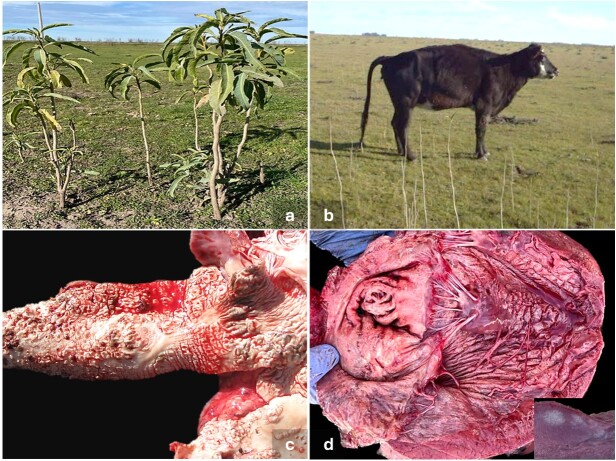 Figure 1
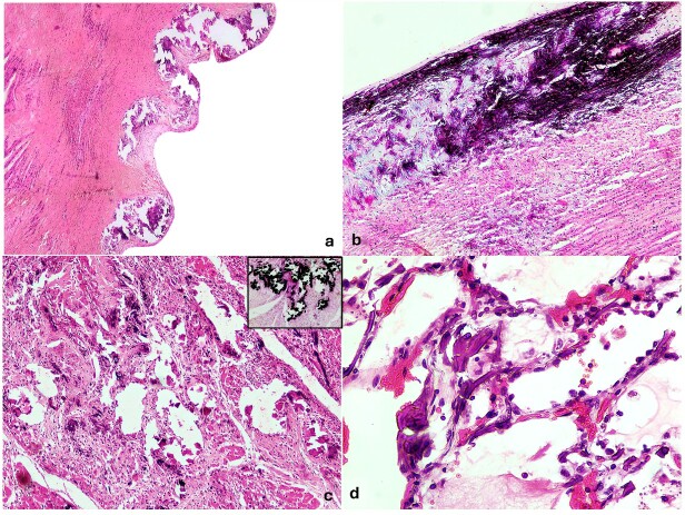 Figure 2
|
|
|
|
|
|
|
|
|
|---|---|---|---|---|---|---|---|
| 1 | Mar Chiquita | June-1993 | Bull | Native grasslands | Breeding | NR | Calcification in large vessels, heart valves and lungs. Ascites |
| 2 | Mar Chiquita | May-1994 | Cows/Heifers | Native grasslands and pastures | Breeding | progressive emaciation enophthalmia, diarrhea, osteophagy | Calcification in large vessels, lungs and tendons |
| 3 | General Madariaga | February-2000 | Cows | Pastures | Breeding | progressive emaciation, stiff gait , xiphosis | Calcification in large vessels, lungs and tendons |
| 4 | General Madariaga | August-2000 | Bulls | Pastures | Breeding | progressive emaciation and diarrhea | Calcification in large vessels, endocardium and lungs, ascites |
| 5 | Magdalena | April-2000 | Calves | Pastures | Wintering | progressive emaciation, difficulty getting up , stiff gait | Calcification in large vessels and endocardium |
| 6 | Mar Chiquita | April-2004 | Cows | Pastures | Breeding | NR | Calcification in large vessels and heart valves |
| 7 | Roque Perez | February-2006 | Heifers | Native grasslands | Breeding | progressive emaciation, difficulty getting up , stiff gait | Calcification in large vessels and heart valves |
| 8 | Mar Chiquita | May-2006 | Cows/Heifers | Native grasslands | Breeding | progressive emaciation, stiff gait | Calcification in large vessels, heart valves and lungs |
| 9 | Maipú | September-2006 | Cows | Native grasslands | Breeding | difficulty getting up | Calcification in large vessels, heart valves and lungs |
| 10 | Maipú | February-2007 | Cows | Native grasslands and pastures | Breeding | progressive emaciation, difficulty getting up , stiff gait | Calcification in large vessels |
| 11 | General Alvarado | April-2007 | Heifers | Native grasslands | Breeding | progressive emaciation, stiff gait , xiphosis | Calcification in large vessels |
| 12 | Las Flores | February-2008 | Heifers | Native grasslands | Breeding | progressive emaciation, stiff gait | Calcification in large vessels, heart valves and lungs |
| 13 | General Madariaga | April-2010 | Steers | Native grasslands and pastures | Wintering | progressive emaciation, stiff gait , enophthalmia, osteophagy | Calcification in large vessels, heart valves and lungs |
| 14 | Saladillo | July-2010 | Steers | Fescue hay and a starter ration | Feedlot | progressive emaciation, osteophagy | Calcification in large vessels, endocardium, heart valves and lungs. Whitish dots in renal cortex. Ascites |
| 15 | Mar Chiquita | May-2011 | Cows | Pasture and ryegrass | Breeding | progressive emaciation, difficulty getting up , stiff gait | Calcification in large vessels, and heart valves |
| 16 | Saladillo | June-2011 | Cows | Native grasslands | Breeding | progressive emaciation, difficulty getting up , stiff gait | Calcification in large vessels |
| 17 | Dolores | November-2011 | Cows/Bulls | Pasture | Breeding | progressive emaciation, diarrhea | Calcification in large vessels, heart valves and lungs |
| 18 | Dolores | May-2013 | Cows | Native grasslands | Breeding | progressive emaciation, xiphosis | Calcification in large vessels and endocardium |
| 19 | General Lavalle | July-2015 | Calves | Native grassland, pasture and balanced feed | Wintering | difficulty getting up | Calcification in large vessels, endocardium and heart valves |
| 20 | Ayacucho | August-2016 | Cows | Native grasslands | Breeding | difficulty getting up , sudden death | Calcification in large vessels. Whitish dots in renal cortex. Ascites. |
| 21 | Maipú | October-2019 | Bulls | Native grasslands | Breeding | progressive emaciation | Calcification in large vessels and lungs. Ascites and hydrothorax |
| 22 | Coronel Suarez | June-2023 | Calves | Native grasslands | Wintering | progressive emaciation, stiff gait , diarrhea, dyspnea | Calcification in large vessels, endocardium, heart valves, lungs, abomasum, rumen, meninges. Whitish dots in renal cortex |
| 23 | Tapalqué | April-2024 | Cows | Native grasslands | Breeding | progressive emaciation, stiff gait , diarrhea | Calcification in large vessels, endocardium, heart valves, lungs and tendons |
Peer Reviews
No public reviews on file for this paper yet. If you reviewed it on a platform where reviews are public (OpenReview, ICLR, NeurIPS, ICML), you can paste yours below so the community can read it here.
Videos
No videos yet. Explain this paper in a talk, walkthrough, or lecture? Add one.
Taxonomy
TopicsMollusks and Parasites Studies · Helminth infection and control · Plant Toxicity and Pharmacological Properties
Introduction
Enzootic calcinosis (EC) is a chronic toxic disease associated with the consumption of calcinogenic plants, affecting ruminants worldwide (Machado et al., 2020a). In South America EC is widely reported associated with the consumption of Solanum glaucophyllum, Solanum stuckertii, Nierembergia veitchii and Nierembergia rivularis (Riet-Correa et al., 2023). S. glaucophyllum, known as “duraznillo blanco”, is an endemic plant present in low-flooded areas, and the main cause of EC in Argentina, Brazil and Uruguay (García et al., 2017; Machado et al., 2020a; Riet-Correa et al., 2023). Particularly in Argentina, EC by S. glaucophyllum is among the most frequently diagnosed toxicities in cattle, producing severe economic losses in beef rearing systems (García et al., 2017). EC is characterized by hypercalcemia, hyperphosphatemia, hypoparathyroidism, hypercalcitoninism, soft tissue calcification, osteonecrosis and osteopetrosis (Machado et al., 2020a). Calcitriol (1,25(OH)2_D_3), an analogue of vitamin D_3_, is the main toxic principle of S. glaucophyllum, causing increased absorption of calcium (Ca) and phosphorus (P), leading mainly to systemic soft tissue mineralization (Jäpelt & Jakobsen, 2013). EC primarily occurs in livestock under extensive grazing systems due to the involuntary consumption of calcinogenic plants (Gimeno, 2000). However, EC outbreaks can also affect fattening animals when they consume hay heavily contaminated with S. glaucophyllum (Micheloud et al., 2012; Ovelar et al., 2024).
This retrospective study analyzes 23 outbreaks of EC in beef cattle caused by S. glaucophyllum consumption in central Argentina. It provides a detailed examination of epidemiological data, clinical presentations, and pathological findings, emphasizing common disease characteristics as well as rarer observations that warrant consideration.
Case description
This retrospective study includes 23 outbreaks of EC caused by S. glaucophyllum in cattle registered from 1990 to 2024 by the Specialized Veterinary Diagnostic Service (SVDS) of INTA Balcarce. Diagnosis criteria was based on anamnesis, clinical signs, compatible gross and/or microscopic lesions, and evidence of consumption of S. glaucophyllum. Epidemiologic data were collected, including geographic location, animal’s age, production system, morbidity and mortality rates. In addition, clinical signs were recorded in all 23 outbreaks. A total of 30 necropsies were performed, though only in 13 tissue samples were fixed in 10% buffered formalin for 48 h and paraffin-embedded for histopathological analysis, including brain, cerebellum, spinal cord, heart, lung, liver, kidney, spleen, lymph nodes, brain, adrenal gland, skeletal muscle, pre-stomachs, abomasum, small and large intestine. Four micrometer sections were prepared routinely and stained with H&E. Selected lung, heart, and aorta sections were von Kossa stained for identification of Ca deposits.
Serum samples were collected from 54 affected animals of 10 outbreaks, to determine Ca (Cseh & Crenovich, 1996) and P levels (Cseh et al., 1994) by atomic absorption spectroscopy (AAS, Perkin Elmer AAnalyst 700, CT, USA). In all cases, the paddocks where the animals were grazing were inspected for identification of S. glaucophyllum. Also, the identification of other potential calcinogenic toxic plants from south America (S. stuckertii, S. torvum, Stenotaphrum secundatum, Cestrum diurnum, Nierembergia riograndensis and N. rivularis) were evaluated.
Clinical EC outbreaks occurred throughout the whole year; however, a marked seasonality with 56.52% (13/23) outbreaks was registered between February and May (mid-summer to mid-autumn) (Table 1). All 23 EC outbreaks occurred in beef cattle (Table 1), affecting mainly breeding systems (78.2%; 18/23), 5 outbreaks in pasture fattening cattle (17.39%; 4/23) and one outbreak in feedlot cattle (4.34%; 1/23). Affected cattle were grazing mainly on native grasslands (21/23) and forage crops when the clinical signs were registered. The feedlot outbreak was previously reported by Micheloud et al. (2012) and affected steers that were consuming fescue hay contaminated with abundant amounts of leaves and whole plants of S. glaucophyllum. Adult cattle (greater than 2 years-old), mainly multiparous cows (11/23) were the category more frequently affected, followed by bulls (4/23). Young cattle (younger than 2 years-old) including heifers (5/23), steers (2/23) and calves (3/23; up to 9 months-old) were also affected (Table 1). In all outbreaks, S. glaucophyllum was identified as part of the grazing diet (Figure 1A), while in the animals confined in feedlots, the presence of S. glaucophyllum was observed in the fescue hay. In addition, other potential calcinogenic toxic plants from south America were not found. The herds affected in the outbreaks have an average of 221 animals (40 to 650), with a mean morbidity of 13.75% ± 9.33% (1.46% to 32.37%) and a mean mortality of 4.22% ± 3.53% (1.25% to 10.68%).
Epidemiological, clinical and macroscopic findings in an outbreak of enzootic calcinosis in cattle. a) Solanum glaucophyllum plants found in the pasture where the animals were grazing (outbreak #22). b) Heifer with progressive emaciation and notable kyphosis (outbreak #16). c) Severe diffuse mineralization of the aorta (outbreak #22). d) Rough, raised, whitish, protruding plaques, measuring from 0.1 to 6 cm long and 0.1 to 4 cm wide, due to mineral deposits in endocardium and heart valves (outbreak #14) Inset: multiple myocardial white delimited area in right ventricle papillary muscle.
Clinical signs were recorded in all 23 outbreaks, mainly characterized by progressive emaciation, in 100% of the outbreaks (23/23) (Figure 1B). In addition, stiff gait (50%) or difficulty getting up (46,15%) were frequently recorded. Other less frequent signs were kyphosis (4/23) (Figure 1B), diarrhea (6/23), dehydration (2/23), osteophagy (3/23), and dyspnea (1/23). In one outbreak cows were found dead without apparent previous signs.
Grossly in all 30 cases, systemic mineralization of the large vessels was observed, characterized by loss of elasticity and multiple to coalescing, elevated, white, rough plaques (Figure 1C). The carotid artery and aorta were the most severely affected. The heart was affected in 14 cases, with mineralization in valves (12/14) and/or endocardium characterized by multifocal to diffuse plaques (7/14) (Figure 1D). The atrioventricular and semilunar valves had focalized and well-defined mineralization characterized by the presence of hard and rough plaques. Rarely, mineralized foci were observed throughout the myocardium, with tendency to be more evident in papillary muscles (Figure 1D). Pulmonary calcification affecting the diaphragmatic lobes was observed in 8 cases, with the presence of multiple formations with a rough surface, measuring 1.5 to 2 cm in diameter, covering up to 50% of the affected lobe. Multifocal white mineralization streaks were observed throughout the renal cortex in three autopsies. Tendons were visibly affected in three cases, while only in one case transmural calcification in ruminal and abomasal mucosa and meninges was observed. Other gross findings were abundant ascites (5/21) and hydrothorax (1/21).
Histological analysis was performed in 13 cases. Microscopically, severe diffuse mineralization of the intima and media layers (13/13) in the aorta was observed in all cases, while muscular arteries like carotid had nonmineralized hyperplasia of the intima layer and tunica media mineralization (8/13), including osteochondroid metaplasia (Figure 2A, B). Mineralization was characterized by irregular basophilic granular deposits. The multifocal-to-coalescent mineralization resulted in protruding plaques towards the lumen (Figure 2A). In the heart, diffuse endocardial-subendocardial (3/13) and pericardial (1/13) mineralization and of adjacent vessels were observed (2/13), with mineralization of medium-caliber vessels in their media and intimal layers. Rarely (2/13), severe mineralization in the connective tissue of tendinous cords was observed. Additionally, multiple-to-coalescent areas of mineralization were present in the myocardium (papillary muscle), replacing cardiac myofibrils and were intermingled with fibrous tissue (Figure 2C), with myosatellite cells and angiogenesis were observed in one case. In the lung, intra-alveolar mineralization lining the wall of the alveoli and throughout alveolar septa most associated to capillary and small blood vessels walls (10/13) (Figure 2D) was present. This finding was rarely accompanied by multifocal alveolar hemorrhage (2/13), edema (2/13) and emphysema (3/13). In addition, segmental to complete mineralization of bronchial (2/13) and tracheal (1/13) cartilages were observed. Mineralization in other tissues including the renal arteries (intima and media layers) (1/13), multifocally the tubular epithelium, of the renal cortex (2/13) and medulla (4/13), the diaphragm serosa (1/13), spleen capsule (2/13), and the middle and intima layers of medium-caliber vessels of the spleen was present. The abomasum presented extensive mineralization in blood vessels (1/13) and muscular layer (2/13), with necrosis and ulceration of the mucosa (2/13). In one case, in rumen and reticulum, muscular arteries mineralization with severe necrosis of the serous and muscular layers, and necrotizing vasculitis was observed. In one case, the blood vessels of the meningeal membranes were mineralized. In lung, heart, and aorta sections, mineral deposits were confirmed as calcium deposits by von Kossa stains (Figure 2C).
Microscopic findings in outbreaks of enzootic calcinosis in cattle. a) Aorta: severe multifocal-to-coalescent mineralization of the intima and media layers, resulted in protruding plaques towards the lumen. H&E, 40×. b) Aorta: severe diffuse mineralization of the intima and media layers. H&E, 200×. c) Heart: multiple areas in the myocardium (papillary muscle), extensive with a tendency to coalesce, of mineralization of myofibrils and replacement by severe fibrous tissue. H&E, 200×. Inset: abundant mineral deposits confirmed as calcium by Von Kossa stains, 200×. d) Lung: intra-alveolar mineralization lining the wall of the alveoli and alveolar septa with multifocal alveolar hemorrhage and edema. H&E, 400×.
From the 54 serum samples collected including affected animals of 10 outbreaks, mean Ca levels were within normal values (10.20 ± 1.19 mg/dl; range 4.97 to 13.7 mg/dl; reference value 8 to 12 mg/dl), and P serum levels (6.64 ± 1.96 mg/dl; range 2.7 to 12.89 mg/dl; reference value 3.5 to 7.5 mg/dl) were slightly to moderately elevated in 20 of the 54 samples (37.03%) (Kaneko et al., 2008).
Discussion
This study describes the clinical and pathological findings in 23 outbreaks of EC due to S. glaucophyllum consumption in cattle in Buenos Aires province, Argentina. However, these outbreaks correspond to consultations by private veterinarians to the SVDS, being a much important problem, because the diagnosis is usually made in the field by practitioners or farmers and most cases are not reported to diagnostic laboratories. These EC outbreaks mainly affected beef cattle grazing in extensive production systems (García et al., 2017; Machado et al., 2020a). Though outbreaks were distributed throughout the year given its chronic characteristic, a seasonal pattern was observed from mid-summer to mid-autumn, possibly since the consumption of this plant mostly occurs in dry seasons or with little forage availability during winter and/or summer, leading the animals to consume on low grounds, being exposed to the plant or when leaves fall and mix with forage (Furlan et al., 2012; Gimeno, 2000). Multiparous cows were the most affected category, since they constitute the predominant population in the problem areas (Gimeno, 2000) and given by the prolonged stay in the farms, increasing exposition and consumption throughout their productive life could lead to the occurrence of clinical EC. In one case, poisoning occurred due to the consumption of contaminated hay, highlighting the toxicity of S. glaucophyllum for long periods after drying and with a subacute onset (Micheloud et al., 2012; Machado et al., 2020a; Ovelar et al., 2024). In most cases, progressive emaciation, difficulty getting up and stiff gait were observed (Gimeno, 2000; Machado et al., 2020a). EC is characterized by a chronic course with very noticeable nutritional and postural changes, being secondary to systemic mineralization (Machado et al., 2020a). In this sense, animals tend to remain recumbent and have a difficult time standing up and death can occur from extreme malnutrition and cachexia if animals are not removed from grazing paddocks with S. glaucophyllum (Furlan et al., 2012). These clinical signs only represent the last stage of the disease, having suffered the stages of metabolic alterations and anatomopathological lesions (Gimeno, 2000). The consumption of S. glaucophyllum produces an increase of intestinal absorption of Ca and P, and further blood increase within a few hours or up to 24 h after plant consumption (Majak, 2001; Machado et al., 2020a). In our study, the mean Ca and P levels were within the reference values, most probably because S. glaucophyllum consumption ceased days to weeks before the final clinical assessment. However, 37% of animals exhibited hyperphosphatemia, which may be linked to EC presentation. Another possible explanation is the naturally higher phosphorus levels in growing calves, as 12 of the 20 animals with hyperphosphatemia were calves. Additionally, the variance in phosphorus or calcium serum levels, is associated with transient changes of both minerals, rarely resulting in high levels during natural EC outbreaks (Machado et al., 2020a; Schild et al., 2021; Brown et al., 2024). Based on this, the diagnostic utility of serological values of Ca and P in cases of natural poisoning by S. glaucophyllum seems very limited (Gimeno, 2000).
In all the outbreaks analyzed, pathological lesions produced by mineralization in soft tissues are similar to those reported in the literature (Furlan et al., 2012; Micheloud et al., 2012; Gimeno, 2000; Machado et al., 2020a). In our experience, mineralizations is a common incidental findings mainly in the aorta of many necropsied animals that have consumed S. glaucophyllum, probably affecting subclinical productive and reproductive parameters, though not severe enough to cause clinical signs and/or death. For the latter, widespread soft tissue mineralization is important. Aortic mineralization was a consistent finding in this study, along with medium- and large-caliber blood vessels throughout body, similar to previous reports in EC cases (Machado et al., 2020a; Schild et al., 2021). Less commonly, mineralization of small-caliber vessels in various organs has also been documented (Machado et al., 2020a), as observed in the spleen, kidney, pre-stomachs, lungs and heart. Additionally, calcification can extend beyond the arteries, affecting alveolar septa, renal tubules and cardiomyocytes (Barros & Rissi 2024). A direct action of the toxic principle of S. glaucophyllum on vascular smooth muscle cells was proposed to produce cellular dedifferentiation, bone and cartilage metaplasia and systemic mineralization of soft tissues (Machado et al., 2020a). Beyond this, decreases in the amount of collagen and elastic fibers present in the aorta were also observed in relation to the continuous consumption of S. glaucophyllum (Portiansky et al., 2002). All these changes may have an adverse influence on the cardiovascular function of the animals, resulting in hemodynamic changes like pulmonary edema and hydrothorax, and sudden death, commonly observed in sheep during management practices (Machado et al., 2020b). Additionally, it was shown that the extract of plants produces adverse effects on chondrocyte cultures from the epiphysis of the long bones of newborn rats (Bertassoli et al., 2023). Stiff gait was frequently observed in our study, a change that could be related to degenerative damage at the osteoarticular level.
It is recommended to avoid consumption of S. glaucophyllum based on rational grazing management and increase care in the preparation of silage or hay (Gimeno, 2000; Machado et al., 2020a). In addition, affected animals should not be exposed to stressful handling because they may develop cardiovascular failure (Machado et al., 2020a).
Conclusions
Despite being a widely known problem, S. glaucophyllum-EC is still an endemic problem in the livestock industry in Argentina, generating great economic losses. For this reason, more studies are needed to increase in the understanding of the pathophysiological mechanisms to treat and/or prevent the adverse effects of EC. Also, further studies mainly focusing to subclinical and/or reproductive losses are needed, since the clinical observations of this study only represents the tip of the iceberg of the impact of EC to the economy of this sector.
The reference list from the paper itself. Each links out to its DOI / PubMed record.
- 1Barros C. S. Rissi D. R. 2024 Ovine enzootic calcinosis by ingestion of Nierembergia veitchii.Brazilian Journal of Veterinary Pathology 173226229 https://10.0.94.6/bjvp.1983-0246.v 17i 3p 226-229
- 2Bertassoli B. M. Melo M. M. Ocarino N. M. Souza Félix I. C. Araújo F. R. Sena Reis A. M. Leonel Alves E. G. Gimeno E. J. Massone A. R. Serakides R. 2023 Effect of the addition of different concentrations of Solanum glaucophyllum desf. extract on chondrocyte cultures from the growth cartilage of newborn rats Toxicon : Official Journal of the International Society on Toxinology 23010715810.1016/j.toxicon.2023.10715837172829 · doi ↗ · pubmed ↗
- 3Brown S. E. Collett M. G. Matthews Z. M. Marshall J. C. Dittmer K. E. 2024 Enzootic calcinosis in Toggenburg goats in New Zealand New Zealand Veterinary Journal 721455210.1080/00480169.2023.226339937752886 · doi ↗ · pubmed ↗
- 4Cseh S. Fay P. Sueldo R. Drake M. 1994 Una microtécnica simple para el dosaje de fósforo inorgánico en suero bovino Revista Argentina de Producción Animal 14137138
- 5Cseh S. B. Crenovich H. 1996 Hipomagnesemia en el sudeste de la provincia de Buenos Aires, Argentina Archivos de Medicina Veterinaria 28111116
- 6Furlan F. H. Colodel E. M. Lemos R. A. A. Castro M. B. Mendoça F. S. Riet-Correa F. 2012 Poisonous plant affecting cattle in Central-Western Brazil International Journal of Poisonous Plant Research 21113
- 7García J. A. Cantón G. J. García B. L. Micheloud J. F. Campero C. M. Späth E. J. A. Odriozola E. R. 2017 Retrospective analysis of cattle poisoning in Argentina (2000 - 2013)Pesquisa Veterinária Brasileira 37321021410.1590/s 0100-736x 2017000300002 · doi ↗
- 8Gimeno E. 2000 Calcinosis enzoótica en rumiantes: Un problema vigente de la ganadería nacional Anales de la Academia Nacional de Agronomía y Veterinaria 54201234