Clinical and translational study of ivosidenib plus nivolumab in advanced solid tumors harboring IDH1 mutations
Matthew K Nguyen, Mark Jelinek, Arjun Singh, Brian Isett, Erica S Myers, Steven J Mullett, Yvonne Eisele, Jan H Beumer, Robert A Parise, Julie Urban, Amy Rose, Lorenzo Sellitto, Krishna Singh, Rose Doerfler, Rebekah E Dadey, Carl Kim, John C Rhee, Diwakar Davar, Liza C Villaruz

TL;DR
A clinical trial tested combining ivosidenib and nivolumab in IDH1-mutated tumors, finding limited clinical benefit but suggesting immune-modulatory effects.
Contribution
This study explores the safety and preliminary efficacy of combining IDH1 inhibition with immunotherapy in IDH1-mutated tumors.
Findings
Only 20% of patients met the primary endpoint of six-month progression-free survival or response rate.
Treatment reduced plasma (R)-2HG levels, especially in patients with clinical benefit.
Exploratory analyses suggested immune-modulatory effects from the combination therapy.
Abstract
Cancers that do not respond to immunotherapy typically harbor a non-T cell-inflamed tumor microenvironment (TME), characterized by the absence of type I/II interferon signaling and CD8+ T cell infiltration. We previously reported IDH1 somatic mutations were enriched in this phenotype across histologies. Mutant IDH1 (mIDH1) drives immune exclusion via metabolic reprogramming of the TME, and preclinical models show that IDH inhibition can restore anti-tumor immunity. We conducted a Phase II study assessing the preliminary activity of ivosidenib, an IDH1 inhibitor, plus nivolumab, an anti-PD1 antibody, in patients with mIDH1 advanced solid tumors (NCT04056910). Patients with advanced or refractory mIDH1 solid tumors and no prior exposure to IDH1 inhibitor were administered ivosidenib 500 mg by mouth daily with nivolumab 480 mg intravenously every 4 weeks. A composite primary endpoint…
Genes, proteins, chemicals, diseases, species, mutations and cell lines named across the full text — each resolved to its canonical identifier and authoritative record.
Click any figure to enlarge with its caption.
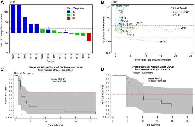 Figure 1
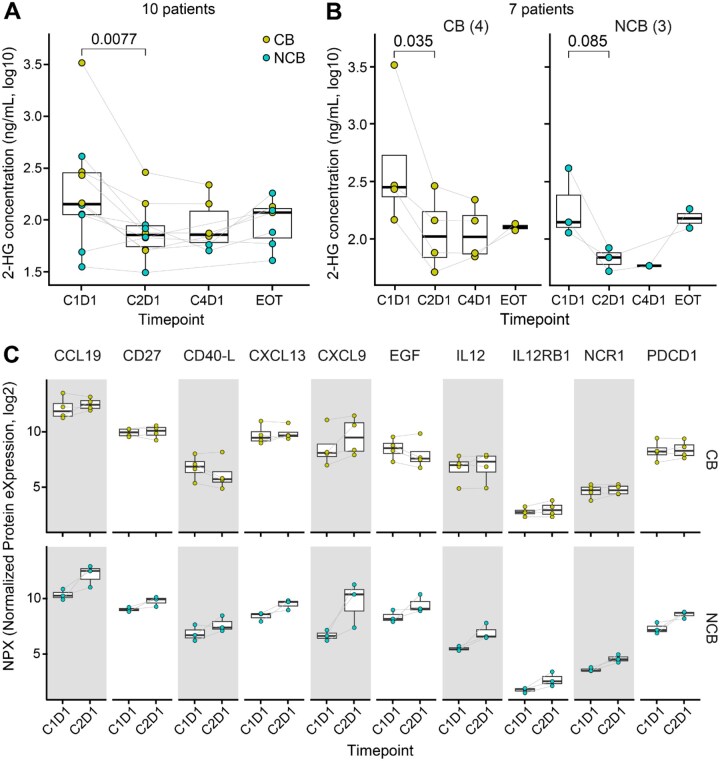 Figure 2
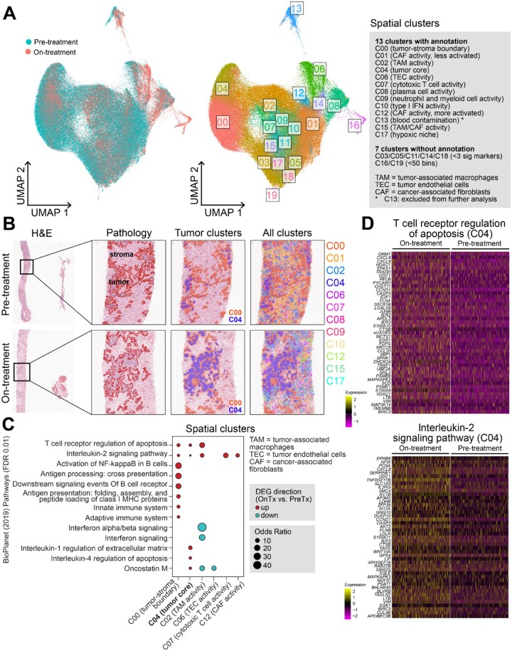 Figure 3
| Number of Participants, n = 15 | |
|---|---|
|
| 54 [50,60] |
|
| |
|
| 8 (53.3) |
|
| 7 (46.7) |
|
| |
|
| 1 (6.7) |
|
| 14 (93.3) |
|
| |
|
| 1 (6.7) |
|
| 14 (93.3) |
|
| |
|
| 6 (40.0) |
|
| 9 (60.0) |
|
| |
|
| 4 (26.7) |
|
| 3 (20.0) |
|
| 7 (46.7) |
|
| 1 (6.7) |
|
| |
|
| 5 (33.3) |
|
| 6 (40) |
|
| 4 (27.7) |
|
| 13 (86.7) |
|
| 2 (0 - 3) |
|
| 1.5 (1 - 3) |
|
| 0 (0 - 3) |
|
| 2 (1 - 4) |
|
| 2 (0) |
|
| 11 (73.3) |
|
| 9 (60) |
| Treatment Related Adverse Events | All Grades | Grade ≥ 3 |
|---|---|---|
|
| 10 (67) | 2 (13) |
|
| 10 (67) | 0 (0) |
|
| 5 (33) | 0 (0) |
|
| 4 (27) | 0 (0) |
|
| 4 (27) | 1 (7) |
|
| 3 (20) | 0 (0) |
|
| 3 (20) | 0 (0) |
|
| 3 (20) | 1 (7) |
|
| 3 (20) | 0 (0) |
|
| 2 (13) | 0 (0) |
|
| 2 (13) | 0 (0) |
|
| 2 (13) | 0 (0) |
|
| 1 (7) | 0 (0) |
|
| 1 (7) | 0 (0) |
|
| 1 (7) | 0 (0) |
|
| 1 (7) | 0 (0) |
|
| 1 (7) | 1 (7) |
|
| 1 (7) | 0 (0) |
|
| 1 (7) | 0 (0) |
|
| 1 (7) | 0 (0) |
|
| 1 (7) | 0 (0) |
|
| 1 (7) | 0 (0) |
|
| 1 (7) | 0 (0) |
| Patient ID | Histology | Lines of SystemicTherapy | IDH1 Mutation | Other Biomarkers | PFS (months) | Time on treatment (months) | Best Overall Response | Extent of Disease Prior to Enrollment |
|---|---|---|---|---|---|---|---|---|
|
| Intrahepatic Cholangiocarcinoma | 1 | R132C | BRAF K601E, ARID1A R1202Q, TMB 3.83 mut/Mb, MSS | 26.76 | 18.44 | SD | Liver |
|
| Intrahepatic Cholangiocarcinoma | 3 | R132C | TP53 H179Q, ERCC2 L461V, KIT D496N, 15.7 Muts/Mb, MSS | 4.6 | 4.47 | SD | Liver, gastro-hepatic lymph node, peri-portal and porto-caval lymph node, retro-caval and aorta-caval lymph nodes, right cardio-phrenic lymph node, perotineal fluid, pericardial fluid |
|
| Intrahepatic Cholangiocarcinoma | 1 | R132G | KRAS G12D, ATRX G1219A, CREBBP M866I, 8.9 Muts/Mb, MSS | 7.2 | 7.82 | SD | Liver, Porta-caval lymph node, retroperitoneal lymph nodes, lung, cardio-phrenic lymph nodes. |
|
| Chondrosarcoma | 0 | R132G | N/A (Only IDH mutations tested) | 16.41 | 11.01 | PR | Right scapula, lung, left adrenal, spleen, right supraclavicular lymph nodes |
- —National Institutes of Health (NIH)
- —UPMC Hillman Cancer Center CCSG
- —NIH10.13039/100000002
- —NIH10.13039/100000002
- —The University of Pittsburgh Center for Research Computing and Data (CRCD)
- —HTC clusters
- —NIH10.13039/100000002
Peer Reviews
No public reviews on file for this paper yet. If you reviewed it on a platform where reviews are public (OpenReview, ICLR, NeurIPS, ICML), you can paste yours below so the community can read it here.
Videos
No videos yet. Explain this paper in a talk, walkthrough, or lecture? Add one.
Taxonomy
TopicsCancer Immunotherapy and Biomarkers · CAR-T cell therapy research · Lung Cancer Treatments and Mutations
Background
Immune checkpoint inhibitors (ICI) have transformed cancer treatment. However, patients with a non-T cell-inflamed tumor microenvironment (TME), characterized by the absence of interferon signaling and tumor infiltration by CD8^+^ T cells, experience limited benefit.1–3 Our group identified that isocitrate dehydrogenase 1 (IDH1) mutations (mIDH1; R132H and R132C) were associated with a non-T cell-inflamed phenotype across cancers.4 mIDH1 confers gain-of-function activity, converting α-ketoglutarate to the oncometabolite (R)-2-hydroxyglutarate [(R)-2HG], which accumulates in tumor cells and subsequently released into the TME.5–7 (R)-2HG inhibits α-ketoglutarate-dependent enzymes, promotes DNA hypermethylation, silences cGAS-STING-IRF3 signaling, and suppresses cytokine/chemokine releases, leading to a reduced CD8^+^ T-cell recruitment and an immunosuppressive TME.8–12 In preclinical tumor models, mIDH1 inhibition reversed these processes, restored anti-tumor immunity, and significantly reduced tumor growth.9
Based on these mechanistic insights and strong correlation of mIDH1 with the non-T cell-inflamed phenotype across tumor types (Supplemental Figure S1), we hypothesized that IDH1 inhibition combined with ICI may be an effective treatment strategy for patients with mIDH1 tumors. Here we report a Phase II study evaluating the safety and preliminary anti-tumor activity of the mIDH1 inhibitor ivosidenib plus the anti-PD1 antibody nivolumab in patients with mIDH1 advanced solid tumors, as well as associated pharmacodynamic, serum and tumor based translational correlates.
Methods
Patient eligibility
Adult men and women were eligible for enrollment into this trial if they had a histopathologic diagnosis of an advanced solid tumor with documented IDH1 gene mutation (R132C/L/G/H/S) by sequencing. Patients had to have progressed on appropriate standard of care treatment or for which no curative treatment was available. Patients had to have at least one evaluable and measurable lesion by RECIST v1.1 (solid tumors) or by Response Assessment in Neuro-Oncology (RANO) criteria (glioma). Patients with glioma were required to have diseases that were both WHO 2016 grade ≥ 2 and contrast enhancing. Patients were required to have a good performance status (ECOG PS of 0 or 1) and adequate end organ function. Toxicities associated with prior anticancer therapy must have been resolved to baseline or ≤ grade 1.
Key exclusion criteria included, but were not limited to, having received a prior IDH1 inhibitor; having received systemic anticancer therapy or an investigational agent less than 2 weeks prior to day 1; active autoimmune disease requiring systemic treatment in the past 2 years; or a diagnosis of immunodeficiency. Patients with non-glioma solid tumors must not have received radiotherapy to metastatic sites of disease < 2 weeks prior to day 1 and glioma patients must not have received radiation within 3 months prior. Non-glioma patients must not have undergone hepatic radiation, chemoembolization, or radiofrequency ablation < 4 weeks prior to day 1. Patients with non-glioma solid tumors were ineligible if they had known symptomatic brain metastases requiring steroids. Patients with previously diagnosed brain metastases were eligible for enrollment if they had completed their treatment and had recovered from the acute effects of radiation therapy or surgery prior to study entry, had discontinued corticosteroid treatment for these metastases for at least 1 week, and had radiographically stable disease for at least 1 month prior to being enrolled on the study.
Clinical trial design
This single-center Phase II clinical trial was designed to assess the preliminary activity of ivosidenib in combination with nivolumab in patients with mIDH1 advanced solid tumors (NCT04056910, Supplemental Figure S2). Participants were administered ivosidenib 500 mg by mouth daily in combination with nivolumab 480 mg intravenously every 4 weeks. Based on the tumor spectrum of IDH1 mutations in cancer, it was observed that some tumor types are rarely associated with RECIST responses, for example chondrosarcoma. Therefore, a composite primary endpoint was designed to include either of six-month progression free survival (PFS6) or overall response rate (ORR). Utilizing PFS6 as an endpoint was proposed to allow capture of clinical benefit in the form of “cytostatic” effect previously observed and proposed as a priority primary endpoint for soft tissue sarcomas.13^,^14 Thus, a participant was considered to meet the primary endpoint if they had PFS6 (scored as either yes or no) or they had at least a partial response (PR; based on RECIST v1.1 in solid tumors or RANO in glioma) after the week 8 scan. This study was conducted in compliance with the Declaration of Helsinki and the International Conference on Harmonization Guidelines for Good Clinical Practice. The University of Pittsburgh institutional review board (IRB) approved this protocol (IRB19-096). Participants gave informed consent to participate in the study before taking part. All samples have written informed patient consent.
We proposed the co-primary endpoint of PFS6 in addition to ORR. This was based on the concern that response alone may not adequately capture the clinical benefit of the study treatments within certain tumor histologies. It has been recognized in the treatment of soft tissue sarcomas that tumors responding to therapy may be replaced with fibrotic tissue that can confound RECIST measurements.14 Additionally, in the Phase 1 study of ivosidenib in chondrosarcoma, a majority of patients achieved stable disease as their best overall response. This suggests a possible cytostatic effect of ivosidenib13 where RECIST response may not reflect the therapeutic effect of the study treatments. Given the heterogeneity of the tumor histologies involved, it is difficult to know if ivosidenib would have similar cytostatic effect across all tumor types, especially when combined with nivolumab. PFS has the potential to capture the benefit of the study treatments across these tumor types since the most commonly IDH1 mutant tumors included have a PFS of < 4 months. This is based the following historical controls:1 Cholangiocarcinoma: A median PFS of 3.2 months2^,^15 Advanced Chondrosarcoma: A median PFS of 3.5 months3^,^16 Enhancing glioma: median duration on ivosidenib or AG-881 treatment of 1.9-3 months.17^,^18 This study has selected diverse, aggressive tumor histologies unified by their low anticipated PFS. With this in mind, we see that the proposed a doubling of expected PFS to 6 months with combination ivosidenib and nivolumab to be a meaningful measure of the efficacy of these drugs.
This trial employed an Optimal Simon two-stage design where a probability of a positive outcome < 0.1 would not be promising and a probability of a positive outcome ≥ 0.3 would warrant further interest. In the first stage, 18 participants were proposed to be accrued however the study was eventually discontinued due to lack of clear clinical activity and changes in the landscape of treatment. This included approval of anti-PD1/L1 antibody in the first line of therapy for cholangiocarcinoma as well as the launch of a competing industry sponsored clinical trial of IDH inhibition with dual checkpoint blockade. Trial accrual occurred from March 29, 2021 to October 25, 2023.
Translational analysis of human specimens from trial
To understand treatment-induced changes in patients, we conducted analyses of longitudinal 2-hydroxyglutarate (2-HG) levels and proteomics in peripheral blood, as well as high-resolution gene expression by spatial transcriptomics in tumor tissues. A full description of the materials and methods is provided in Supplementary Methods. In brief: (1) Plasma 2-HG quantification: Serial plasma samples from 10 patients (C1D1, C2D1, C4D1, EOT) were analyzed using liquid chromatography tandem mass spectrometry (LC-MS/MS). Four patients without paired C1D1 and C2D1 samples were excluded. Calibration curves were generated with 2-HG-[^1^³C_5_] surrogate analyte in blank plasma, using linear regression (1/y^2^ weighting) of surrogate/IS ratios. Reduction in 2-HG served as a pharmacodynamic marker of IDH1 inhibition. (2) Serum proteomics: Paired serum samples (C1D1, C2D1) from seven patients (cholangiocarcinoma or chondrosarcoma) were profiled using the Olink Target 96 Immuno-Oncology panel. Normalized protein abundance was compared between patients with clinical benefit (CB, PFS ≥4 m) and those with no benefit (NCB) using limma regression,19 focusing on exploratory immune-related changes. (3) Tumor spatial transcriptomics: Pre- and on-treatment FFPE tumor biopsies from one patient with CB were analyzed using the 10x Genomics Visium HD platform. The pre-treatment biopsy was obtained prior to C1D1, and the on-treatment biopsy was at C2D1. H&E-guided regions of viable tumor were selected by pathology review and profiled by Illumina sequencing. Data was processed with SpaceRanger (10x Genomics) and Seurat,20 with quality filters applied, followed by clustering and Uniform Manifold Approximation and Projection21 (UMAP) visualization and neighborhood annotation. Tumor and stroma compartments were further analyzed for treatment-induced differential gene expression (DEG) and pathway enrichment, and pseudobulk expression was assessed using xCell digital cytometry22 to evaluate immune cell subsets.
Survival analysis
PFS and OS were estimated with the Kaplan-Meier method using PROC LIFETEST and plotted with PROC SGPLOT in SAS 9.4 (SAS Institute Inc. Cary, NC). Patient 008 had no tumor measurements due to clinical progression prior to follow up scans and was not included in analysis.
Statistical analysis
For 2-HG data from plasma samples, its abundance was transformed into log_10_ scale and compared between different timepoints of 10 patients by paired t-test. For Olink targeted proteomics (NPX values at log_2_ scale), differentially abundant proteins were identified using limma regression models. For spatial transcriptomics of pre/on-treatment tumors from patient 016, on- versus pre-treatment DEGs were detected by Wilcoxon rank-sum test on normalized, unintegrated read counts per Seurat’s best practice. Pathway enrichment was identified using hypergeometric test. All tests are two-sided unless otherwise noted. P-values were adjusted by Benjamini-Hochberg (BH)-False Discovery Rate (FDR) procedure when appropriate. Statistical analysis was performed in R (v4.4.2).
Results
Baseline patient characteristics
A total of 15 patients were enrolled, with baseline characteristics described in Table 1. The median age was 54 years (interquartile range: 50–60) with 46.7% male and 53.3% female, predominantly of white race (93.3%). 60% of patients had an ECOG performance status of 1 (capable of performing most daily activities with some limitations). Tumor histologies were glioma (46.7%), cholangiocarcinoma (26.7%), chondrosarcoma (20.0%) and colorectal adenocarcinoma (6.7%). mIDH1 included R132H (40%), R132C (33.3%), and R132G (27.7%). Median lines of systemic therapy amongst all disease histologies were 2, and most patients received prior surgery or radiotherapy. All gliomas included in this study were high-grade (at least grade 3), contrast enhancing, and had undergone a median of 2 lines of systemic therapy.
Treatment-Related adverse events
Adverse events (AE) are described in Supplemental Tables S1 and S2. All patients enrolled experienced an adverse event of any grade, with 13 (87%) experiencing a treatment-related adverse event (TRAE) of any grade. Of these TRAEs, 27% were grade 3 or higher. No dose-limiting toxicities (DLT) were observed and there were no fatal TRAEs. The most common TRAEs were leukopenia and rash. The most common grade 3 or higher TRAEs were leukopenia, QTC prolongation, anemia, and hyperthyroidism (Table 2; Supplemental Table S3).
Treatment outcome
Out of 15 patients, 14 had tumor measurements with an ORR of 6.67%. One patient had no responses recorded due to clinical progression prior to any follow up imaging. We observed 1 PR (6.67%), 6 stable disease (SD; 40.00%), and 8 progressive disease (PD; 53.33%) 8 weeks after initiation of treatment. One patient with chondrosarcoma (013) had PR. All four patients with intrahepatic cholangiocarcinoma had SD (Figure 1A–B). Three patients met the composite primary endpoint of ORR or PFS6 (3/15; 20.00%). The median PFS was 1.94 months (95% CI 1.61–3.68) and median OS was 10.26 months (95% CI 5.10–19.66) (Figure 1C–D).
*Clinical outcome of ivosidenib plus nivolumab in 14 patients with mIDH1 advanced solid tumors. (A) waterfall plot showing best response and best percentage of tumor size change from baseline. Color indicates best response groups. Dotted lines denote cutoffs for PD and PR at +20 and −30%. Asterisk denotes: two patient labeled as PD, one had progression of NT5 lesion and withdrew consent at that time; the other had clinical progression. Both came off treatment. (B) spider plot showing percentage of tumor size change from baseline over time. Colored lines indicate clinical benefiters (CB, PFS ≥ 4m), and non-clinical benefiters (NCB). n = 14 in A and B. One patient had no responses recorded due to clinical progression prior to any follow up imaging and not shown (C-D). Kaplan-Meier estimates of (C) Progression-free survival and (D) Overall survival. Red dotted lines indicate the median and shaded indicates the 95% Hall-Wellner confidence band.
Despite the primary endpoint including PFS6, we were interested in detailing the outcomes of any patients who might be considered to derive clinical benefit. Therefore, we specifically call out those with at least PFS ≥ 4 months as possible CB (n = 4, 1 chondrosarcoma and 3 cholangiocarcinoma). Patient-level characteristics, including histology, IDH1 mutation type, prior lines of therapy, and clinical outcomes, are summarized in Table 3. All four patients harbored either an R132C or R132G IDH1 mutation.
The PR observed during this trial was the only patient with chondrosarcoma (013) that clinically benefitted. This was a patient with conventional chondrosarcoma of the right scapula. Pathology demonstrates a grade 3 chondrosarcoma with IDH1 R132G mutation identified. In contrast, the other two patients with chondrosarcoma had dedifferentiated histology. This patient enrolled on the trial after recurrence in the bilateral lungs and right proximal upper extremity/shoulder. The patient received 12 cycles of treatment on protocol. Ultimately, the patient was discontinued from the trial due to intolerable grade 2 arthralgias/arthritis. Restaging scans 2 months after the end of trial demonstrated ongoing partial response with a 51% decrease of the target lesions compared to baseline.
Ivosidenib plus nivolumab suppresses (R)-2-hydroxyglutarate in plasma
Prior pharmacodynamic studies have shown that plasma (R)-2HG levels were inhibited by as much as 98% compared to baseline after one week of continuous ivosidenib dosing and persisted at C2D1.5 To confirm that ivosidenib and nivolumab decreased (R)-2HG concentration levels, we collected plasma specimens at C1D1, C2D1, C4D1 as well as EOT (Supplemental Figure S2). In 10 patients with paired C1D1 and C2D1 plasma (3 chondrosarcoma, 4 cholangiocarcinoma, and 3 glioma), (R)-2HG was significantly reduced at C2D1 relative to C1D1 (*P *= 0.0077) (Figure 2A). In 7 patients with cholangiocarcinoma or chondrosarcoma, this pattern was observed in CB (n = 3; *P *= 0.035) but not in NCB (n = 4; *P *= 0.085) (Figure 2B). A higher reduction in C2D1-C1D1 (R)-2HG levels was detected in CB compared to NCB (69.70%±8.45 and 60.56%±11.8, respectively; mean±S.E.M.). At the time of progression or EOT, a trend toward increasing concentrations of (R)-2HG back to baseline was observed (Figure 2B). Our study shows that the combination of ivosidenib and nivolumab significantly reduced (R)-2HG levels in patients, with an even greater reduction observed in patients who experienced a clinical benefit. However, this reduction was not as pronounced as the near-98% inhibition of (R)-2HG previously reported when ivosidenib was used alone in earlier studies.5
Peripheral blood marker changes upon Ivosidenib plus nivolumab. (A) Plasma (R)-2HG levels. N = 10 patients shown. (B) Plasma (R)-2HG concentration stratified by clinical benefiters versus non-benefiters. N = 7 patients shown. (C) Serum protein expression in clinical benefiters and non-clinical benefiters. Ten proteins at nominal P < 0.05 are shown. Two-sided Welch’s two-sample t-test was used in A on log10 transformed data. Limma regression models were used in B. C1D1 = cycle 1 day 1. C2D1 = cycle 2 day 1. C4D1 = cycle 4 day 1. EOT = end of treatment. Color denotes clinical benefiters (CB, PFS ≥ 4m) and non-clinical benefiters (NCB).
Proteomic analysis identifies pro-inflammatory protein expression with mIDH1 inhibition plus ICI
To determine longitudinal proteomic changes in peripheral blood, C1D1 and C2D1 serum samples from 7 patients with cholangiocarcinoma (n = 4) or chondrosarcoma (n = 3) were analyzed using Olink 96-plex targeted immuno-biology panel. We identified ten proteins with treatment-induced abundance differences between CB and NCB, including CCL19, CD27, CD40L, CXCL13, CXCL9, EGF, IL-12, IL-12RB1, NCR1, and PDCD1 (nominal *P *< 0.05; Figure 2C). Contrary to our initial hypothesis that these pro-inflammatory markers would increase in patients with clinical benefit, these proteins were consistently upregulated in C2D1 relative to C1D1 specifically in NCB. Conversely, CB showed no distinct trend of increase in these same proteins. Among these, CXCL9 showed the largest increases in protein expression in both groups, albeit with different magnitudes or patterns between CB and NCB (average log_2_(fold change)=1.01 and 3.00, respectively; Figure 2C).
Spatial transcriptomics reveal immune gene expression changes upon treatment
To investigate treatment-induced TME changes, we analyzed paired pre- and on-treatment specimens from one patient (016) who experienced CB and had sufficient tumor tissues for Visium HD spatial transcriptomics. This patient had intrahepatic cholangiocarcinoma, a R132C IDH1 mutation, and an overall best response of SD. Due to low gene detection at the original 2 μm bin size, we aggregated data into 8 μm bins to improve expression capture. UMAP-based clustering revealed 20 spatially distinct neighborhoods (C00-C19; Figure 3A), with 12 clusters retained for biological interpretation describing malignant, immune, endothelial, or fibroblast activity (Supplemental Figure S3). Both samples were assayed on the same Visium HD slide and showed similar cluster distribution by UMAP (Figure 3A, left) and bin proportions (Supplemental Figure S4A-B), suggesting minimal batch effect. Tumor cell-rich neighborhoods were predominantly in clusters 00 and 04, with others as stromal neighborhoods (Supplemental Figures S5 and S6). Of interest, C00 were enriched at tumor-stroma boundaries (Figure 3B, middle, orange) and C04 in tumor core (Figure 3B, middle, blue).
TME changes by spatial transcriptomics upon Ivosidenib plus nivolumab. (A) Clusters by UMAP showing distinct cellular neighborhoods with biological interpretation. (B) Representative examples of pathology tumor/stroma annotation and spatial clusters from pre- and on-treatment samples. (C) Immune-related pathway enrichment in each spatial cluster (FDR-adjusted P < 0.01). Clusters without significant pathways are not shown. (D) Gene expression heatmap of two immune pathways from C, showing that in tumor core (C04), on-treatment sample has significantly higher pathway gene expression compared to pre-treatment sample (FDR-adjusted P < 0.01). Two-sided hypergeometric test was used in C, two-sided Wilcoxon rank-sum test was used in D. BH-FDR adjustment of p-values was used in multiple comparisons.
We and others have shown that IDH activation is associated with immune exclusion,4 and that IDH inhibition enhances anti-tumor immunity in preclinical models.9 To investigate the immune impact of IDH inhibition plus anti-PD1 in human tissue, we identified DEGs between on- and pre-treatment tissues within each cluster (FDR-adjusted *P *< 0.01), and performed pathway analysis with curated gene sets (BioPlanet 2019). Immune-related pathways, including T-cell receptor regulation of apoptosis and IL-2 signaling, were upregulated in both tumor clusters (C00, C04) as well as in cytotoxic T cell (C07) and cancer-associated fibroblasts (CAF; C12) neighborhoods (Figure 3C-D). In contrast, tumor-associated macrophage (TAM)-rich neighborhoods (C02) showed downregulation of IFN-α/β signaling genes (e.g., IFITM3, OAS1-3, MX1, ISG15), suggesting potential immunosuppression.
Using an orthogonal approach by xCell on pseudobulked gene expression from tumor and stromal compartments, we found higher enrichment scores for T cell and dendritic cell (DC) subsets in the on-treatment sample, alongside an increase in M2-like macrophages (Supplemental Figure S7). These findings indicate that ivosidenib plus nivolumab enhances anti-tumor immunity, but this may be offset by increased immunosuppressive macrophages, potentially contributing to the SD rather than PR observed in this patient.
Discussion
Here we report a phase II clinical trial investigating the combination of ivosidenib, an IDH1 inhibitor, with the anti-PD1 antibody nivolumab in patients with IDH1 mutant advanced solid tumors. We observed a safety profile consistent with the known toxicity spectrum of the individual agents but no clear suggestion of combinatorial benefit. Three of 15 patients met the composite primary endpoint of ORR or PFS6, with a single PR.
We and others have observed pan-cancer associations between IDH1 mutations, the non-T cell-inflamed TME and immunotherapy resistance. mIDH1 leads to production of the oncometabolite (R)-2HG which alters DNA methylation and suppresses interferon signaling.11^,^12 (R)-2HG in the TME may impair production of IFNγ and IL-2 by CD4^+^ and CD8^+^ T cells as well as impacting myeloid cell populations, for example immunosuppressive glioma-associated macrophages generated due to altered tryptophan metabolism.23^,^24 In human glioma, an inverse relationship between (R)-2HG concentrations and expression of interferon and antigen presentation signaling pathways, as well as CD3^+^ and CD8^+^ T-cells, has been observed.25 In cholangiocarcinoma, inhibition of mIDH1 correlates with induction of interferon responsive molecules, such as PD-L1, PD1, and VISTA/BY-H5, on tumor infiltrating immune cells and lower lymphocytes counts within mIDH1 versus wildtype tumors.26 Overall, these studies emphasize that mIDH1 likely has an immunomodulatory role and inhibition may therefore have potential as a combinatorial partner for ICI. Despite this rationale, the clinical outcomes from our study are more aligned with those seen in clinical trials evaluating the efficacy of ivosidenib monotherapy. The ClarIDHy trial assessed the use of ivosidenib in patients with advanced mIDH1 cholangiocarcinoma and reported a median PFS of 2.7 months and ORR of 2%.27 In gliomas, ivosidenib demonstrated a median PFS of 1.4 months (95% CI, 1.0–1.9 months) in the enhancing by MRI cohort vs. 13.6 months (95% CI, 9.2 to 33.2 months) in the non-enhancing cohort across all dosage levels. ORR was 0% in the enhancing glioma cohort vs. 2.9% in the non-enhancing cohort.28 The gliomas treated in our study were predominately enhancing and had undergone more lines of systemic therapy as compared to the approved setting where vorasidenib is used for Grade 2 astrocytoma or oligodendroglioma.29 Considering that our combination treatment demonstrated a median PFS was 1.94 months (95% CI 1.61–3.68) with an ORR of 6.7%, these data suggest that ivosidenib in combination with nivolumab did not outperform ivosidenib monotherapy.
Noting the heavily pre-treated nature of our cohort, we were interested in further investigating the pharmacodynamic and translational impact of mIDH1 inhibition with anti-PD1. Consistent with prior studies, we observed significant suppression of (R)-2HG from C1D1 to C2D1 and that clinical benefit was associated with greater degrees of (R)-2HG suppression after the first cycle. In contrast with prior studies, the degree of (R)-2HG suppression observed was substantially lower than that previously reported.5 The reason for this is unclear and may contribute to the minimal clinical efficacy observed. Despite this lower (R)-2HG suppression, serum proteomic analyses did reveal upregulation of ten proteins involved in immune signaling and regulation, however this was not associated with clinical benefit. Further studies correlating these proteomic changes with TME analyses and more extensive longitudinal sampling will be important to delineate the immunologic impact of mIDH1 inhibition.
Despite an initial plan for pre- and on-treatment tumor sample collection, the ability to actualize this was limited. Spatial omic characterization of the TME prior to and during therapy for one patient with cholangiocarcinoma, who had clinical benefit, was obtained. Analysis of the TME demonstrated increased gene expression of immune-related pathways, such as T cell receptor regulation of apoptosis and IL-2 signaling, in multiple spatially distinct tumor and stroma clusters. Meanwhile, immunosuppressive signals including M2-like macrophages were observed. Collectively, these data suggest that the combination of mIDH1 inhibition and anti-PD1 may result in revitalization of the T-cell inflamed TME, though further work is needed to validate this observation.
Our study’s small sample size, lack of a comparator arm, and inclusion of heterogenous tumor histologies limit broader interpretation. Based on the observation for correlation of mIDH1 and the non-T-cell-inflamed TME across tumor types, an underlying hypothesis for the study was that mIDH1 may be an immunotherapy combination target independent of cancer histology. In retrospect, including any tumor harboring IDH1 mutation may have hindered the study, especially due to the inclusion of gliomas (47% of the study population) where ivosidenib has minimal activity in enhancing disease. Due to limited biopsy tissue available, we were unable to perform extensive immunohistochemistry (IHC) validation on immune cell findings from the Visium HD studies. With this limitation, we analyzed the immune impact of IDH1 inhibition with anti-PD1 via two computational methods including DEG pathway enrichment analysis and digital cytometry. The translational findings from this study are limited due to sample availability as well as the inclusion of stable disease as and endpoint, therefore may not generalize across populations. In this regard, these data are exploratory and hypothesis-generating for the field.
In conclusion, this phase II trial of ivosidenib with nivolumab demonstrated an expected toxicity spectrum and modest clinical activity, with no obvious clinical benefit over ivosidenib monotherapy. Exploratory translational analysis suggests that ivosidenib plus nivolumab may have immuno-modulatory impact, which can be hypothesis-generating for future studies assessing other novel combinatorial designs. Further translational and clinical studies will be required to better assess the utility of mIDH1 inhibition with immune checkpoint blockade and/or emerging immune-based technologies such as CD3 multispecifics or adoptive cellular transfer. A clinical trial evaluating the efficacy of mutant IDH1 inhibition with dual checkpoint blockade in cholangiocarcioma is underway.30
Supplementary Material
oyaf362_Supplementary_Data
The reference list from the paper itself. Each links out to its DOI / PubMed record.
- 1Gajewski TF , Louahed J, Brichard VG. Gene signature in melanoma associated with clinical activity: a potential clue to unlock cancer immunotherapy. Cancer J. 2010;16:399-403.20693853 10.1097/PPO.0b 013e 3181 eacbd 8 · doi ↗ · pubmed ↗
- 2Gajewski TF , Woo SR, Zha Y, et al Cancer immunotherapy strategies based on overcoming barriers within the tumor microenvironment. Curr Opin Immunol. 2013;25:268-276.23579075 10.1016/j.coi.2013.02.009 · doi ↗ · pubmed ↗
- 3Harlin H , Meng Y, Peterson AC, et al Chemokine expression in melanoma metastases associated with CD 8+ T-cell recruitment. Cancer Res. 2009;69:3077-3085.19293190 10.1158/0008-5472.CAN-08-2281 PMC 3886718 · doi ↗ · pubmed ↗
- 4Bao R , Stapor D, Luke JJ. Molecular correlates and therapeutic targets in T cell-inflamed versus non-T cell-inflamed tumors across cancer types. Genome Med. 2020;12:90.33106165 10.1186/s 13073-020-00787-6PMC 7590690 · doi ↗ · pubmed ↗
- 5Fan B , Mellinghoff IK, Wen PY, et al Clinical pharmacokinetics and pharmacodynamics of ivosidenib, an oral, targeted inhibitor of mutant IDH 1, in patients with advanced solid tumors. Invest New Drugs. 2020;38:433-444.31028664 10.1007/s 10637-019-00771-x PMC 7066280 · doi ↗ · pubmed ↗
- 6Pirozzi CJ , Yan H. The implications of IDH mutations for cancer development and therapy. Nat Rev Clin Oncol. 2021;18:645-661.34131315 10.1038/s 41571-021-00521-0 · doi ↗ · pubmed ↗
- 7Wu MJ , Shi L, Merritt J, Zhu AX, Bardeesy N. Biology of IDH mutant cholangiocarcinoma. Hepatology. 2022;75:1322-1337.35226770 10.1002/hep.32424 · doi ↗ · pubmed ↗
- 8Greten TF , Schwabe R, Bardeesy N, et al Immunology and immunotherapy of cholangiocarcinoma. Nat Rev Gastroenterol Hepatol. 2023;20:349-365.36697706 10.1038/s 41575-022-00741-4PMC 12468729 · doi ↗ · pubmed ↗