Multinucleate cell angiohistiocytoma
Connor A. Sheehan, Jayci G. Rhein, Jordan T. Hyde, Jason B. Lee, Sylvia Hsu

Abstract
Genes, proteins, chemicals, diseases, species, mutations and cell lines named across the full text — each resolved to its canonical identifier and authoritative record.
Click any figure to enlarge with its caption.
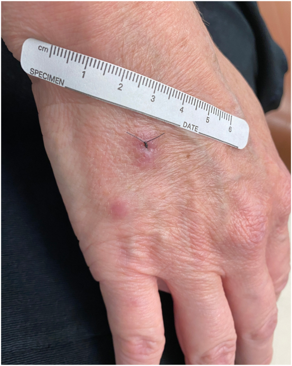 Figure 1
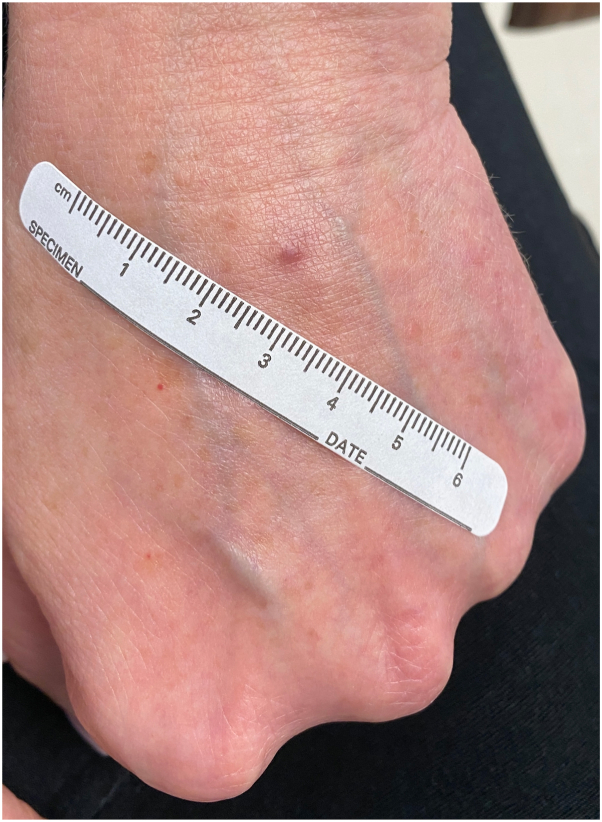 Figure 2
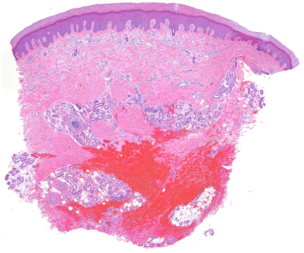 Figure 3
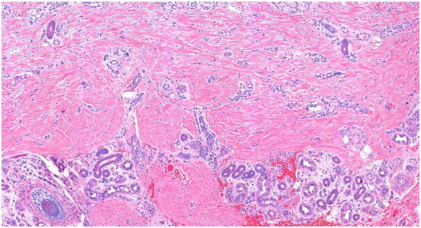 Figure 4
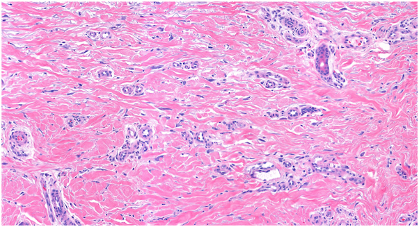 Figure 5
Peer Reviews
No public reviews on file for this paper yet. If you reviewed it on a platform where reviews are public (OpenReview, ICLR, NeurIPS, ICML), you can paste yours below so the community can read it here.
Videos
No videos yet. Explain this paper in a talk, walkthrough, or lecture? Add one.
Taxonomy
TopicsVascular Tumors and Angiosarcomas · Sarcoma Diagnosis and Treatment · Cardiac tumors and thrombi
Case presentation
A 50-year-old woman presented with a 6-month history of papules on her hands. She had been evaluated by a previous dermatologist for these lesions and was given the diagnosis of scar. The patient had been vigilant about avoiding trauma to the hands yet noted the formation of new lesions. The papules were asymptomatic. On exam, there were 3 firm, nontender, nonscaly, erythematous papules on the bilateral dorsal hands (Figs 1 and 2).Fig 1. Two erythematous papules on the right dorsal hand.Fig 2. Erythematous papule on the left dorsal hand.
Question: Besides the dorsal hands, what is the most common location for this condition?
- A.Dorsal feet
- B.Extensor surfaces
- C.Face
- D.Legs
- E.Upper back and chest
Answer discussion
The correct answer is D. Legs. Multinucleate cell angiohistiocytoma (MCAH) is a benign condition characterized clinically by red or violaceous papules or plaques.1^,^2 It is a rare entity that has been described fewer than 200 times in the literature and is most commonly found on the dorsal hands or lower extremities of middle-aged women.2^,^3 The clinical appearance of MCAH can resemble that of angiofibroma, granuloma annulare, or Kaposi sarcoma, and biopsy is standard for making the diagnosis.2 Histologically, MCAH shows dermal fibrosis, histiocytosis, dilated vasculature, and stellate multinucleated cells (Fig 3, Fig 4, Fig 5). These giant cells are characteristic of this condition, although they are not pathognomonic.1 CD10 staining highlights the multinucleated cells but is not diagnostic.1 The pathogenesis is debated but MCAH is proposed to be a reactive fibroblast proliferation, possibly secondary to trauma, given distribution on trauma-prone sites.1^,^4^,^5 Some authors have proposed that this lesion may be a variant of dermatofibroma.1 There is some reported association of MCAH with estrogen receptor overexpression, which may explain the disproportionate incidence in female patients.5Fig 3. Dermal fibrosis and dilated vasculature with incidental procedural hemorrhage (H&E 5.6×).Fig 4. Thickened collagen bundles and dilated vessels with increased interstitial fibroblasts (H&E 15.6×).Fig 5. Characteristic multinucleated cells with scalloped borders (H&E 29.1×).
The clinical behavior of MCAH is benign with some enlargement over time before either spontaneous regression or persistence. While there is no consensus treatment, case reports suggest efficacy of pulsed dye laser or surgical excision.3 Other reports include Nd:YAG, CO2 laser, intense pulsed light, and cryotherapy.3 Once treated, lesions are not expected to recur.3
Conflicts of interest
None disclosed.
The reference list from the paper itself. Each links out to its DOI / PubMed record.
- 1Ross C.L.Chung J.Ross N.A.Lee J.B.Generalized multinucleate cell angiohistiocytoma: histopathological and immunohistochemical analyses of 10 lesions Am J Dermatopathol 4312202197697910.1097/DAD.000000000000195433899769 · doi ↗ · pubmed ↗
- 2Shapiro P.E.Nova M.P.Rosmarin L.A.Halperin A.J.Multinucleate cell angiohistiocytoma: a distinct entity diagnosable by clinical and histologic features J Am Acad Dermatol 303199441742210.1016/s 0190-9622(94)70049-48113454 · doi ↗ · pubmed ↗
- 3Taibo A.Padin C.P.Losada J.D.P.Fonseca E.Treatment options for multinucleate cell angiohistiocytoma: a case series and review of the literature J Cutan Med Surg 273202321922510.1177/1203475423116353936927096 · doi ↗ · pubmed ↗
- 4Jia Q.N.Qiao J.Qu T.Generalized multinucleate cell angiohistiocytoma with possible origin from fibroblasts: a clinicopathological study of 15 cases J Dermatol 481202111411910.1111/1346-8138.1561033180332 · doi ↗ · pubmed ↗
- 5Frew J.W.Multinucleate cell angiohistiocytoma: clinicopathological correlation of 142 cases with insights into etiology and pathogenesis Am J Dermatopathol 373201522222810.1097/DAD.000000000000007525140659 · doi ↗ · pubmed ↗