Linear syringocystadenoma papilliferum: an unusual presentation in the inguinal region
Christian Robles-Silva, Valentina Ross, Javier González, Alex Castro, Constanza Del Puerto

Abstract
Genes, proteins, chemicals, diseases, species, mutations and cell lines named across the full text — each resolved to its canonical identifier and authoritative record.
Click any figure to enlarge with its caption.
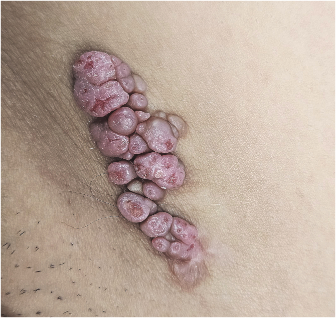 Figure 1
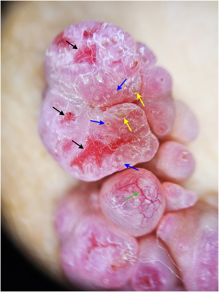 Figure 2
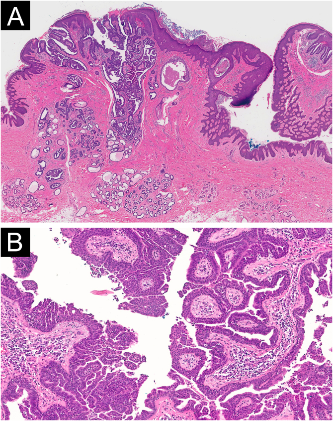 Figure 3
Peer Reviews
No public reviews on file for this paper yet. If you reviewed it on a platform where reviews are public (OpenReview, ICLR, NeurIPS, ICML), you can paste yours below so the community can read it here.
Videos
No videos yet. Explain this paper in a talk, walkthrough, or lecture? Add one.
Taxonomy
TopicsCancer and Skin Lesions · Sympathectomy and Hyperhidrosis Treatments · Genetic and rare skin diseases.
Dear Editor,
Syringocystadenoma Papilliferum (SCAP) is a benign adnexal tumor characterized by apocrine differentiation.1 It often manifests at an early age, with approximately 50% of cases identified at birth, and 15%‒30% emerging during puberty.2 While most SCAPs develop de novo, up to 40% have been reported to arise from a pre-existing sebaceous nevus.3
The histogenesis mechanisms underlying SCAP remain unclear. Genetic studies have identified mutations in the HRAS gene in SCAPs associated with sebaceous nevi, and RAS and BRAF mutations in sporadic lesions.1
SCAP occurs predominantly on the head and neck region, accounting for 75% of reported cases.3 Other less common sites include the arms, chest, axilla, scrotum, perineal, and inguinal regions.2 Clinically, SCAP manifests in three forms: solitary nodule, plaque, and linear type, with the latter being extremely rare.2 This paper presents an unusual case of a congenital, de novo SCAP with a linear configuration located in the inguinal region.
A 29-year-old female presented with a history of several pink lesions on the right inguinal region since birth. Physical examination revealed multiple pink dome-shaped papules with central umbilication and some exhibiting surface erosions, arranged in a linear configuration (Fig. 1). Dermoscopy showed pink papillomatous structures with erosions, white rosettes, white circles, yellowish crusts, and telangiectasias (Fig. 2). Suspecting an adnexal tumor based on clinical and dermoscopic findings, an incisional biopsy was performed. Histopathological analysis revealed hyperkeratosis and acanthosis of the epidermis, with invaginations of tubular-papillary structures lined by a bilayer of cuboidal epithelium, exhibiting focal stratification and some evidence of apical secretion by decapitation. Numerous plasma cells were noted in the stroma. No evidence of cellular atypia was observed (Fig. 3).Fig. 1. Clinical image. Multiple pink dome-shaped papules with central umbilication and some with erosions on the surface, arranged in a linear plaque.Fig. 1. Fig. 2Dermoscopy image (×10): Pink papillomatous structures with erosions (black arrows), white circles (blue arrows), yellowish crusts (yellow arrows), and telangiectasias (green arrow).Fig. 2. Fig. 3(A) Histopathological image (Hematoxylin & eosin, 20×): Epidermal and dermal tumor. The epidermis shows hyperkeratosis and acanthosis with invaginations composed of papillary projections. (B) Histopathological image (Hematoxylin & eosin, 100×): Tubulo-papillary structures lined by a bilayer cuboidal epithelium with focal stratification and some evidence of apical secretion by decapitation. The tumoral stroma contains numerous plasma cells. No cellular atypia was observed.Fig. 3
The diagnosis of linear syringocystadenoma papilliferum was made on a comprehensive assessment of clinical, dermoscopic, and histopathologic findings. The patient subsequently underwent a complete tumor excision, and the diagnosis was confirmed by the definitive histopathological results.
The linear presentation of SCAP is extremely infrequent, with only 22 cases reported in the literature.2, 3, 4 The head and neck are the most commonly affected regions, accounting for 8 of these 22 cases. Notably, only two cases have been documented in the inguinal region.2, 3
Most cases of linear SCAP are de novo occurrences. Only two cases have been associated with Jadassohn's sebaceous nevus,2, 5 two with hidrocystoma and cystadenoma,2, 6 and one with apocrine tubular adenoma.2
Dermoscopic findings described in SCAP include red exophytic papillary structures with central umbilication, ulceration, hairpin vessels, polymorphous vessels, and comma vessels. Additional features may include white circles, crusts, yellowish scales, and pink-white globular structures.2, 3 The circle umbilicated structures might correspond to the open pseudocystic spaces of SCAP, but further studies are needed to validate these findings.
The differential diagnosis includes molluscum contagiosum, warts, epidermal nevi, lymphangioma circumscriptum, basal cell carcinoma, and other adnexal neoplasms. Therefore, clinical and dermoscopic features are crucial techniques to identify this tumor.
Malignant transformation of SCAP is rare. Most documented cases correspond to syringocistoadenocarcinoma papilliferum,7 although other malignancies such as ductal carcinoma have also been reported.8 There are no reports of malignant transformation specifically associated with lineal SCAP.3
This report presents a new and exceptional case of linear SCAP located in an atypical region, with distinctive clinical and dermoscopic findings. Although the malignant transformation of linear SCAP has not been described in the literature, it is prudent to perform a complete excision. This approach is warranted as non-linear SCAP has the potential to develop secondary malignancies.
ORCID ID
Christian Robles-Silva: 0000-0002-0263-8011
Valentina Ross: 0009-0009-3651-5536
Javier González: 0009-0008-8625-0399
Alex Castro: 0000-0003-4431-5293
Constanza Del Puerto: 0000-0003-4667-1873
Financial support
This research did not receive any specific grant from funding agencies in the public, commercial, or not-for-profit sectors.
Authors' contributions
Christian Robles-Silva: The study concept and design, data collection, writing of the manuscript or critical review of important intellectual content, data collection, analysis and interpretation, effective participation in the research guidance; intellectual participation in the propaedeutic and/or therapeutic conduct of the studied cases; critical review of the literature; final approval of the final version of the manuscript.
Valentina Ross: Writing of the manuscript or critical review of important intellectual content; data collection, analysis and interpretation; effective participation in the research guidance; critical review of the literature; final approval of the final version of the manuscript.
Javier González: Writing of the manuscript or critical review of important intellectual content; data collection, analysis and interpretation; effective participation in the research guidance; critical review of the literature; final approval of the final version of the manuscript.
Alex Castro: Data collection, or analysis and interpretation of data; writing of the manuscript or critical review of important intellectual content; data collection, analysis and interpretation; critical review of the literature; final approval of the final version of the manuscript.
Constanza Del Puerto: The study concept and design; data collection, or analysis and interpretation of data; writing of the manuscript or critical review of important intellectual content; data collection, analysis and interpretation; effective participation in the research guidance; intellectual participation in the propaedeutic and/or therapeutic conduct of the studied cases; critical review of the literature; final approval of the final version of the manuscript.
Research data availability
Does not apply.
Conflicts of interest
None declared.
The reference list from the paper itself. Each links out to its DOI / PubMed record.
- 1Alegría-Landa V.Jo-Velasco M.Santonja C.Eraña I.Vergara-Sanchez A.Kutzner H.Syringocystadenoma papilliferum associated with verrucous carcinoma of the skin in the same lesion: report of four cases J Cutan Pathol.47202012163144966510.1111/cup.13572 · doi ↗ · pubmed ↗
- 2Chauhan P.Chauhan R.K.Upadhyaya A.Kishore S.Dermoscopy of a rare case of linear syringocystadenoma papilliferum with review of the literature Dermatol Pract Concept.8201833382944557210.5826/dpc.0801 a 07PMC 5808369 · doi ↗ · pubmed ↗
- 3Monaco M.González V.M.Vigovich F.A.Larralde M.Syringocystadenoma papilliferum in the scalp, with a linear presentation An Bras Dermatol.9820234064093662140410.1016/j.abd.2021.11.011PMC 10173062 · doi ↗ · pubmed ↗
- 4Tayyebi Meibodi N.Nahidi Y.Izanlu M.Davoodi N.Davoodi S.Perianal linear syringocystadenoma papilliferum: a case report with review of the literature Iran J Pathol.1820234834873802454710.30699/IJP.2023.1999659.3094 PMC 10646747 · doi ↗ · pubmed ↗
- 5Dawn G.Gupta G.Linear warty papules on the neck of a young woman: syringocystadenoma papilliferum (SP) in a sebaceous nevus (SN)Arch Dermatol.13820021091109610.1001/archderm.138.8.1091-b 12164752 · doi ↗ · pubmed ↗
- 6Martorell-Calatayud A.Sanz-Motilva V.Garcia-Sales M.A.Calatayud-Blas A.Linear syringocystadenoma papilliferum: an uncommon event with a favorable prognosis Dermatol Online J.172011521906485 · pubmed ↗
- 7Zhang Y.Kong Y.Y.Cai X.Shen X.X.Kong J.C.Syringocystadenocarcinoma papilliferum: clinicopathologic analysis of 10 cases J Cutan Pathol.4420175385432833777710.1111/cup.12934 · doi ↗ · pubmed ↗
- 8Hügel H.Requena L.Ductal carcinoma arising from a syringocystadenoma papilliferum in a nevus sebaceus of Jadassohn Am J Dermatopathol.2520034904931463119010.1097/00000372-200312000-00006 · doi ↗ · pubmed ↗