Cardiovascular magnetic resonance as an initial screening tool in individuals with SLE and chest pain
Isak Samuelsson, Simon Thalén, Giorgia Grosso, Magnus Lundin, Henrik Engblom, Peder Sörensson, Iva Gunnarsson, Martin Ugander, Elisabet Svenungsson

TL;DR
This study shows that cardiovascular magnetic resonance (CMR) can help identify heart issues like coronary artery disease in people with lupus who experience chest pain.
Contribution
The study demonstrates that CMR can detect cardiac ischaemia more frequently than pericarditis or myocarditis in SLE patients with chest symptoms.
Findings
CMR identified coronary artery disease (CAD) and coronary microvascular dysfunction (CMD) in 35% of SLE patients with chest symptoms.
CMR findings led to changes in medical treatment for 32% of participants.
No cases of myocarditis were found among the 19 individuals studied.
Abstract
Individuals with SLE commonly report chest pain or discomfort. We performed cardiovascular magnetic resonance (CMR) to differentiate coronary artery disease (CAD), coronary microvascular dysfunction (CMD), pericarditis and myocarditis in individuals with SLE who presented with chest symptoms. We also assessed the clinical utility of CMR. Adults with SLE were included if reporting chest pain or dyspnoea suggestive of cardiac involvement to a rheumatologist between 2018 and 2023. Individuals underwent CMR, including quantitative myocardial perfusion mapping at rest and during adenosine stress if not contraindicated. CAD, CMD, pericarditis and myocarditis were identified by CMR. Confirmatory investigations were performed when indicated. We reviewed medical files to assess if CMR led to altered medical treatment or invasive interventions. Nineteen individuals with SLE (84% female) with a…
Genes, proteins, chemicals, diseases, species, mutations and cell lines named across the full text — each resolved to its canonical identifier and authoritative record.
Click any figure to enlarge with its caption.
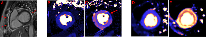 Figure 1
| Variable | Measure | Number of observations with complete data |
|---|---|---|
| Female, n (%) | 16 (84) | 19/19 |
| Age, years | 39 (31–55) | 19/19 |
| Disease duration, years | 11 (4–13) | 19/19 |
| SLICC/ACR DI | 1 (0–1) | 19/19 |
| Previous lupus nephritis | 7 (37) | 19/19 |
| Sjögren’s syndrome | 1 (5) | 19/19 |
| APS | 1 (5) | 19/19 |
| Previous VTE | 4 (21) | 19/19 |
| Prednisolone equivalent dose, mg | 5 (5–15) | 19/19 |
| HCQ treatment, n (%) | 15 (79) | 19/19 |
| Symptoms prompting inclusion | 19/19 | |
| Pleuritic chest pain | 10 (53) | |
| Angina-like chest pain | 2 (11) | |
| Other types of chest pain | 5 (26) | |
| Dyspnoea suggestive of cardiac involvement | 2 (11) | |
| SLEDAI-2K | 5 (4–6) | 19/19 |
| C-reactive protein, mg/L | 3 (1–4) | 17/19 |
| Sedimentation rate, mm | 27 (15–50) | 17/19 |
| Plasma albumin, g/L | 34 (30–37) | 17/19 |
| Platelet count, ×109/L | 320 (220–360) | 17/19 |
| Leucocyte count, ×109/L | 6 (4–7) | 17/19 |
| Lymphocyte count, ×109/L | 1 (1–1.5) | 17/19 |
| Plasma creatinine, µmol/L | 75 (61–88) | 19/19 |
| Albumin-creatinine ratio in urine, mg/mmol | 3 (1–19) | 15/19 |
| ANA, n (%) | 12 (100) | 12/19 |
| Anti-dsDNA, n (%) | 10 (59) | 18/19 |
| Anti-nucleosome, n (%) | 14 (82) | 17/19 |
| Anti-Sm, n (%) | 4 (24) | 17/19 |
| Anti-SmRNP, n (%) | 7 (41) | 17/19 |
| Anti-RNP68, n (%) | 3 (18) | 17/19 |
| Anti-ribosomal P, n (%) | 2 (12) | 17/19 |
| Anti-CL IgG, n (%) | 2 (12) | 17/19 |
| Anti-CL IgM, n (%) | 0 (0) | 16/19 |
| Anti-β2GP1 IgG, n (%) | 2 (12) | 17/19 |
| Anti-β2GP1 IgM, n (%) | 0 (0) | 16/19 |
| Lupus anticoagulant, n (%) | 2 (14) | 14/19 |
| Anti-SSA/Ro52, n (%) | 9 (53) | 17/19 |
| Anti-SSA/Ro60, n (%) | 12 (71) | 17/19 |
| Anti-SSB, n (%) | 6 (35) | 17/19 |
| C1q, mg/L | 100 (72–121) | 14/19 |
| C3, g/L | 0.8 (0.6–1) | 16/19 |
| C4, g/L | 0.1 (0.1–0.2) | 13/19 |
| C3d, mg/L | 7 (6–8) | 15/19 |
| Variables | Measure | Number of observations with complete data |
|---|---|---|
| Risk of CVD according to ref, | 17/19 | |
| Low to moderate risk | 10 (59) | |
| High risk | 1 (6) | |
| Very high risk | 6 (35) | |
| CVD | 5 (26) | 19/19 |
| CAD | 3 (16) | 19/19 |
| Creatine-based eGFR | 77 (69–93) | 19/19 |
| Type 1 or type 2 diabetes, n (%) | 0 (0) | 19/19 |
| Smoking status, n (%) | 18/19 | |
| Current | 0 (0) | |
| Previous | 9 (50) | |
| Never | 9 (50) | |
| Systolic BP, mm Hg | 120 (110–130) | 19/19 |
| Diastolic BP, mm Hg | 79 (71–83) | 19/19 |
| Antihypertensive treatment, n (%) | 9 (47) | 19/19 |
| BMI, kg/m2 | 25 (20–28) | 19/19 |
| Total cholesterol, mmol/L | 4.7 (4.2–5.0) | 14/19 |
| LDL, mmol/L | 2.6 (2.2–2.7) | 13/19 |
| HDL, mmol/L | 1.5 (1.2–1.7) | 14/19 |
| Triglycerides, mmol/L | 1.3 (1.0–1.9) | 14/19 |
| Cholesterol-lowering treatment, n (%) | 4 (21) | 19/19 |
| CMR variable | Measure | Number of observations with complete data |
|---|---|---|
| LVEDV, mL | 148 (125–157) | 19/19 |
| LVEDVI, mL/m2 | 78 (68–87) | 19/19 |
| LVESV, mL | 57 (50–71) | 19/19 |
| LVESVI, mL/m2 | 32 (27–53) | 19/19 |
| SV, mL | 79 (71–93) | 19/19 |
| SVI, mL/m2 | 44 (36–51) | 19/19 |
| LVM, g | 103 (93–121) | 19/19 |
| LVMI, g/m2 | 60 (53–67) | 19/19 |
| LVEF, % | 59 (53–64) | 19/19 |
| ECV, % | 28 (25–29) | 19/19 |
| Myocardial T1, ms | 1316 (1286–1357) | 17/19 |
| Myocardial T2, ms | 43 (43–46) | 11/19 |
| Rest perfusion, mL/min/g | 0.9 (0.9–1.1) | 14/19 |
| Stress perfusion, mL/min/g | 2.7 (2.5–3.0) | 14/19 |
| Myocardial LGE, % | 4 (22) | 18/19 |
| Pericardial LGE, % | 1 (6) | 18/19 |
| Pericardial effusion >5 mm, % | 3 (17) | 19/19 |
| Index | Age group (years) | Sex | SLE duration (years) | Previous CAD | CMR findings | Global perfusion defect | Regional perfusion defect | Coronary angiography findings | Clinical conclusion | Medical intervention |
|---|---|---|---|---|---|---|---|---|---|---|
| 1 | 20–25 | Female | 12 | No | CMD | Yes | No | Not performed | CMD | Medications altered |
| 2 | 40–45 | Male | 6.5 | CMR-verified MI at inclusion, but no CAD at coronary angiography 4 months prior to inclusion | Previous MI and CMD or balanced CAD in all three major coronary arteries | Yes | Yes | Complete blockage in a side branch of the LCx and CMD in the main LCx | CAD | Medications altered |
| 3 | 55–60 | Female | 0.1 | No CAD | Pericardial effusion | Not tested | Not tested | Not performed | Pericarditis | Medications altered |
| 4 | 55–60 | Female | 12 | Previous MI with PCI of the LAD | Reduced LVEF, general hypokinesia and a subendocardial perfusion rest defect. Questionable stress effect. | Inconclusive | Yes | Significant atherosclerosis affecting all major coronary arteries | CAD | CABG and medications altered |
| 5 | 55–60 | Female | 27 | No | Pericarditis with active inflammation. Stress-induced ischaemia in the RCA territory. | No | Yes | Not performed | CAD and pericarditis | Medications altered |
| 6 | 55–60 | Female | 2.3 | No | Pericarditis with active inflammation | No | No | Not performed | Pericarditis | No change |
| 7 | 55–60 | Female | 13 | No | CMD | No | Yes | Not performed | CMD | Medications altered |
- —http://dx.doi.org/10.13039/501100004047Karolinska Institutet
- —http://dx.doi.org/10.13039/501100007687Svenska Läkaresällskapet
- —http://dx.doi.org/10.13039/501100007857Stiftelsen Konung Gustaf V:s 80-årsfond
- —http://dx.doi.org/10.13039/501100007949Reumatikerförbundet
- —http://dx.doi.org/10.13039/501100011727Stockholm läns landsting
- —http://dx.doi.org/10.13039/501100004359Vetenskapsrådet
- —http://dx.doi.org/10.13039/501100003793Hjärt-Lungfonden
Peer Reviews
No public reviews on file for this paper yet. If you reviewed it on a platform where reviews are public (OpenReview, ICLR, NeurIPS, ICML), you can paste yours below so the community can read it here.
Videos
No videos yet. Explain this paper in a talk, walkthrough, or lecture? Add one.
Taxonomy
TopicsPericarditis and Cardiac Tamponade · Viral Infections and Immunology Research · Cardiac Imaging and Diagnostics
Introduction
Chest pain or discomfort is frequently reported by individuals with SLE.1 These symptoms may originate from various lung and musculoskeletal conditions. Chest symptoms may also be due to heart disease, which has been associated with a twofold to threefold increased risk of mortality in SLE.2 Early identification of cardiac causes of chest symptoms is thus important to initiate cardioprotective treatment to ameliorate symptoms and improve long-term prognosis when possible.
In clinical practice, chest symptoms are often considered to be due to pleuropericarditis, a clinical criterion for SLE classification.3 4 Although rare, lupus myocarditis is another possible cause of chest symptoms in this population.5 Additionally, SLE increases risk of ischaemic heart disease (IHD) independent of traditional cardiovascular risk factors.69 This underscores the importance of considering pericarditis, myocarditis and IHD in individuals with SLE who present with chest symptoms.
The high risk of IHD in SLE is generally assumed to be caused by accelerated coronary artery disease (CAD). This view is supported by several observations, including a meta-analysis of coronary CT angiography (CCTA) investigations.10 This study reported that coronary calcium scores greater than 0, that is, the cut-off for asymptomatic CAD, were more common in individuals with SLE compared with controls.10 Another study supporting this view studied individuals with SLE and matched comparators, who underwent invasive coronary angiography on clinical indications.11 Although individuals with SLE were on average 21 years younger than their controls, both groups had similar extent of CAD.11
Coronary microvascular dysfunction (CMD), another subtype of IHD, has recently gained increased attention in the general population.12 However, CMD is less studied in SLE. Few have investigated individuals with SLE and chest symptoms using advanced imaging techniques like stress cardiovascular magnetic resonance (CMR) or positron emission tomography (PET).13 14 These modalities detect CMD by measuring myocardial perfusion at rest and during pharmacological hyperaemic stress, with excellent agreement between the two.15 Previous studies using CMR or PET suggest that individuals with SLE have a higher risk of CMD compared with those without SLE,13 14 but more studies are warranted.
In conclusion, chest pain or discomfort is common in SLE and may originate from undetected heart disease, which is associated with increased mortality in this population.2 Pericarditis, CAD, CMD and myocarditis are important heart diseases to consider in individuals with SLE presenting with chest symptoms.3510 11 13 14 However, data on the relative prevalence of these cardiac conditions are limited, making diagnostic work-up harder. Therefore, we assessed the frequencies of pericarditis, CAD, CMD and myocarditis in individuals with SLE presenting with chest symptoms, using CMR as a screening modality. We also evaluated the clinical utility of CMR, as indicated by whether it led to altered medical management or not.
Methods
The frequencies of pericarditis, CAD, CMD and myocarditis were estimated in individuals with SLE and chest symptoms. These heart diseases were diagnosed using CMR together with clinical assessments performed by rheumatologists and cardiologists according to clinical routine. We assessed whether CMR results altered medical management or not, using medical records from follow-up visits.
Study participants
Individuals aged ≥18 years with SLE were included if reporting chest pain or dyspnoea suggestive of cardiac involvement to a rheumatologist at Karolinska University Hospital in Stockholm, Sweden, between April 2018 and May 2023. SLE was defined according to the 1982 revised American College of Rheumatology (ACR)3 and/or the Systemic Lupus International Collaborating Clinics (SLICC) classification criteria.16
Included individuals underwent CMR. We excluded individuals with prior allergic reactions to gadolinium-based contrast agents, severe claustrophobia, an estimated glomerular filtration rate (GFR) ≤30 mL/min/1.73 m^2^ or any other contraindication to CMR related to in-dwelling ferromagnetic material or implants where CMR was otherwise contraindicated.
Contraindications to pharmacological stress with adenosine, including atrioventricular (AV) blocks II–III before or during pharmacological stress, asthma or chronic obstructive pulmonary disease, did not constitute exclusion criteria. These individuals underwent CMR but without pharmacological stress. Thus, study participants were not selected for adenosine stress based on chest pain type or clinical presentation; rather, the presence of contraindications to adenosine stress.
CMR protocol
CMR was performed at 3 or 1.5 T according to a predefined protocol outlined in online supplemental figure S1. The CMR protocol included full-coverage cine imaging to assess ventricular volumes and function. Late gadolinium enhancement (LGE) was used to assess the presence of myocardial scarring and enhancement of the pericardium. Quantitative stress first-pass myocardial perfusion images were screened for perfusion defects. Quantitative stress perfusion maps were assessed for regional and global myocardial perfusion defects. Native T1 and T2 maps and extracellular volume maps were assessed for diffuse myocardial fibrosis, myocardial oedema and infiltrative cardiomyopathies. CMR image interpretation was performed in clinical routine by at least one experienced physician with European Association of Cardiovascular Imaging level III certification not blinded to the investigated individual’s medical history.
Pericarditis was evaluated according to Adler et al,17 with pericardial thickening or inflammation, as evidenced by LGE and/or oedema, considered positive. Myocarditis was evaluated according to the 2018 Lake Louise criteria with at least one T1-based criterion of non-ischaemic myocardial injury as well as T2-based signs of myocardial oedema considered positive.18
CAD and CMD were evaluated according to Kotecha et al.19 A regional perfusion defect identified by quantitative perfusion mapping with a regional stress myocardial blood flow (MBF) ≤1.94 mL/g/min in a coronary territory, or, in the absence of a discrete regional defect, the presence of a visual perfusion defect, together with a global stress MBF ≤2.25 mL/g/min, was considered indicative of CAD. CMD was deemed present either when no regional or visual perfusion defect was observed but the global stress MBF was ≤2.25 mL/g/min, or when a regional perfusion defect was accompanied by a corresponding regional stress MBF >1.94 mL/g/min. CMR findings indicative of pericarditis, CAD and CMD are illustrated in figure 1.
Three examples of pathologies in three different individuals with SLE. Late gadolinium enhancement imaging in short axis (A) reveals hyperenhancement of the pericardium (red arrows) indicative of pericarditis. Quantitative perfusion images in short axis at rest (B) and stress (C) reveal a regional perfusion defect (red arrow) indicative of obstructive coronary artery disease. Quantitative perfusion images in short axis at rest (D) and stress (E) with a modest perfusion increase during stress indicative of microvascular dysfunction.
Definitions of the study outcomes
Individuals with CMR findings suggestive of CAD or CMD were referred to a cardiologist, while those with findings supporting pericarditis or myocarditis were followed up by a rheumatologist. Individuals with neither pericarditis, CAD, CMD nor myocarditis at CMR were classified as having chest symptoms of non-cardiac origin.
Presence of pericarditis, CAD, CMD and/or myocarditis was determined according to the treating cardiologist or rheumatologist at follow-up. The treating physician was informed of the CMR results and performed additional testing, such as invasive coronary or CT angiography, when clinically indicated rather than per protocol. We also assessed whether medical management, defined as invasive interventions or altered drug prescriptions, was changed during follow-up because of the CMR results. Retrospective medical file review was used for data collection on the study outcomes.
Other variables
Retrospective medical file review was used to collect data on symptoms prompting inclusion categorised into angina-like chest pain, pleuritic chest pain, other types of chest pain and dyspnoea suggestive of cardiac involvement. Angina-like chest pain was defined as chest pain triggered by exercise and/or relieved by nitrates or rest. Pleuritic chest pain was defined as becoming worse with breathing or positional changes. Chest pain not meeting any of the previous two definitions was classified as other types of chest pain. Dyspnoea suggestive of cardiac involvement was defined as exercise-induced dyspnoea suggestive of cardiac ischaemia.
SLE disease duration, smoking status and comorbidities were collected using retrospective medical file review up until CMR. Reported comorbidities included previous cardiovascular disease, defined as previous CAD, stroke, transient ischaemic attack, aortic aneurysm or peripheral arterial disease. These comorbidities were also reported separately. Previous CAD was defined as a medical history of myocardial infarction (MI), unstable angina or chronic coronary syndrome. MI was defined as medical files consistent with the fourth universal definition of MI20 or CMR findings of previous MI at inclusion in those without any documented MI in their medical records. Further definitions of comorbidities are provided in online supplemental table S1.
Furthermore, we reported prevalence of previous venous thromboembolism and diabetes defined in online supplemental table S1, lupus nephritis according to kidney biopsies or Tan et al,3 secondary antiphospholipid syndrome (APS) as defined by Miyakis et al21 and secondary Sjögren’s syndrome as defined by Vitali et al.22 The time between inclusion and CMR was calculated using data from medical records.
Blood and urine samples were collected adjacent to CMR and analysed as per clinical routine at the Department of Clinical Chemistry, Karolinska University Hospital. Creatinine-based estimated GFRs were calculated according to the revised Lund-Malmö GFR estimating equation.23 ANA screening was performed using indirect immunofluorescence in Hep-2 cells (Immunoconcepts, Sacramento, California, USA). Antibodies towards double‐stranded DNA, nucleosomes, Smith (Sm), ribonucleoprotein (RNP) 68, Sm/RNP, ribosomal P, cardiolipin immunoglobulin G (IgG)/IgM, β_2_ glycoprotein 1 IgG/IgM, Sjögren’s syndrome antigen A (SSA)/Ro52, SSA/Ro60 and Sjögren’s syndrome antigen B were analysed in serum by BioPlex 2200 (Bio-Rad Laboratories, Hercules, California, USA).
Laboratory data and retrospective medical file review were used to assess disease activity according to the Systemic Lupus Erythematosus Disease Activity Index 2000 (SLEDAI-2K)24 and damage accrual according to the Systemic Lupus International Collaborating Clinics/American College of Rheumatology Damage Index (SLICC/ACR DI).25 Data on laboratory results, comorbidities and blood pressure were used to estimate traditional cardiovascular risk according to the European Society of Cardiology (ESC) and Visseren et al.26 Blood pressure and body mass index were measured at the time of CMR.
Statistical analysis
The distributions of study variables were described using descriptive statistics, that is, median and IQR, or frequencies, as appropriate. No statistical testing was performed, as this study had limited power and was descriptive.
Patient and public involvement
Neither individuals with SLE nor the public were involved in the design, conduct, reporting or dissemination plans of this study.
Results
Nineteen individuals with SLE were included, of whom 16 (84%) were females. Median age and SLE disease duration at CMR were 39 (IQR 31–55) and 11 (IQR 4–13) years, respectively. Included individuals had a median SLICC/ACR DI of 1 (IQR 0–1) and a median SLEDAI-2K score of 5 (IQR 4–6). Seven out of 19 (37%) had a history of lupus nephritis, while secondary APS and secondary Sjögren’s syndrome were less common, corresponding to 1 of 19 (5%) and 1 of 19 (5%) individuals, respectively (table 1).
Pleuritic chest pain was the most common symptom at inclusion, reported by 10 of 19 (53%) individuals. Other symptoms prompting inclusion were less common. Angina-like chest pain was present in 2 of 19 (11%) individuals, while other types of chest pain were reported by 5 of 19 (26%) individuals. Dyspnoea suggestive of cardiac involvement was present in 2 of 19 individuals (11%). Further baseline characteristics are presented in table 1.
Assessment of traditional cardiovascular risk according to ESC and Visseren et al26 followed a bimodal distribution. Out of 17 of the 19 (89%) individuals with complete data, 10 (59%) were classified as low to moderate risk. One (6%) individual was classified as high risk. Six (35%) individuals were classified as very high risk, of whom three (16%) had CAD prior to inclusion. Traditional cardiovascular risk factors at inclusion are presented in more detail in table 2.
The median time between inclusion and CMR was 10 (IQR 4–23) days. Pharmacological stress was used in 14/19 (74%) individuals. Reasons for not performing or interrupting pharmacological stress in enrolled individuals included asthma (n=2), AV block during infusion (n=1), miscommunication in the referral for CMR (n=1) and failure to obtain vascular access (n=1), which also hindered the administration of the gadolinium-based contrast agent.
CAD and CMD were together more common than pericarditis and myocarditis, corresponding to 3 of 14 (21%), 2 of 14 (14%), 3 of 19 (16%) and 0 of 19 (0%) individuals, respectively. Collectively, pericarditis, CAD and/or CMD were found in 7 of 19 (37%) individuals. Note that these diseases were not mutually exclusive, as one individual had both CAD and pericarditis. More CMR-related estimates not reported as the primary outcome are presented in table 3.
Two of the three individuals with prior CAD were diagnosed with recurrent CAD after being investigated with stress CMR. One had a previous MI successfully treated with percutaneous coronary intervention (PCI) of the left anterior coronary artery. Despite previous PCI and medical treatment, coronary angiography prompted by stress CMR revealed significant coronary obstruction affecting all three major coronary arteries. This finding resulted in coronary bypass grafting and adjusted medical therapy (individual 4, table 4).
A second individual was classified as having prior CAD based on CMR findings which indicated a previous MI, despite no documented MI in the medical records and no significant atherosclerosis on a coronary angiography performed 4 months earlier. Myocardial perfusion mapping showed decreased perfusion, and repeat coronary angiography revealed an index of microcirculatory resistance of 25 in the main left circumflex artery (LCx) and a chronically occluded marginal branching from the LCx. This individual was diagnosed with CAD and prescribed several new medications due to this finding (individual 2, table 4).
We also sought to evaluate CMR as a screening modality, which led to changed medical management in six of seven individuals with CAD, CMD and/or pericarditis. All individuals with CAD or CMD underwent changes in medical therapy due to altered drug prescriptions, but also coronary bypass grafting in one case. Two out of three individuals with pericarditis underwent changes in prescribed medical treatment (table 4).
Twelve individuals (83% female) with a median age of 37 (IQR 30–46) years and a median SLE duration of 11 (IQR 7–14) years showed no signs of CAD, CMD or perimyocarditis on CMR. Consequently, they did not undergo any further diagnostic work-up or therapeutic interventions. Although T2 mapping was not performed in all individuals, none of those with missing T2 data had elevated T1 values nor missing data regarding T1 measurements. Myocarditis was therefore excluded in accordance with the 2018 Lake Louise criteria. Four of the 12 individuals did not undergo adenosine stress testing due to asthma (n=1), AV block during adenosine infusion (n=1), dysfunctional peripheral lines (n=1) or referral error (n=1). As a result, CAD and CMD could not be completely ruled out in these individuals.
Discussion
This study assessed the frequencies of pericarditis, CAD, CMD and myocarditis in 19 individuals with SLE who presented with chest pain or dyspnoea suggestive of cardiac involvement. We performed CMR and used sequential clinical follow-up data obtained from visits with cardiologists or rheumatologists. Notably, cardiac ischaemia due to CAD or CMD was more common than pericarditis and myocarditis in our study sample. The CMR results, together with clinically indicated follow-up, led to altered medical management in 6 of 19 individuals, supporting CMR as a screening modality in individuals with SLE presenting with chest symptoms.
In clinical practice, chest symptoms in individuals with SLE are often attributed to pleuropericarditis or musculoskeletal conditions. However, the present study highlights cardiac ischaemia due to CAD or CMD, rather than pericarditis, as an important cardiac cause of chest symptoms in SLE. Our results align with previous studies in which SLE has been identified as a risk enhancer for CAD10 11 and CMD,13 14 further emphasising the need to consider cardiac ischaemia in individuals with SLE. In this limited sample, we observed no cases of myocarditis. This is not surprising since myocarditis is fairly rare among adults with SLE.5
This study is the first to use fully quantitative stress CMR to assess CAD and CMD in SLE. However, a few studies are available for comparison.13 14 Ishimori et al14 used semiquantitative stress CMR and CCTA to investigate 20 women with SLE and chest pain, as well as 10 asymptomatic female comparators. The average age (41±11 years) and gender distribution in the SLE group were like the present study. However, in contrast to Ishimori et al,14 who excluded all individuals with established CAD or arrhythmias, we included individuals with SLE and chest symptoms regardless of previous diagnoses. Furthermore, their study participants were living in the USA, while ours were Swedish residents, enrolled approximately a decade later. Thus, differences in genetics and environmental exposures such as medical treatments, which have evolved substantially during the last decade,27 may account for substantial variation between study samples.
Ishimori et al14 reported that SLE was associated with CMD, which was diagnosed in 8 of 18 (44%) females with SLE. None of these had >50% stenosis on CCTA.14 In our study, 2 of 14 (14%) individuals had CMD, out of which none had previous CAD. This represents a lower frequency compared with Ishimori et al.14 However, given the differences in target populations, study designs and the limited sample sizes in both studies, a quantitative comparison is not applicable. Nevertheless, both studies support CMD as a considerable cause of chest symptoms in SLE.
Weber et al13 also studied CMD in SLE. These authors included individuals with SLE and matched comparators, all of whom had chest pain or dyspnoea prompting semiquantitative PET myocardial perfusion imaging. Matching was based on age, sex and traditional cardiovascular risk factors. After the exclusion of individuals with obstructive CAD and left ventricular systolic dysfunction, 42 individuals with SLE and 69 comparators were included. Subjects with SLE were similar regarding gender distribution (97% female) but were on average older (61±0.5 years) relative to the present study. CMD was diagnosed in 24 of 42 (57%) individuals with SLE and was significantly more common compared with controls.13 Collectively, the present study, together with previous studies,13 14 supports CMD as a considerable cause of chest symptoms in SLE.
It is not surprising that we identified individuals with symptomatic CAD. Accelerated atherosclerosis, including CAD, has been studied in SLE for more than 20 years, but most commonly in asymptomatic subjects.10 11 28 Although our study is too small to draw any conclusions regarding cardiovascular risk factors, SLE is an established risk factor for IHD independent of traditional cardiovascular risk.69 Disease-specific factors matter, including long8 29 and short8 9 disease duration and older age at diagnosis.7 8
Subsets of individuals with SLE carry specific risk factors that confer additional cardiovascular risk, such as antiphospholipid antibodies8 30 and nephritis with reduced GFR.31 32 Therapies for SLE also play a role. Long-term corticosteroids raise cardiovascular risk,33 whereas hydroxychloroquine appears protective in some studies.34 Because SLE is heterogeneous, cardiovascular findings from any single small cohort—including ours—should not be extrapolated to all individuals; each individual’s pericarditis, CAD, CMD and myocarditis risk profile is unique.
Diagnosing chest pain or discomfort in SLE may be clinically challenging because there is yet no established way to determine pretest probability of pericarditis, CAD, CMD and myocarditis in this population. We demonstrate that using stress CMR as a screening modality in individuals with SLE presenting with chest symptoms can aid clinicians in their assessments and guide further investigations. While transthoracic echocardiography excels in availability, provides a rapid haemodynamic assessment and excludes significant pericardial effusion, CMR is more informative. It is increasingly available and offers a single, non-invasive, radiation-free examination that can differentiate between major cardiac complications of SLE. CMR enables comprehensive evaluation of IHD,35 pericarditis36 and myocarditis18 and has shown promise in differentiating CAD and CMD.19 However, in ambiguous cases, invasive coronary angiography providing measurements of epicardial stenoses/occlusions and microcirculatory pathology may be necessary to separate CAD from CMD.12
Moreover, in the diverse and heterogeneous SLE population, the high negative predictive value of CMR significantly helps rule out these complications,37 thus preventing individuals from unnecessary downstream testing and invasive procedures. In conclusion, due to its advantages, it is not surprising that CMR emerged as an important screening tool, altering medical management in approximately one-third of the individuals in our study. The long-term prognostic impact of early CMR in SLE remains to be determined in future studies.
Strengths and limitations
This study is the first real-life clinical evaluation of individuals with SLE and chest symptoms using a comprehensive, state-of-the-art CMR protocol with fully quantitative stress myocardial perfusion. The CMR protocol provides non-invasive, radiation-free, robust and operator-independent assessments of pericarditis, CAD, CMD and myocarditis. In addition, all individuals with SLE met the 1982 ACR3 or the SLICC classification criteria for SLE,16 making the SLE diagnosis robust.
The present study is limited by the small sample size, which impacts the precision and generalisability of the observed outcome data. Assessment of SLE duration as a risk stratifier was not performed, as subdivision by disease duration and outcome would have yielded strata too small for robust statistical analysis. Individuals with SLE were not randomly assigned to CMR or standard of care, which would have been ideal for assessing the clinical value of CMR in this group.
CAD and CMD could not be assessed in 5 of 19 individuals due to asthma (n=2), AV block during adenosine infusion (n=1), malfunctioning peripheral lines (n=1) and referral error (n=1). Instead of excluding these individuals, they underwent CMR without adenosine stress to preserve precision and generalisability of pericarditis and myocarditis estimates, as our study was already impacted by a small sample size. This approach provided a realistic reflection of the real-world clinical utility of CMR in SLE, where similar practical limitations frequently occur.
The study has also limitations related to potential misclassification of the study outcomes. While the individuals with SLE were recruited and included prospectively, most data were collected retrospectively, which inherits risks regarding accuracy. Furthermore, performing invasive coronary angiography or CCTA at follow-up per protocol, rather than only when clinically indicated, would have been preferable to differentiate CMD and CAD. Additionally, CMR was performed at a median of 10 days after referral. During this time, some individuals had already begun corticosteroid treatment for suspected pericarditis, potentially leading to an underestimation of the frequency of pericarditis detected in the present study.
Conclusions
This study highlights the importance of considering cardiac ischaemia, namely CAD and CMD, in individuals with SLE and chest symptoms. In this study, CMR facilitated early identification and guided follow-up investigations and treatment of these cardiac conditions, known to be major causes of morbidity and mortality in SLE. Larger studies are needed to confirm our findings and to prospectively evaluate the long-term prognostic impact of early CMR in symptomatic individuals with SLE.
Supplementary material
10.1136/lupus-2025-001652online supplemental file 1
The reference list from the paper itself. Each links out to its DOI / PubMed record.
- 1Svenungsson E Gunnarsson I Illescas-Bäckelin V et al Quick Systemic Lupus Activity Questionnaire (Q-SLAQ): a simplified version of SLAQ for patient-reported disease activity Lupus Sci Med 20218 e 00047110.1136/lupus-2020-00047133972457 PMC 8112425 · doi ↗ · pubmed ↗
- 2Lee YH Choi SJ Ji JD et al Overall and cause-specific mortality in systemic lupus erythematosus: an updated meta-analysis Lupus (Los Angel)2016257273410.1177/096120331562720226811368 · doi ↗ · pubmed ↗
- 3Tan EM Cohen AS Fries JF et al The 1982 revised criteria for the classification of systemic lupus erythematosus Arthritis & Rheumatism 1982251271710.1002/art.17802511017138600 · doi ↗ · pubmed ↗
- 4Aringer M Costenbader K Daikh D et al European League Against Rheumatism/American College of Rheumatology Classification Criteria for Systemic Lupus Erythematosus Arthritis Rheumatol 2019711400123138546210.1002/art.40930 PMC 6827566 · doi ↗ · pubmed ↗
- 5du Toit R Karamchand S Doubell AF et al Lupus myocarditis: review of current diagnostic modalities and their application in clinical practice Rheumatology (Sunnyvale)2023625233410.1093/rheumatology/keac 40935861382 · doi ↗ · pubmed ↗
- 6Esdaile JM Abrahamowicz M Grodzicky T et al Traditional Framingham risk factors fail to fully account for accelerated atherosclerosis in systemic lupus erythematosus Arthritis Rheum 2001442331710.1002/1529-0131(200110)44:10<2331::aid-art 395>3.0.co;2-i 11665973 · doi ↗ · pubmed ↗
- 7Manzi S Meilahn EN Rairie JE et al Age-specific incidence rates of myocardial infarction and angina in women with systemic lupus erythematosus: comparison with the Framingham Study Am J Epidemiol 19971454081510.1093/oxfordjournals.aje.a 0091229048514 · doi ↗ · pubmed ↗
- 8Nived O Ingvarsson RF Jöud A et al Disease duration, age at diagnosis and organ damage are important factors for cardiovascular disease in SLE Lupus Sci Med 20207 e 00039810.1136/lupus-2020-00039832587062 PMC 7319716 · doi ↗ · pubmed ↗