Brown remodeling of white adipose tissue protects against abdominal aortic aneurysm via batokine FSTL1
Chunling Huang, Yuna Huang, Boshui Huang, Lei Yao, Zenghui Zhang, Luoxiao Dong, Chang Guan, Junping Li, Zhaoqi Huang, Sixu Chen, Yuan Jiang, Yuling Zhang, Jingfeng Wang, Yangxin Chen, Zhaoyu Liu

TL;DR
Brown remodeling of fat tissue protects against aortic aneurysms by secreting a protective protein called FSTL1.
Contribution
The study identifies FSTL1 as a novel protective batokine that inhibits vascular cell death in AAA.
Findings
AAA patients have reduced adipose tissue browning levels.
FSTL1 inhibits vascular smooth muscle cell apoptosis via DIP2A/AKT signaling.
FSTL1 supplementation reduces aortic dilation and AAA progression in mice.
Abstract
Abdominal aortic aneurysm (AAA) is a life-threatening vascular disease without effective medical therapies. Emerging evidence have suggested a crosstalk between adipose tissue and vascular cells. Besides, brown adipose tissue is considered beneficial for cardiovascular health. Nevertheless, whether brown remodeling of white adipose tissue would protect against AAA remains unclear. Here, we showed that patients with AAA had a decreased browning level of adipose tissue, and induction of adipose tissue browning significantly reduced AAA incidence and attenuated AAA development in mice. Using LC-MS/MS and proteomic analysis, we further identified Follistatin-like 1 (FSTL1) as a novel vessel-protective adipokine secreted by browning adipocytes. Mechanistically, FSTL1 inhibited VSMC apoptosis through DIP2A/AKT signaling. Furthermore, we demonstrated that adipocyte-specific deficiency of FSTL1…
Genes, proteins, chemicals, diseases, species, mutations and cell lines named across the full text — each resolved to its canonical identifier and authoritative record.
Click any figure to enlarge with its caption.
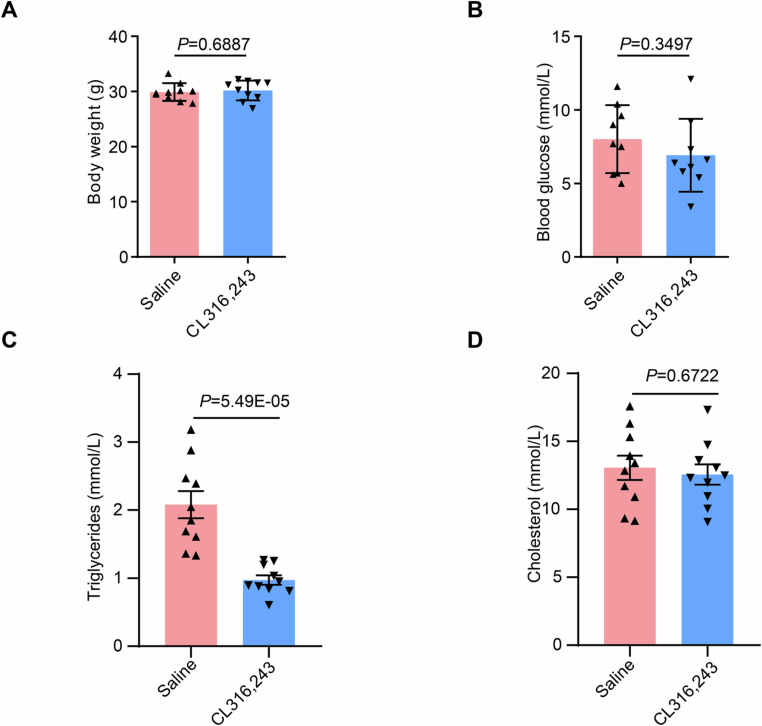 Figure 10
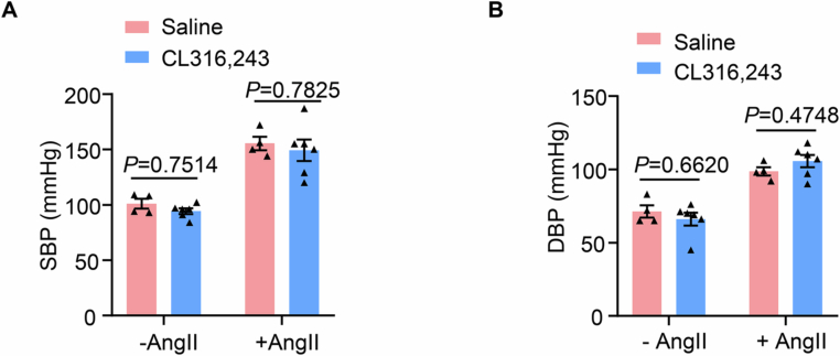 Figure 11
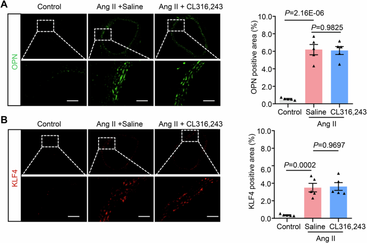 Figure 12
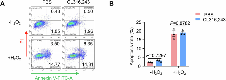 Figure 13
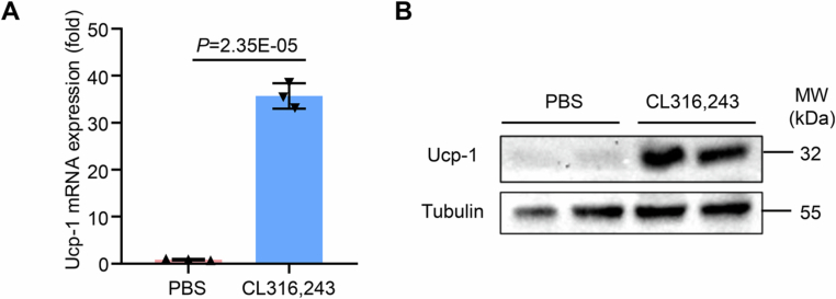 Figure 14
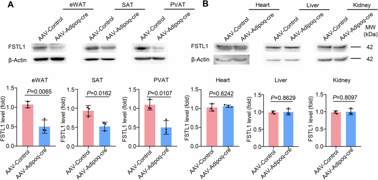 Figure 15
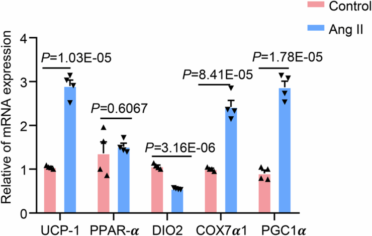 Figure 16
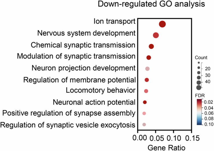 Figure 17
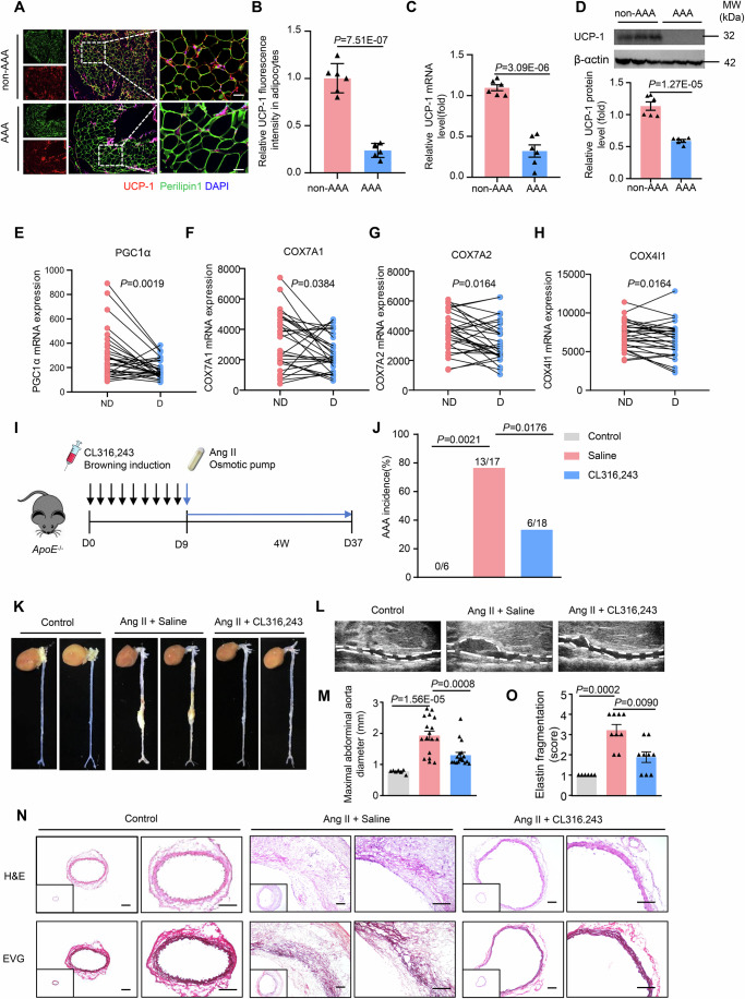 Figure 1
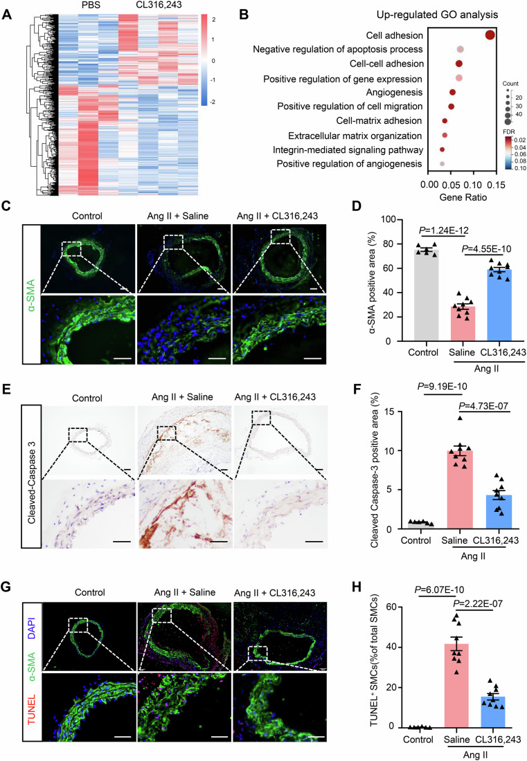 Figure 2
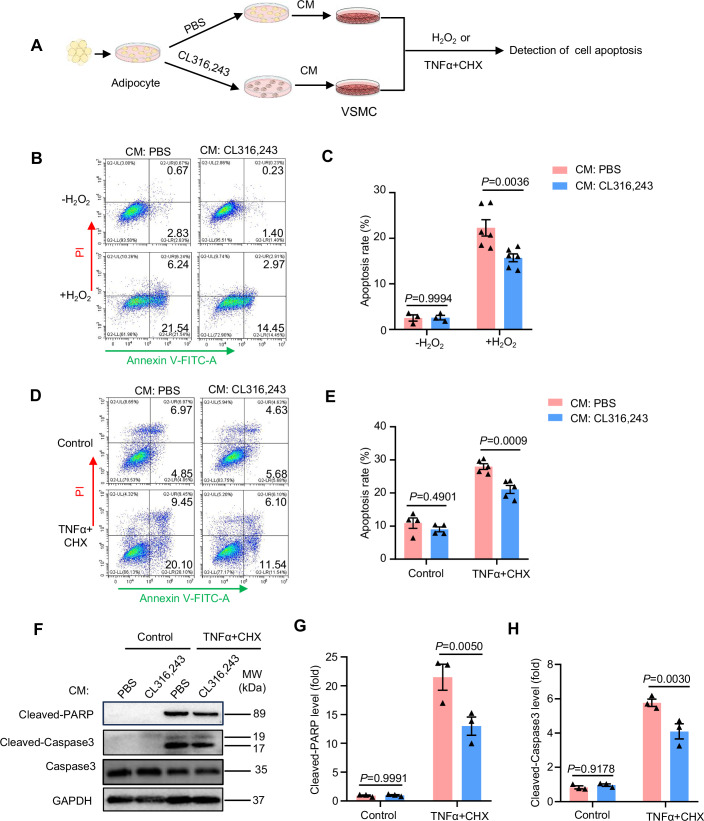 Figure 3
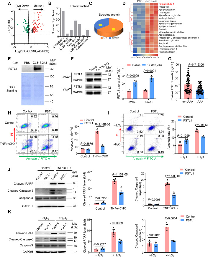 Figure 4
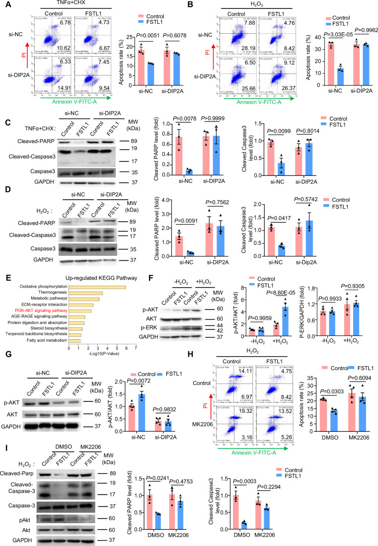 Figure 5
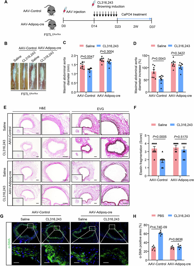 Figure 6
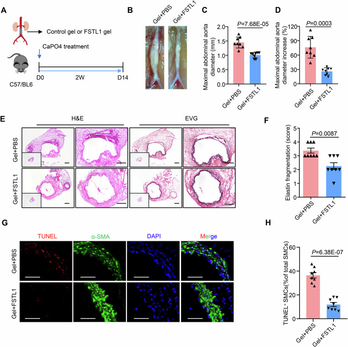 Figure 7
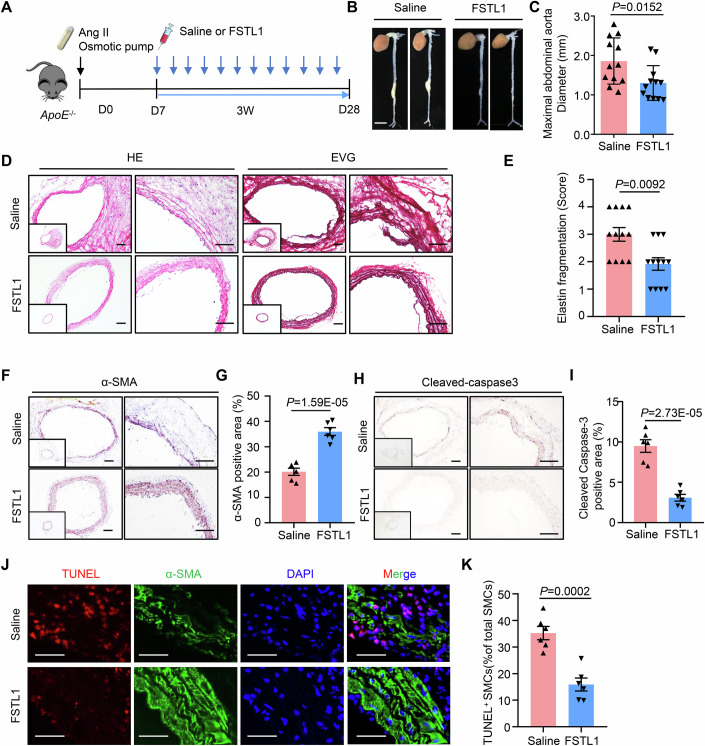 Figure 8
 Figure 9
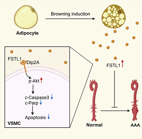 Figure 18
 Figure 19
- —http://dx.doi.org/10.13039/501100001809MOST | National Natural Science Foundation of China (NSFC)
- —http://dx.doi.org/10.13039/100017369Scientific and Technological Planning Project of Guangzhou City (广 州 市 科 技 规 划 项 目)
- —http://dx.doi.org/10.13039/501100021171GDSTC | Basic and Applied Basic Research Foundation of Guangdong Province (廣 東 省 基 礎 與 應 用 基 礎 研 究 專 項 資 金)
- —http://dx.doi.org/10.13039/501100012245GDSTC | Science and Technology Planning Project of Guangdong Province (S&T Project of Guangdong Province)
Peer Reviews
No public reviews on file for this paper yet. If you reviewed it on a platform where reviews are public (OpenReview, ICLR, NeurIPS, ICML), you can paste yours below so the community can read it here.
Videos
No videos yet. Explain this paper in a talk, walkthrough, or lecture? Add one.
Taxonomy
TopicsAdipose Tissue and Metabolism · Aortic aneurysm repair treatments · Cardiovascular Disease and Adiposity
The paper explainedProblemThe impact of adipose tissue on abdominal aortic aneurysm (AAA) development has received much attention, but whether brown remodeling of white adipose tissue would protect against AAA remains unclear.ResultsPatients with AAA had a decreased browning level of adipose tissue, and browning induction in mice attenuated AAA development. Browning adipocytes secreted a vessel-protective adipokine, Follistatin-like 1 (FSTL1), that inhibited vascular smooth muscle cell apoptosis. Adipocyte-specific FSTL1 deficiency abrogated the protective effect of browning induction, and supplementation of FSTL1 inhibited AAA development.ImpactOur study suggests the therapeutic potential of adipose tissue browning and FSTL1 supplementation for treating AAA.
Introduction
Abdominal aortic aneurysm (AAA) is a life-threatening vascular disease characterized by abnormal focal dilation of the abdominal aorta, which is mostly asymptomatic but can lead to sudden death due to aortic rupture (Kent, 2014). Current management of AAA is limited to surgical repair (Golledge, 2019), whereas clinical trials indicated that early elective surgical intervention for people with small AAAs does not reduce mortality (United Kingdom Small Aneurysm Trial et al, 2002). Most small AAAs continue to expand, and the risk of rupture increases with increasing aneurysm diameter (1998). Despite extensive research, there are no approved pharmacological therapies that can effectively prevent or decelerate AAA progression.
Adipose tissue is an important organ not merely for lipid storage and energy homeostasis but also an important source of bioactive molecules that regulate cardiovascular physiology (Polkinghorne et al, 2024). Several clinical and animal studies have suggested that excessive adipose tissue, mostly white adipose tissue accumulation, heightens the risk of developing AAA (Golledge et al, 2007; Kugo et al, 2019; Stackelberg et al, 2013). However, surgical removal of the adipose tissue is not clinically feasible for AAA intervention. Therefore, to remodel those adipose tissues to reduce their adverse effects while enhancing their beneficial effects may offer a better strategy. Recently, a large clinical study has revealed a potential role of brown adipose tissue in promoting cardiometabolic health (Becher et al, 2021). In mouse studies, brown adipose tissue has been found to protect against pathological cardiac remodeling and decrease myocardial damage (Lin et al, 2022; Marti-Pamies et al, 2023; Pinckard et al, 2021; Ruan et al, 2018). Besides, brown adipose tissue and brown remodeling of perivascular adipose tissue have also been demonstrated to protect from vascular dysfunction such as atherosclerosis (Adachi et al, 2022; Berbee et al, 2015; Shi et al, 2022). However, whether brown remodeling of white adipose tissue would protect against AAA development is largely unknown.
The loss of vascular smooth muscle cells (VSMC) is one of the major features in the pathogenesis of AAA (Lu et al, 2020). The reduced VSMC number in the aortic wall impairs their ability to produce connective tissue and repair elastin breaks, which leads to a weakened vessel and subsequent dilatation of the aorta (Lu et al, 2021). The apoptosis of VSMCs is a major cause of VSMC depletion in AAA development (Rowe et al, 2000; Thompson et al, 1997). Multiple studies have indicated an interplay between adipose tissue and the vascular system, in an endocrine or paracrine manner, through secreting adipokines and other factors (Akoumianakis et al, 2017; Wang et al, 2009; Zhang et al, 2018). Nevertheless, whether browning adipocytes secrete VSMC-protective factors remains to be elucidated.
In this study, we investigated the functional role of adipose tissue browning in AAA development. We found that browning induction significantly enhanced vessel integrity and attenuated AAA development in mice. In addition, browning adipocytes secreted Follistatin-like 1(FSTL1) and inhibited VSMC apoptosis. Supplementation of recombinant FSTL1 markedly inhibited AAA progression, indicating its therapeutic potential for AAA intervention.
Results
Decreased browning level correlates with AAA development in human
To investigate the association between the browning level of adipose tissue and AAA development, we first analyzed the expression of classic browning marker UCP-1 (uncoupling protein 1) in perivascular adipose tissue (PVAT) in patients with/without AAA. Compared to non-AAA patients, there was a significant reduction in UCP-1 levels in adipocytes of PVAT from patients with AAA, as determined by immunofluoresence staining (Fig. 1A,B). This decrease in UCP-1 expression was further confirmed at both the mRNA and protein levels by quantitative real-time PCR and western blot analysis, respectively (Fig. 1C, D). These data suggested a decreased browning level with AAA development. To further confirm this association, we carried out paired comparison analysis of adipose tissue surrounding the dilated abdominal aorta and non-dilated aortic neck in AAA patients using data from the Gene Expression Omnibus (GEO) database (GSE119717) (Piacentini et al, 2019). The results indicated that browning-associated genes such as PGC1α, COX7A1, COX7A2, and COX4I1 were significantly decreased in dilated PVAT compared to non-dilated PVAT (Fig. 1E–H). These data suggested that decreased browning level correlates with aortic dilation and possibly AAA progression.Figure 1. Browning of adipose tissue protects against AAA in vivo.(A) Representative immunofluorescent staining of uncoupling protein 1 (UCP-1, red) and Perilipin 1 (green) in perivascular adipose tissue (PVAT) in abdominal aortic aneurysm (AAA) patients or healthy control (n = 6). Scale bar = 50 μm. (B) Quantification of UCP-1 staining in (A). (C) Q-PCR analysis of UCP-1 mRNA level in PVAT of AAA patients and control (n = 6). (D) Western blot analysis of UCP-1 protein level in PVAT of AAA patients and control (n = 6). (E–H) Expression of browning-related genes in perivascular adipose tissues of dilated vs non-dilated AAA patients (GSE119717, n = 30). ND non-dilated, D dilated. (I) Experimental design. Twenty-week-old male ApoE^−/−^ mice were injected with saline or CL316,243 and then infused with Ang II (1000 ng/kg/min) for 28 days. (J) AAA incidence. (K) Representative photographs of the abdominal aorta taken during sacrifice. (L) Representative ultrasound images of abdominal aortas 4 weeks after Ang II infusion. (M) Quantification of maximal abdominal aortic diameter (n = 6, 17, 18). (N) Hematoxylin and eosin (H&E) and Elastica Van Gieson (EVG) staining of abdominal aortic sections. Scale bar = 50 μm. (O) Quantification of elastin fragmentation (n = 6, 9, 9). Paired or unpaired Student’s t test was used for comparing differences between two groups. Fisher’s exact test was used to analyze differences in AAA incidence. One-way ANOVA and Kruskal–Wallis H test were used for multiple group comparisons. Source data are available online for this figure.
Brown remodeling of white adipose tissue protects against AAA in vivo
To determine the role of brown remodeling of white adipose tissue in AAA development, we administered CL316,243, a drug widely used for browning induction, or saline control to 5-month-old ApoE^−/−^ mice and then constructed an angiotensin II (Ang II)-induced AAA model (Fig. 1I). After 9 days of administration of CL316,243, brown remodeling was successfully induced in both epididymal white adipose tissues (eWAT) and subcutaneous white adipose tissues (sWAT), as indicated by a more brownish gross appearance (Fig. EV1A), more adipocytes with small lipid droplets in a multilocular pattern (Fig. EV1B), and a much higher Ucp-1 expression (Fig. EV1C). In addition, perivascular adipose tissue around the abdominal aorta also showed a higher Ucp-1 expression as indicated by immunohistochemical staining (Fig. EV1D). However, mice's body weight and blood glucose levels were not changed (Fig. EV2A,B). Further analysis of serum lipids revealed a significant reduction in triglyceride levels but unchanged cholesterol levels (Fig. EV2C,D). Notably, CL316,243 administration significantly reduced AAA incidence (76.5% vs 16.7%, Fig. 1J) and decreased the maximal diameter of the abdominal aorta compared with saline control after Ang II infusion, as indicated by ex vivo analyses and ultrasound imaging (Fig. 1K–M). Histological analysis revealed that CL316,243 administration reduced elastin fragmentation in the abdominal aortas, as determined by Elastica van Gieson staining (Fig. 1N,O). Collectively, these results suggested that browning remodeling of white adipose tissue protects against AAA development in vivo. As hypertension has been suggested as one of the most important risk factors for AAA (Iribarren et al, 2007), we further examined the blood pressure in CL316,243-treated mice. Nevertheless, CL316,243 administration did not significantly change systolic or diastolic blood pressures with or without Ang II infusion, as compared to saline control (Fig. EV3A,B), suggesting that the protective role of CL316,243 during AAA is independent of blood pressure regulation.
Brown remodeling of white adipose tissue inhibits VSMC apoptosis in AAA
To gain insight into the mechanisms underlying the protective effects of adipose tissue browning on AAA, we performed a high-throughput RNA-sequencing of aortic tissues from mice after saline or CL316,243 treatment and Ang II modeling. A total of 911 genes were differentially expressed in mice after CL316,243 treatment, with 355 upregulated and 556 downregulated (Fig. 2A). Gene Ontology analysis revealed that the upregulated genes were significantly enriched in cell matrix adhesion and negative regulation of apoptotic process (Fig. 2B). As smooth muscle cells (SMCs) are the major component of the arterial wall and their apoptosis contributes to degeneration of aortic extracellular matrix and subsequent AAA formation and progression (Lu et al, 2020; Quintana et al, 2019a), we thus went on to investigate the content of SMCs. The results showed that Ang II treatment significantly decreased SMC content in the aortic wall, whereas CL316,243 administration significantly reversed this decrease as revealed by immunofluorescence staining of alpha-smooth muscle actin (SMA) (Fig. 2C,D). However, the markers for VSMC phenotypic switch, such as krüppel-like factor 4 (KLF4) and osteopontin (OPN), were unchanged (Fig. EV4). In addition, the apoptosis marker cleaved-Caspase3 was significantly decreased (Fig. 2E,F). Co-staining of TUNEL (terminal deoxynucleotidyl transferase dUTP nick end labeling) and SMA further indicated that apoptosis of VSMCs was significantly decreased by CL316,243 administration (Fig. 2G,H). All these results suggested that brown remodeling of white adipose tissue inhibits VSMC apoptosis in AAA.Figure 2. Browning of adipose tissue inhibits VSMC apoptosis in AAA.(A) Heatmap of differentially expressed genes in aortic tissue. (B) Gene ontology (GO) analysis. (C) Representative immunofluorescent staining of alpha smooth muscle actin (α-SMA) in suprarenal abdominal aortic sections (scale bar, 50 μm). (D) Quantification of α-SMA positive area as in (C) (n = 6, 9, 9). (E) Representative immunohistochemical staining of Cleaved-Caspase 3 in suprarenal abdominal aortic sections (scale bar, 50 μm). (F) Quantification of Cleaved-caspase 3-positive area as in (E) (n = 6, 9, 9). (G) Representative terminal deoxynucleotidyl transferase dUTP nick end labeling (TUNEL) and α-SMA staining of suprarenal aortic sections (scale bar, 50 μm). (H) Quantification of TUNEL-positive SMCs (n = 6, 9, 9). One-way ANOVA was used to determine statistical difference. Source data are available online for this figure.
Secretome of browning adipocytes inhibits VSMC apoptosis
Next, we investigated whether browning agent CL316,243 has a direct effect on VSMC apoptosis. Annexin V/PI staining combined with flow cytometry analysis indicated that direct CL316,243 treatment did not change H_2_O_2_-induced VSMC apoptosis (Fig. EV5A,B), suggesting that CL316,243 has an indirect effect on VSMCs. We further cultured human VSMCs with conditioned medium obtained from adipocytes that have been treated with PBS or CL316,243, and then examined VSMC apoptosis (Fig. 3A). As shown in Fig. EV6A,B, CL316,243 treatment robustly induced adipocyte browning, as indicated by dramatical increase of Ucp-1 expression. Notably, conditioned medium derived from CL316,243-treated adipocytes significantly inhibited VSMC apoptosis induced by either H_2_O_2_ or TNFα plus CHX, as measured by Annexin-V/PI assay (Fig. 3B–E). To further investigate the apoptotic process, we detected the level of selected apoptosis markers- the cleavage of poly ADP-ribose polymerase (PARP) and Caspase 3. The results showed that conditioned medium derived from CL316,243-treated adipocytes significantly decreased the cleavage of both PARP and Caspase 3 (Fig. 3F–H). Collectively, these results indicated that the secretome of browning adipocytes inhibits VSMC apoptosis.Figure 3. Secretome of browning adipocytes inhibits VSMC apoptosis.(A) Experimental design. Primary adipocytes were treated with PBS or CL316,243 for 24 h, and the culture medium (CM) was then collected and incubated with vascular smooth muscle cells (VSMCs), which further underwent H_2_O_2_ or TNFα plus cycloheximide (CHX) treatment to induce apoptosis. (B) Flow cytometric analysis of Annexin V-FITC staining in VSMCs treated with adipocyte CM and H_2_O_2_. (C) Quantification of apoptosis as in B (without H_2_O_2_, n = 3; with H_2_O_2_, n = 6). (D) Flow cytometric analysis of Annexin V-FITC staining in VSMCs treated with adipocyte CM and TNFα plus CHX. (E) Quantification of apoptosis as in (D) (without TNFα + CHX, n = 4; with TNFα + CHX, n = 5). (F) Western blot analysis of cleaved-PARP and cleaved-Caspase 3 in VSMCs treated with adipocyte CM and TNFα plus CHX. (G) Quantification of cleaved-PARP level (n = 3). (H) Quantification of cleaved-Caspase 3 level (n = 3). Two-way ANOVA was used to determine statistical difference. Source data are available online for this figure.
Identification of FSTL1 as a novel batokine inhibiting VSMC apoptosis
To explore the potential protective factors secreted by browning adipocytes, conditioned medium from PBS or CL316,243-treated adipocytes was subjected to liquid chromatography with tandem mass spectrometry (LC-MS/MS) analysis. A total of 106 proteins were identified to be differentially expressed in the medium of CL316,243-treated adipocytes, with a criterion of 1.5-fold change and P < 0.05, of which 64 were upregulated and 42 were downregulated (Fig. 4A). After subcellular localization classification of these proteins using UniProt database, 28 proteins classified as “secreted” were selected, with 18 upregulated and 10 downregulated after CL316,243 treatment (Fig. 4B,C). The upregulated secreted proteins were then subjected to cluster analysis (Fig. 4D). Among the 18 proteins that were upregulated in browning adipocytes, Follistatin-like 1 (FSTL1) emerged as a protein of particular interest because of its anti-apoptotic function in multiple cell types (Liang et al, 2014; Ogura et al, 2012; Ouchi et al, 2008; Wei et al, 2015). By western blot analysis, we confirmed that FSTL1 was dramatically increased in the conditioned medium of CL316,243-treated adipocytes (Fig. 4E). We further analyzed FSTL1 expression in adipose tissues in vivo. As shown in Fig. 4F, administration of browning agent CL316,243 significantly increased FSTL1 expression in both epididymal white adipose tissues (eWAT) and subcutaneous white adipose tissues (sWAT). In addition, we found that FSTL1 levels were significantly decreased in the plasma of AAA patients (Fig. 4G). After adjusting for age, sex, smoking status, drinking status, body mass index, systolic and diastolic blood pressure, heart rate, LDL, HDL, hypertension, and diabetes, plasma FSTL1 levels remained significantly associated with AAA after adjustment (OR 0.94, 95% CI 0.90–0.96, P < 0.001). Together, all these data suggested that FSTL1 may be a novel vessel-protective batokine.Figure 4. Identification of FSTL1 as a novel batokine inhibiting VSMC apoptosis.(A) Volcano plot of the proteins identified by liquid chromatography with tandem mass spectrometry (LC-MS/MS) (n = 3). (B) Subcellular localization categorization of differentially expressed proteins based on subcellular localization. (C) Pie-chart of the number of upregulated and downregulated secreted proteins. (D) Heatmap of the upregulating secreted proteins. (E) Western blot analysis of FSTL1 in CM of adipocytes treated with PBS or CL316,243 for 24 h. Coomassie brilliant blue (CBB) staining was used as a loading control. (F) Western blot analysis of FSTL1 expression in epididymal white adipose tissue (eWAT) and subcutaneous white adipose tissue (sWAT) (n = 3). (G) ELISA analysis of FSTL1 in the plasma of control (n = 68) and AAA patients (n = 55). Student’s t test was used to determine statistical difference. (H, I) Flow cytometric analysis of Annexin V-FITC staining in VSMCs treated with recombinant FSTL1 (100 ng/mL) and stimulated with TNFα plus CHX (H) or H_2_O_2_ (I) (n = 3). (J,** K**) Western blot analysis of cleaved-PARP and cleaved-Caspase 3 in VSMCs treated with recombinant FSTL1 and stimulated with TNFα plus CHX (J) (n = 4) or H_2_O_2_ (K) (n = 3). Two-way ANOVA was used to determine statistical difference. Source data are available online for this figure.
Next, we investigated whether FSTL1 inhibits VSMC apoptosis. The results showed that recombinant FSTL1 treatment significantly inhibits either TNFα plus CHX or H_2_O_2_-induced VSMC apoptosis, as measured by Annexin-V/PI staining and flow cytometry analysis (Fig. 4H,I). Consistently, recombinant FSTL1 treatment significantly decreased the cleavage of PARP and Caspase 3 (Fig. 4J,K). Together, our results suggested that the secreted FSTL1 inhibits VSMC apoptosis.
FSTL1 inhibits VSMC apoptosis via receptor DIP2A-mediated AKT activation
DIP2A is a cell surface receptor for FSTL1, which has been reported to mediate the protective effects of FSTL1 in endothelial cells (Ouchi et al, 2010). To investigate whether DIP2A mediates the protective effects of FSTL1 in VSMCs, we silenced DIP2A in VSMCs and then treated the cells with recombinant FSTL1. The results showed that DIP2A deficiency abrogated the inhibitory effect of FSTL1 on VSMC apoptosis induced by either TNFα plus CHX or H_2_O_2_ (Fig. 5A,B). In addition, decreased cleavage of PARP and Caspase 3 mediated by FSTL1 was also reversed in DIP2A-deficient VSMCs (Fig. 5C,D).Figure 5FSTL1 inhibits VSMC apoptosis via receptor DIP2A-mediated AKT activation.(A,** B**) Flow cytometric analysis of Annexin V-FITC staining in VSMCs silenced with DIP2A and stimulated with TNFα plus CHX (A) or H_2_O_2_ (B) in the absence or presence of FSTL1 (n = 3). (C, D) Western blot analysis of cleaved-PARP and cleaved-Caspase 3 in VSMCs silenced with DIP2A and stimulated with TNFα plus CHX (C) or H_2_O_2_ (D) (n = 3). (E) Kyoto Encyclopedia of Genes and Genomes (KEGG) analysis of differentially expressed genes in VSMCs treated with/without recombinant FSTL1 and stimulated with H_2_O_2_. (F) Western blot analysis of p-AKT and p-ERK in VSMCs treated with/without recombinant FSTL1 and stimulated with H_2_O_2_ (n = 4). (G) Western blot analysis of p-AKT in VSMCs silenced with DIP2A and treated with/without FSTL1 (n = 4). (H) Flow cytometric analysis of Annexin V-FITC staining in VSMCs treated with AKT inhibitor MK2206 and stimulated with H_2_O_2_ (n = 4). (I) Western blot analysis of cleaved-PARP and cleaved-Caspase 3 in VSMCs treated with AKT inhibitor MK2206 and stimulated with H_2_O_2_ (n = 3). Two-way ANOVA was used to determine statistical difference. Source data are available online for this figure.
To elucidate the mechanism underlying the effects of FSTL1 on VSMCs, we conducted RNA sequencing in H_2_O_2_-induced VSMCs treated with or without FSTL1. Kyoto Encyclopedia of Genes and Genomes (KEGG) analysis showed that the upregulated genes in FSTL1-treated VSMCs were enriched in the PI3K-AKT signaling pathway (Fig. 5E). We further confirmed the activation of AKT signaling by western blot analysis. As shown in Fig. 5F, FSTL1 treatment significantly enhanced H_2_O_2_-induced AKT phosphorylation, whereas it had no effect on ERK phosphorylation. In addition, FSTL1-mediated AKT phosphorylation was abolished in DIP2A-deficient VSMCs (Fig. 5G). Moreover, pharmacologic inhibition of AKT signaling by MK-2206, an allosteric AKT inhibitor, abrogated the inhibitory effect of FSTL1 on VSMC apoptosis (Fig. 5H). In addition, decreased cleavage of PARP and Caspase 3 mediated by FSTL1 was also abrogated by AKT inhibition (Fig. 5I). Together, these data suggested that the protective effect of FSTL1 in VSMCs is through the DIP2A/AKT pathway.
Adipocyte-specific FSTL1 deficiency abrogates the protective effect of browning induction against AAA
To determine whether the protective effect of browning induction against AAA is through FSTL1 secreted by adipocytes, we constructed adipocyte-specific FSTL1-deficient mice. Briefly, FSTL1^flox/flox^ mice were injected with AAV-Adipoq-Cre or AAV-control and then administered with CL316,243 to induce adipose tissue browning, followed by CaPO4 application to induce AAA formation (Fig. 6A). We first confirmed that FSTL1 expression after CL316,243 administration was significantly repressed by AAV-Adipoq-Cre injection in adipose tissues such as eWAT, sWAT, and PVAT (Fig. EV7A), but not in other tissues, such as heart, liver, and kidney (Fig. EV7B). Notably, we found that browning induction by CL316,243 significantly reduced aortic dilation and decreased the maximal diameter of the abdominal aorta in the AAV-Control group, whereas it showed minimal effects in adipocyte-specific FSTL1-deficient mice (Fig. 6B–D). In addition, browning induction significantly decreased arterial wall thickness and elastin fragmentation in AAV-control mice, whereas adipocyte FSTL1 deficiency abrogated these effects (Fig. 6E,F). Moreover, the increased SMC content by browning induction was also abrogated (Fig. 6G,H). Together, these results suggested that the protective effect of browning induction is through adipocyte-derived FSTL1.Figure 6. Adipocyte-specific FSTL1 deficiency abrogates the protective effect of CL316,243 against AAA.(A) Experimental design. AAV-Control or AAV-Adipoq-Cre was first injected into FSTL1^fl/fl^ mice, followed by CL316,243 treatment for 9 days and CaPO4 treatment for another 2 weeks to induce AAA formation. (B) Representative photographs of abdominal aortas (scale bar, 1 mm). (C) Quantification of maximal abdominal aortic diameter (n = 8). (D) Quantification of maximal abdominal aortic diameter increase (n = 8). (E) Representative H&E and EVG staining images of abdominal aortic sections (scale bar, 50 μm). (F) Quantification of elastin fragmentation (n = 8). (G) Representative immunofluorescent staining of α-SMA in suprarenal abdominal aortic sections (scale bar, 25 μm). (H) Quantification of α-SMA positive area as in (C) (n = 5). Two-way ANOVA was used to determine statistical differences. Source data are available online for this figure.
Hydrogel-patched recombinant FSTL alleviates AAA development
To further determine whether local FSTL1 treatment impacts AAA development, Pluronic F-127 Hydrogel containing recombinant FSTL1 was applied around the abdominal aorta after CaPO4 treatment (Fig. 7A). The results showed that FSTL1-containing hydrogel treatment significantly decreased CaPO4-induced aortic dilation and the maximal diameter of the abdominal aorta (Fig. 7B–D). Histological analysis further indicated that FSTL1-containing hydrogel treatment significantly decreased elastin fragmentation (Fig. 7E,F). In addition, TUNEL staining revealed that apoptosis of SMC in the abdominal aorta was significantly decreased by FSTL1-containing hydrogel treatment (Fig. 7G,H). All these data suggested that local treatment of recombinant FSTL1 around the abdominal aorta could alleviate AAA development.Figure 7. Hydrogel-patched recombinant FSTL alleviates AAA development.(A) Experimental design. Pluronic F-127 hydrogel containing recombinant FSTL1 was applied around the abdominal aorta after CaPO4 treatment in C57/BL6 mice. (B) Representative photographs of the abdominal aorta (scale bar, 1 mm). (C) Quantification of maximal abdominal aortic diameters (n = 8). (D) Quantification of maximal abdominal aortic diameter increase (n = 8). (E) Representative H&E and EVG staining images of abdominal aortic sections (scale bar, 50 μm). (F) Quantification of elastin fragmentation (n = 8). (G) Representative TUNEL and α-SMA staining of abdominal aortic sections (scale bar, 25 μm). (H) Quantification of TUNEL-positive SMCs (n = 8). Student’s t test and Mann–Whitney U tests were used to determine statistical difference. Source data are available online for this figure.
Systemic FSTL1 infusion attenuates AAA development
We next explored the therapeutic potential of FSTL1 infusion in attenuating AAA development. Starting from the 7th day after Ang II modeling, ApoE−/− mice were intraperitoneally injected with recombinant FSTL1 protein at a dose of 100 μg/kg or saline control every other day (Fig. 8A). After 3 weeks of treatment, the results showed that FSTL1 infusion significantly decreased the maximal diameter of the abdominal aorta (Fig. 8B,C). In addition, FSTL1 infusion significantly decreased Ang II-induced elastin fragmentation and increased vessel integrity (Fig. 8D,E). Moreover, FSTL1 infusion significantly increased SMC content as indicated by more SMA staining (Fig. 8F,G). The apoptosis of SMC was also significantly inhibited, as indicated by decreased cleaved-caspase 3 staining and co-staining of TUNEL and SMA (Fig. 8H–K). These results suggested that systemic FSTL1 infusion effectively inhibited VSMC apoptosis and restricted AAA development.Figure 8. Systemic FSTL infusion attenuates AAA development.(A) Experimental design. FSTL1 was injected intraperitoneally into Ang II-infused ApoE^−/−^ male mice every other day for 3 weeks, starting on day 7 of AAA induction. (B) Representative photographs of the abdominal aorta (scale bar, 4 mm). (C) Quantification of maximal abdominal aortic diameters (n = 12). (D) Representative H&E and EVG staining images of abdominal aortic sections (scale bar, 50 μm). (E) Quantification of elastin fragmentation (n = 12). (F) Representative immunohistochemical staining of α-SMA in abdominal aortic sections (scale bar, 50 μm). (G) Quantification of α-SMA positive area as in (F) (n = 6). (H) Representative immunohistochemical staining of cleaved-Caspase 3 in abdominal aortic sections. (I) Quantification of cleaved-Caspase 3-positive area as in (H) (n = 6). (J) Representative TUNEL and α-SMA staining of abdominal aortic sections (scale bar, 25 μm). (K) Quantification of TUNEL-positive SMCs (n = 6). Student’s t test and Mann–Whitney U tests were used to determine statistical difference. Source data are available online for this figure.
Discussion
Abdominal aortic aneurysm (AAA) is a life-threatening aortic disease without pharmacological approaches. Adipose tissue is composed of white adipose tissue (WAT) and brown adipose tissue (BAT), which differ morphologically and functionally. Multiple lines of evidence demonstrate that there is a significant expansion of white adipose tissue with abdominal aortic aneurysm development (Cronin et al, 2013; Stackelberg et al, 2013), whereas the prevalence and amount of brown adipose tissue declines with advancing age (Cypess et al, 2009), which is known to increase the risk of AAA(Quintana et al, 2019b). Although adipose tissue has been implicated in AAA development, little attention has been paid to its remodeling in AAA progression. Here, we found that AAA patients have a reduced browning level in perivascular adipose tissue, and browning induction in white adipose tissue significantly attenuated AAA development in mice. Our study suggested that remodeling the white adipose tissue by “re-browning” may be beneficial to prevent or slow down AAA progression.
Interestingly, while we observed significantly decreased browning levels in AAA patients, the expression of browning-associated genes was increased in Ang II-induced mouse models (Fig. EV8). Since Ang II is known to induce inflammatory responses that require increased energy expenditure, this elevated browning may represent a compensatory mechanism against Ang II-induced inflammation and metabolic stress. However, as AAA progresses, chronic inflammation and metabolic dysfunction may eventually overwhelm this protective response, leading to a decline in browning levels.
In the present study, β3 adrenergic receptor agonist treatment was used to induce white adipose tissue browning. Indeed, there are some other classical ways for browning induction, such as cold exposure and exercise, during which norepinephrine will be released by the activated sympathetic nervous system and binds to β3 adrenergic receptor on adipocytes to induce browning (Cannon et al, 2004; Seals et al, 1991; van Marken Lichtenbelt et al, 2009). However, it is worth noting that increased sympathetic excitability can also cause accelerated heartbeat and high blood pressure, which are important risk factors for AAA. Therefore, we should be cautious about cold exposure and exercise training as browning induction methods for practical use against AAA. Recently, some special dietary patterns such as intermittent fasting and caloric restriction have also been demonstrated to induce adipose tissue browning (Fabbiano et al, 2016; Lin et al, 2021). Interestingly, a previous study demonstrated that calorie restriction protects against experimental AAA in mice (Liu et al, 2016). Therefore, it is possible that calorie restriction may also exert its protection via adipose tissue browning.
Browning adipocytes are known to regulate energy metabolism and have thus been implicated for therapeutic exploitation for metabolic diseases, including Type 2 diabetes mellitus (T2DM) and obesity (Liu et al, 2013; Stanford et al, 2013). However, in our current study, although we observed a decrease in AAA incidence and development by browning induction, there was no significant change in metabolic parameters such as body weight and blood glucose levels, suggesting that adipose tissue browning may protect against AAA in a metabolism-independent manner. Interestingly, we found that adipocytes after brown induction dramatically increase the secretion of Follistatin-like 1 (FSTL1), which directly inhibits VSMC apoptosis and AAA development. Importantly, adipocyte-specific FSTL1 deficiency abrogates the protective effect of browning induction against AAA These studies suggested that the secretory function of browning adipocytes may play an essential role in protecting vascular cells and suppressing AAA development. However, a limitation of the current in vitro study is the use of human VSMCs to investigate the effects of mouse adipocyte-derived secretory factors. Future studies should validate these findings in a syngeneic system (e.g., mouse adipocytes co-cultured with primary mouse aortic VSMCs).
FSTL1 is a secreted glycoprotein and has previously been implicated to be a potential prognostic marker for cardiovascular diseases (Widera et al, 2012; Widera et al, 2009). Mark Gorelik et al found that the plasma levels of FSTL1 were elevated in toddler patients with acute Kawasaki disease (KD) (Gorelik et al, 2012), suggesting its level may be associated with acute arterial injury and coronary artery aneurysms (CAA) formation. However, considering the relatively small number of patients in the CAA group (only 7 cases) and the difference between CAA and AAA, the relationship between FSTL1 level and AAA development remains unclear. In this study, we found that plasma levels of FSTL1 were significantly lower in AAA patients, suggesting that reduced levels of FSTL1 may be associated with AAA development. However, whether the FSTL1 level could predict AAA development still needs further investigation.
Many recent studies indicated that FSTL1 not only serves as a molecular marker but also possesses important functions in the development of cardiovascular diseases. FSTL1 insufficiency could result in deformed mitral valves (Prakash et al, 2017), atrial and venous wall fibrosis (Jiang et al, 2020), and pulmonary vasculature defects (Tania et al, 2017). In addition, FSTL1 deletion exacerbates hypoxia-induced pulmonary hypertension(Zhang et al, 2017), wire-induced arterial injury (Miyabe et al, 2014), pressure overload-induced hypertrophy and cardiac failure (Shimano et al, 2011), etc. Importantly, FSTL1 overexpression or reconstitution has been reported to prevent myocardial infarction and cardiomyopathy (Lu et al, 2024; Oshima et al, 2008; Wei et al, 2015), reduce neointimal formation (Miyabe et al, 2014), and prevent against aortic dissection (Li et al, 2025). Many of the functions of FSTL1 in cardiovascular diseases are associated with its roles in cardiomyocytes and vascular cells. However, accumulating evidence also suggests a role of FSTL1 in adipocytes, as its deficiency could impair brown adipose tissue development and brown adipose tissue thermogenesis(Fang et al, 2019; Guggeri et al, 2024). Hereby, proteomic analysis of the culture medium from browning adipocytes, we identified FSTL1 as a vessel-protective adipokine. Importantly, supplementation of recombinant FSTL1 either at the beginning or during AAA progression significantly attenuates AAA development, suggesting its great potential for intervening AAA.
Notably, our proteomic analysis revealed some other secretory proteins alongside FSTL1 in the browning adipocyte medium. Periostin is an extracellular matrix protein known to regulate VSMC migration and osteoblastic switch, and has been suggested to promote atherosclerosis and vascular calcification (Alesutan et al, 2022; Li et al, 2006; Sun et al, 2021). Thrombospondin-4 is a member of the extracellular calcium-binding protein family and is linked to cell adhesion and migration (Lv et al, 2016), and has been reported to promote VSMC proliferation, local inflammation, atherogenesis, and restenosis(Frolova et al, 2010; Lv et al, 2016; Stenina et al, 2003). Given our focus on identifying protective mediators of adipocyte-VSMC crosstalk, we prioritized targets with clearer therapeutic potential and thus excluded periostin and thrombospondin-4. It is worth noting that serpin superfamily proteins, Serpin A9 and SerpinA3N, were also detected. Given their anti-apoptotic functions in neurological contexts and tumor milieus (Kummer et al, 2007; Zhang et al, 2022), investigation of their potential protective roles in AAA would be of interest in the future study. Separately, transcriptomic analysis of post-browning aortic tissues also revealed downregulation of genes associated with synaptic transmission (Fig. EV9), suggesting a possible link between browning and neurogenic pathways.
In summary, our study demonstrates a protective role of adipose tissue browning in AAA development. Mechanistically, we found that browning adipocytes secreted FSTL1, which inhibited VSMC apoptosis through DIP2A/AKT signaling. Our findings indicate the therapeutic potential of adipose tissue browning induction and FSTL1 supplementation for treating AAA.
Methods
Reagents and tools tableReagent/resourceReference or sourceIdentifier or catalog number Experimental models ApoE^−/−^ miceGempharmatech Co., LtdCat# T001458-11Fstl1^flox/flox^ miceGempharmatech Co., LtdCat#T018356C57BL/6 J miceGempharmatech Co., LtdCat# C57 N000013HASMCSunn Cell BiotechCat#SNL-683 Antibodies Caspase-3 Antibody (Diluted at 1:1000 for WB)Cell Signaling TechnologyCat#9662Cleaved Caspase-3 (Asp175) Antibody(Diluted at 1:1000 for WB; 1:200 for IHC)Cell Signaling TechnologyCat#9661Cleaved PARP (Asp214) (D6X6X) Rabbit mAb (Diluted at 1:1000 for WB)Cell Signaling TechnologyCat#94885alpha Smooth Muscle Actin antibody (Diluted at 1:200 for IHC and IF)GeneTexCat#GTX100034FSTL1 Polyclonal antibody (Diluted at 1:2000 for WB)ProteintechCat#20182-1-APPerilipin 1 Polyclonal antibody (Diluted at 1:200 for IF)ProteintechCat# 27716-1-APKLF4 Polyclonal antibody (Diluted at 1:200 for IF)ProteintechCat#11880-1-APOsteopontin Recombinant antibody (Diluted at 1:200 for IF)ProteintechCat# 83341-1-RRDIP2A Antibody - BSA Free (1.0 μg/mL for WB)NovusCat#NBP1-56909GAPDH (D16H11) XP® Rabbit mAb (Diluted at 1:2000 for WB)Cell Signaling TechnologyCat #5174Anti-UCP1 Rabbit polyclonal antibody (Diluted at 1:1000 for WB)AbcamCat#ab10983Anti-UCP1 Rabbit antibody (Diluted at 1:400 for IF)Sigma-AldrichCat# U6382Phospho-Akt (Ser473) Antibody (Diluted at 1:1000 for WB)Cell Signaling TechnologyCat #9271Akt Antibody (Diluted at 1:1000 for WB)Cell Signaling TechnologyCat#9272Phospho-p44/42 MAPK (Erk1/2) (Thr202/Tyr204) (D13.14.4E) XP® Rabbit mAb(Diluted at 1:1000 for WB)Cell Signaling TechnologyCat#4370Mouse Actin Monoclonal antibody (Diluted at 1:2000 for WB)Santa Cruz BiotechnologyCat# sc-58673Alpha Tubulin Polyclonal antibody (Diluted at 1:5000 for WB)ProteintechCat#11224-1-APHRP-Affinipure Goat Anti-Mouse IgG(H + L) (Diluted at 1:2000 for WB; 1:200 for IHC)ProteintechCat#SA00001-1HRP-conjugated Goat Anti-Rabbit IgG(H + L) (Diluted at 1:2000 for WB; 1:200 for IHC)ProteintechCat#SA00001-2Goat anti-Rat IgG (H + L) Cross-Adsorbed Secondary Antibody, Alexa Fluor™ 555 (Diluted at 1:400 for IF)InvitrogenCat#A21434Donkey anti-Rabbit IgG (H + L) Highly Cross-Adsorbed Secondary Antibody, Alexa Fluor™ 488 (Diluted at 1:400 for IF)InvitrogenCat#A21206Goat anti-Rabbit IgG (H + L) Cross-Adsorbed Secondary Antibody, Alexa Fluor™ 568 (Diluted at 1:400 for IF)InvitrogenCat#A-11011 Oligonucleotides and other sequence-based reagents PCR PrimersThis study“Methods”siRNA oligosThis study“Methods” Chemicals, enzymes, and other reagents PhosSTOP™RocheCat#4906845001cOmplete™ Mini EDTA-freeRocheCat# 4693159001One-Step TUNEL Apoptosis Detection KitBeyotimeCat#C1090Weigert elastic fiber staining solutionleageneCat#DC0064HematoxylinServicebioCat#G1004-500MLEosinServicebioCat#G1001-500MLBovine serum albuminBiofroxxCat#4240GR100DAB Horseradish Peroxidase Color Development KitZSGB-BIOCat#ZLI-9017Skim milkBiofroxxCat# 1172GR500Human Follistatin Like Protein 1 (FSTL1) ELISA KitbioswampCat#HM11377Osmotic minipumpsAlzetCat#2004Angiotensin II (human), Vasoconstrictor peptideAbcamCat#ab120183VECTASHIELD® Antifade Mounting Medium with DAPI/VECTASHIELDVectorCat#H-1200-10Collagenase IBiofroxxCat# 1904Dulbecco’s Modified Eagle Medium/Nutrient Mixture F-12EBYBIOCat#C11330500BTInsulin (From Bovine)YeasenCat#40107ES253,3’,5-Triiodo-LthyronineSigma-AldrichCat#T2877Fetal bovine serumGIBCOCat#10099141 CPenicillin/streptomycinGIBCOCat# 15140-122IndomethacinSigma-AldrichCat# I7378DexamethasoneSigma-AldrichCat#D49023-Isobutyl-1-methylxanthineSigma-AldrichCat# I5879RosiglitazoneSigma-AldrichCat# R2408CL316,243Sigma-AldrichCat# C5976Phosphate Buffered SalineHyCloneCat# SH30256.01BDMEM basicThermoFisherCat# C11995500BT-1TNFαProSpec-TanyCat#CYT-223CycloheximideGLPBIOCat# GC17198Hydrogen peroxide solution 3%Sigma-AldrichCat#88597Lipofectamine RNAiMAXInvitrogen™Cat# 13778150Trizol reagentInvitrogenCat# 15596018CNHifair® II 1st Strand cDNA Synthesis KitYeasenCat#11121ES60ChamQ Universal SYBR qPCR Master MixVazymeCat# Q711-02Trichloroacetic Acid (TCA) Precipitation KitKeyGEN BiotechCat#KGP5100Annexin V-FITC/PI staining KitKeyGEN BiotechCat#KGA1102-100NEB Next Ultra RNA Library Prep Kit for IlluminaNEBCat#E7760SUltraBio™ Oligo (dT)25 Magnetic BeadsAladdinCat#O751564Agilent 2100 RNA nano 6000 assay kitAgilentCat# 5067-1511Recombinant Human FSTL1PeproTechCat#120-51-50PierceTM Quantitative Colorimetric Peptide AssayInvitrogenCat# 23275PageRuler™ Unstained Protein LadderThermo FisherCat#26616BCA Protein Assay KitThermoFisherCat# 232350.25% Trypsin-EDTAThermoFisherCat#25200-072Pluronic® F-127Sigma-AldrichCat#P2443AAV-adipoq-CreOBiO Technology (Shanghai) Corp., Ltd.Cat#M1797AAV-adipoq-controlOBiO Technology (Shanghai) Corp., Ltd.Cat#H29035CaCl_2_GuangZhou Chemical Reagent FactoryCat# GS0177GlycineSigma-AldrichCat# V900144Trizma baseSigma-AldrichCat# V900483Neutral balsamServicebioCat#WG10004160RIPA Lysis BufferBeyotimeCat#P0013BTotal cholesterol assay kitJianchengCat# A111-1-1Triglyceride assay kitJianchengCat# A110-1-1ACCU-CHEK Performa test stripsRocheCat# 06454011022Trizma baseSigma-AldrichCat# V900483TSA Fluorescence Staining KitServicebioCat# G1226-50TSmooth muscle cell mediumFineTestCat# C490-WPProteomics sample pretreatment kitkefu_techCat#proteomic kit-10MK2206Selleck ChemicalsCat#S1078 Software ImageJ Software https://imagej.net/ij/download.html FlowJo software Version 10.3BD BiosciencesRoche LightCycler® 480 II softwareRoche Other Leica CM3050S cryostatLeicaVisitech SystemsBP-2000Flow cytometerBD BiosciencesAgilent 2100 BioanalyzerAgilent TechnologiesRoche LightCycler 480 IIRocheNanoPhotometer®IMPLENIllumina Novaseq6000Gene Denovo Biotechnology Co.Upright Fluorescence MicroscopeOlympus BX63Multimode ReaderBioTek H1Upright MicroscopeNikon NI-UiBright Imaging SystemsInvitrogen iBright CL1000Orbitrap FusionThermoFisherVevo 2100 echography deviceFujifilm VisualSonicsACCU-CHEK PerformaRocheNoninvasive tail-cuff methodVisitech BP-2000
Human tissue study
The collection of human tissue and plasma samples was conducted in accordance with the approved protocol by the Sun Yat-sen Memorial Hospital Ethics Committee (SYSKY-2023-1135-01). Informed consent was obtained from all participants, and the experiments conformed to the principles set out in the WMA Declaration of Helsinki and the Department of Health and Human Services Belmont Report. Perivascular adipose tissue of abdominal aortic tissue samples was collected from AAA patients undergoing surgical operations, and control perivascular adipose tissues were obtained from donors undergoing liver transplantation. Plasma samples were collected from patients diagnosed with AAA by abdominal ultrasound. Control participants who were confirmed to be free of AAA by abdominal ultrasound and matched to AAA cases by age (±2 years), sex, and date of hospitalization were selected. Individuals with hereditary syndromes known to cause distinct aortic pathology (e.g., Marfan syndrome, Loeys–Dietz syndrome) were excluded. The samples were collected from participants of Chinese ethnicity, both male and female. The detailed baseline characteristics of all patients are presented in Tables 1 and 2. Publicly available normalized gene expression data were obtained from the Gene Expression Omnibus (GEO) database (accession number: GSE119717).Table 1. Baseline characteristics of patients with and without AAA for tissue sampling.VariablesWithout AAA(n = 6)With AAA(n = 6)P valueMen (%)5 (83.3)5 (83.3)0.999Age (years)42.0 ± 10.1263.7 ± 8.20.004Smoker (%)1 (16.7)4 (66.7)0.242Drinker (%)1 (16.7)2 (33.3)0.999Body mass index, kg/m^2^21.7 ± 4.322.3 ± 2.20.700Systolic blood pressure, mmHg120.5 ± 24.4152.2 ± 29.00.093Diastolic blood pressure, mmHg72.7 ± 22.981.5 ± 15.60.300LDL, mmol/L2.3 ± 0.43.1 ± 0.90.075HDL, mmol/L1.3 ± 0.61.0 ± 0.20.185Heart rate, bpm (%)88.5 ± 28.477.8 ± 17.80.589Hypertension (%)1 (16.7)5 (83.3)0.080Diabetes mellitus (%)1 (16.7)2 (33.2)0.999AAA abdominal aortic aneurysmTable 2Baseline characteristics of patients with and without AAA for serum sampling.VariablesWithout AAA(n = 68)With AAA(n = 55)P valueMen (%)61 (89.7%)49 (87.5%)0.919Age (years)69.4 (5.46)70.3 (5.18)0.346Smoker (%)31 (45.6%)26 (46.4%)1.000Drinker (%)14 (20.6%)15 (26.8%)0.550Body mass index, kg/m^2^23.7 (4.60)24.0 (2.85)0.608Systolic blood pressure, mmHg128 (21.0)138 (21.0)0.008Diastolic blood pressure, mmHg76.4 (12.0)78.4 (10.7)0.318LDL, mmol/L2.72 (0.93)2.81 (1.17)0.649HDL, mmol/L1.07 (0.36)0.99 (0.32)0.164Heart rate, bpm (%)80.0 (16.4)76.8 (13.2)0.236Hypertension (%)33 (48.5%)48 (85.7%)<0.001Diabetes mellitus (%)20 (29.4%)21 (37.5%)0.447FSTL1, ng/mL41.3 (21.7)26.1 (11.2)<0.001AAA abdominal aortic aneurysm
Animals
All study protocols for animal experiments were approved by the Institutional Animal Care and Use Committee of Sun Yat-sen Memorial Hospital and conformed to the current National Institutes of Health (NIH) guidelines for Care and Use of Laboratory Animals (AP20220082). ApoE^−/−^ mice, C57BL/6 J mice, and FSTL1^flox/flox^ were purchased from Gempharmatech Co., Ltd. All mice were housed in a controlled environment (20 ± 2 °C, 12-h/12-h light/dark cycle), where they were maintained on a standard chow diet with free access to water. Animals were randomly grouped. All animals used are male due to variability in phenotype. During the feeding period, the body weights of the mice were monitored weekly. In all experiments, mice were anaesthetized by isoflurane inhalation (1.5–2%) followed by euthanasia via cervical dislocation.
Ang II-induced AAA model
In all, 20-week-old ApoE^−/−^ mice were used for Ang II-induced AAA model. Osmotic minipumps containing Ang II were subcutaneously implanted in the dorsum of the neck at a release rate of 1.44 mg/kg/day for four weeks. Blood pressure was monitored by a noninvasive tail-cuff method. At the end of the 28-day Ang II infusion period, mice underwent vascular ultrasonography imaging and were then euthanized, and the aortas were dissected, photographed, and measured for maximal abdominal aortic diameter. AAA was defined as ≥50% dilation of the external diameter of the abdominal aorta compared with the normal abdominal aorta in saline-infused mice of the control group, as previously described (Lu et al, 2020).
CaPO4-induced AAA model
8-week-old C57BL/6 J mice were used for CaPO_4_-induced AAA model. The infrarenal region of the abdominal aorta was isolated, measured, and surrounded with a small piece of gauze soaked in CaCl_2_ (0.5 M) for 10 min, replaced with another piece of PBS-soaked gauze for 5 min. Mice in the sham group received a single treatment of PBS-soaked gauze for 15 min. Fourteen days after PBS or CaPO_4_ treatment, mice were euthanized, the aortic outer diameter was measured, and tissues were collected for analysis.
Induction of adipose tissue browning
Mice were daily intraperitoneally injected with CL316,243 (1 mg/kg) for 9 days. One day following the final injection, mice were sacrificed for investigation of adipose tissue browning or underwent Ang II or CaPO_4_ treatment.
Construction of adipocyte-specific FSTL1-deficient mice
FSTL1^flox/flox^ mice were subcutaneously multipoint injected and intraperitoneally injected with AAV8-adipo-cre or AAV8 control at 5 × 10^10^ vg/mice. Two weeks after AAV injection, FSTL1 expression in different adipose depots and other tissues was investigated.
FSTL1 treatment
For local FSTL1 treatment, recombinant FSTL1 (0.1 mg/mL) was mixed with Pluronic F-127 hydrogel on ice until fully dispersed. C57BL/6J mice were first treated with CaPO_4_ at the infrarenal region of the abdominal aorta, and then FSTL1-hydrogel composite was applied to cover the wound after CaPO_4_ incubation. For systemic FSTL1 treatment, recombinant FSTL1 (100 μg/kg) was intraperitoneally injected into ApoE^−/−^ mice every other day for 3 weeks, starting from day 7 after Ang II induction.
Histological analysis
The abdominal aorta with maximal diameter were embedded in OCT and sectioned to 6 um thickness using a Leica CM3050S cryostat. Tissue sections were prepared for hematoxylin-eosin (H&E), elastin van Gieson (EVG) staining. The criteria for different grades of elastin fragmentation are mainly based on previous studies (Lu et al, 2020; Zhang et al, 2024) and are briefly as: 1, no elastin degradation; 2, mild degradation: degradation of less than 25%; 3, moderate degradation: degradation of 25–50%; 4, moderate to severe degradation: degradation of 50–75%; 5, severe degradation: degradation of more than 75%.
For immunohistochemical staining, aortic sections were blocked with 1% bovine serum albumin (BSA) in PBS for 1 h at room temperature. Primary antibodies were then applied onto sections and incubated overnight at 4 °C, followed by HRP-conjugated secondary antibodies before staining with DAB Kit. Hematoxylin was used to counterstain nuclei. Sections incubated with species-matched IgG alone were used as negative controls. Primary antibodies against Cleaved-caspase 3 (CST) and smooth muscle alpha actin were used.
For immunofluorescent staining, aortic sections were blocked with 1% BSA in PBS for 1 h at room temperature. Primary antibodies were then applied onto sections and incubated overnight at 4 °C, followed by Alexa Fluor secondary antibody incubation for 1 h at room temperature. DAPI was used to stain the nuclei. Sections incubated with species-matched IgG alone were used as negative controls. Primary antibody against α-SMA was purchased from GeneTex. For TUNEL staining, aortic sections were stained with the One-Step TUNEL Apoptosis Detection Kit according to the manufacturer’s protocol. For all immunofluorescent staining, an anti-fluorescence quencher was applied, and the samples were immediately imaged using a confocal microscope. To allow comparative quantification, fluorescent images of each set of experiments were acquired under the same microscope exposure settings.
Quantification of immunostaining was performed using ImageJ Software. For DAB-stained images, color deconvolution was applied to separate the brown DAB signal from hematoxylin counterstain. For fluorescence images, raw images were first converted to grayscale format. Then the aortic wall area was selected and specific staining signals were defined using thresholding tools. To ensure consistency, the same threshold was applied. Then signal intensity normalized to selected aortic wall area was measured.
Evaluation of browning
To assess adipose tissue browning, immunohistochemistry or immunofluorescence staining was performed using antibodies against UCP1 (uncoupling protein 1), a hallmark of browning (Wu et al, 2012). Besides, mRNA expression levels of browning-related genes (e.g., PGC1α, COX7A, COX4I) (Fisher et al, 2012; Hattori et al, 2016) were analyzed by quantitative real-time PCR. Protein levels of UCP1 were also assessed by western blot to validate the browning phenotype.
Primary adipocyte culture and treatment
The inguinal subcutaneous adipose tissue from C57BL/6 J mice was fully cut and digested by collagenase I (1 mg/mL) for 30–40 min at 37 °C, shaking at 150 rpm. Cell suspension was then filtered and centrifuged at 700 × g for 10 min. The pellet was re-suspended in DMEM/F12 complete medium containing 5 μg/mL insulin, 1 nM 3,3’,5-Triiodo-L-thyronine, 10% fetal bovine serum, and 1% Penicillin/Streptomycin. Grow primary adipocytes to 95–97% confluence in complete medium and then induce differentiation by replacing the medium with DMEM/F12 induction medium containing 125 μM Indomethacin, 2 μg/ml Dexamethasone, 0.5 mM 3-Isobutyl-1-methylxanthine, 0.5 μM Rosiglitazone, 10% FBS, and 1% penicillin/streptomycin at 37 °C in 5% CO_2_. Change the induction medium every 2–3 days until the cells are fully differentiated. The induction medium was freshly made each time. After about 6–7 days of differentiation, cells are fully differentiated to mature fat cells and are filled with oil droplets. The differentiated adipocytes were then treated with CL316,243 or PBS for 24 h, and the conditioned medium was collected for further analysis.
Smooth muscle cell culture and treatment
Human aortic smooth muscle cells were purchased from Sunncell Biotech and maintained in DMEM, supplemented with 10% FBS and 1% P/S at 37 °C in 5% CO_2_. Cells have been tested for mycoplasma contamination and were confirmed to be negative. Cells were verified by smooth muscle alpha actin antibody staining and cells between three and eight passages were used. At 80% confluency, cells were treated with H_2_O_2_ for 24 h or with TNF-α and CHX for 6 h to induce apoptosis.
siRNA transfection
Small interfering RNA (siRNA) against human DIP2A was designed and synthesized by Qingke (Beijing, China). The human DIP2A targeting sequences of siRNA oligos were: sense, 5’-GAUUGCUUGGAAUCACGAA(dT)(dT)-3’ and antisense, 5’-UUCGUGAUUCCAAGCAAUC(dT)(dT)-3’. VSMCs were transfected with siRNA (50 nM) via Lipofectamine RNAiMAX according to the manufacturer’s protocol.
RNA isolation and real-time PCR (RT-PCR)
Total RNA was extracted from VSMCs or adipose tissue using Trizol reagent according to the manufacturer’s protocols. Subsequently, cDNA was reverse transcribed from 1 μg of total RNA using Hifair II 1st Strand cDNA Synthesis Kit. SYBR Green 2× PCR mix was used according to the manufacturer’s instructions. The primer for human UCP-1: forward primer: 5’-AGGTCCAAGGTGAATGCCC-3’, reverse primer: 5’-TTACCACAGCGGTGATTGTTC-3’. The primer for human GAPDH: forward primer: 5’-GGAGCGAGATCCCTCCAAAAT-3’, reverse primer: 5’-GGCTGTTGTCATACTTCTCATGG-3’. The primer for mouse UCP-1: forward primer: 5’-GGATTGGCCTCTACGACTCA-3’, reverse primer: 5’-TAAGCCGGCTGAGATCTTGT-3’. The primer for mouse GAPDH: forward primer: 5’-TGACCTCAACTACATGGTCTACA-3’, reverse primer: 5’-CTTCCCATTCTCGGCCTTG-3’.
Secreted protein extraction
Secreted protein from the culture medium of adipocytes was extracted using the Trichloroacetic Acid precipitation Kit. Briefly, 200 μL of the culture medium supernatant was added to 50 μL of 100% TCA and shaken by vortexing. After incubation on ice for 30 min, centrifuge at 12,000 rpm at 4 °C for 15 min. Remove the supernatant and retain the sediment at the bottom. After the precipitation was cleaned with acetone several times, the bottom precipitation was retained by centrifugation and re-suspended in the sample buffer for subsequent western blot analysis.
Western blot analysis
Equal amounts of proteins from each group were separated by SDS-PAGE and transferred to PVDF membranes. The membranes were incubated with primary antibodies at 4 °C overnight and HRP-labeled secondary antibodies for 1 h. Labeled proteins were visualized with an enhanced chemiluminescence system and quantified using the ImageJ software.
Annexin V/propidium iodide (PI) assay
Cell apoptosis was evaluated by using an annexin V-FITC/PI staining Kit. Briefly, VSMCs were digested with 0.25% trypsin, then centrifuged at 1000 rpm for 5 min at 4 °C. Add 300 μl binding buffer into each flow tube and gently shake the tube to form a cell suspension. Add 5 μl annexin V-FITC and 5 μl propidium iodide into each tube and incubate at room temperature for 20 min. The samples were examined immediately on a flow cytometer. Data were analyzed using FlowJo software.
RNA sequencing and DEGs analysis
Aortic tissues from Ang II-infused mice treated with CL316,243 or PBS, and the VSMCs treated with or without recombinant FSTL1 were used for RNA sequencing. Briefly, total RNA was extracted using Trizol reagent kit according to the manufacturer’s protocol. RNA quality was assessed on an Agilent 2100 Bioanalyzer and checked using RNase-free agarose gel electrophoresis. After total RNA was extracted, eukaryotic mRNA was enriched by Oligo(dT) beads. Then the enriched mRNA was fragmented into short fragments using the fragmentation buffer and reverse transcribed into cDNA by using NEB Next Ultra RNA Library Prep Kit for Illumina. The purified double-stranded cDNA fragments were end-repaired, A base added, and ligated to Illumina sequencing adapters. The ligation reaction was purified with the AMPure XP Beads (1.0X) and amplified by polymerase chain reaction. The resulting cDNA library was sequenced using Illumina Novaseq6000 by Gene Denovo Biotechnology Co. Differential expression analysis was performed by DESeq2 software. The genes/transcripts with the parameter of false discovery rate below 0.05 and absolute fold change ≥2 were considered differentially expressed genes.
Sample preparation for secretome
The supernatant of the culture medium of adipocytes was collected and concentrated into dry powder under vacuum at −80 °C. 1 mg of the lyophilized powder was weighed and redissolved by adding 100 μL of 50 mmol/L NH_4_HCO_3_ solution, and the protein was precipitated by acetone overnight. Acetone and ethanol were used alternately to clean the protein precipitation. After centrifugation, the precipitation was freeze-dried and redissolved in urea. The protein was reduced by dithiothreitol for 60 min and alkylated by iodoacetamide for 45 min. After incubation with trypsin for 16 h, the reaction was terminated by trifluoroacetic acid solution. The peptides were eluted with 60% acetonitrile aqueous solution (containing 0.1% formic acid), 80% acetonitrile aqueous solution (containing 0.1% formic acid), and 100% acetonitrile (0.1% formic acid), and concentrated in a vacuum concentrator. The peptide concentration was tested using PierceTM Quantitative Colorimetric Peptide Assay, followed by LC⁃MS/MS analysis.
LC-MS/MS analysis
Label-Free quantitative method was used to analyze the peptides from the cell culture media. The liquid phase was Easy1200 liquid system, packed with an integrated C18 Tip chromatographic analysis column (75 μm × 150 mm, 11.9 μm). Suitable mobile phase A: 0.1% formic acid/water solution; Mobile phase B: 0.1% formic acid, 80% acetonitrile/aqueous solution, flow rate of 300 nL/min, gradient elution for 90 min, elution ratio is as follows: 0–2 min, 4–8% B phase; 2–72 min, 8–28% B phase; 72–82 min, 28–38% B phase; 82–85 min, 38–100% B phase; 85–90 min, 100% B phase. The Proteome Discoverer 2.4 software is used for qualitative statistical analysis of the original mass spectrum data. The software search parameters are set to: The maximum missing site was 3, the mass deviation of parent ion was ±10 ppm, searched using the UniProt protein database.
Statistical analysis
No animal, sample, or data were excluded from the analysis. No blinding was performed. Animals were randomly grouped. The number of samples included in each experiment is included in the results section/figure legends. Continuous and categorical variables were analyzed with appropriate parametric or non-parametric tests based on distributional assumptions. Multivariable conditional logistic regression was performed, adjusting for age, sex, smoking status, drinking status, body mass index, systolic and diastolic blood pressure, heart rate, LDL, HDL, hypertension, and diabetes. Results were reported as odds ratios with 95% confidence intervals. Data are presented as the means ± SEM. All data were assessed for normality using the Shapiro–Wilk normality test. For normally distributed data, Student’s t test was used for comparing differences between groups, and one-way ANOVA or two-way ANOVA was employed for multiple group comparisons. For data not following a normal distribution, Mann–Whitney U tests were used for two independent samples, and the Kruskal–Wallis H test was applied for multiple independent samples. Fisher’s exact test was employed to analyze differences in percentages (e.g., AAA incidence). Statistical analyses were performed using GraphPad Prism.
Supplementary information
Peer Review File Source data Fig. 1 Source data Fig. 2 Source data Fig. 3 Source data Fig. 4 Source data Fig. 5 Source data Fig. 6 Source data Fig. 7 Source data Fig. 8 Expanded View Figures
The reference list from the paper itself. Each links out to its DOI / PubMed record.
- 1The UK Small Aneurysm Trial Participants (1998) Mortality results for randomised controlled trial of early elective surgery or ultrasonographic surveillance for small abdominal aortic aneurysms. Lancet 352:1649–16559853436 · pubmed ↗