Observation and cholangioscopic biopsy of resectable gallbladder cancer using a new ultra-slim cholangioscope
Tatsuya Kurokawa, Hirotsugu Maruyama, Yoshinori Shimamoto, Yuki Ishikawa-Kakiya, Kojiro Tanoue, Masanori Shiohara, Yasuhiro Fujiwara

Abstract
Genes, proteins, chemicals, diseases, species, mutations and cell lines named across the full text — each resolved to its canonical identifier and authoritative record.
Click any figure to enlarge with its caption.
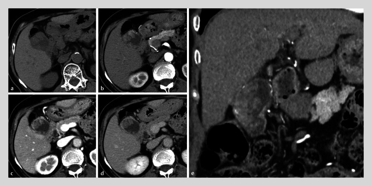 Fig. 1
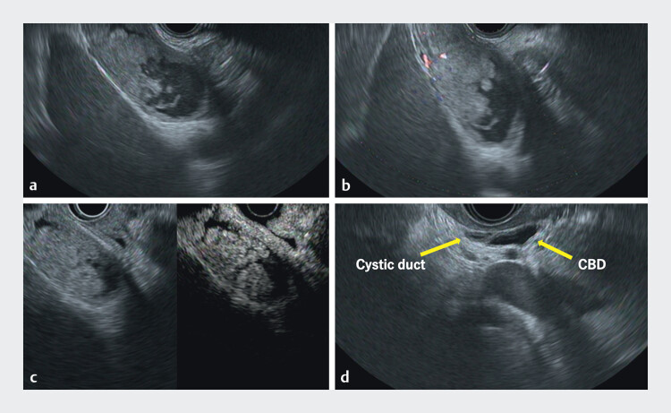 Fig. 2
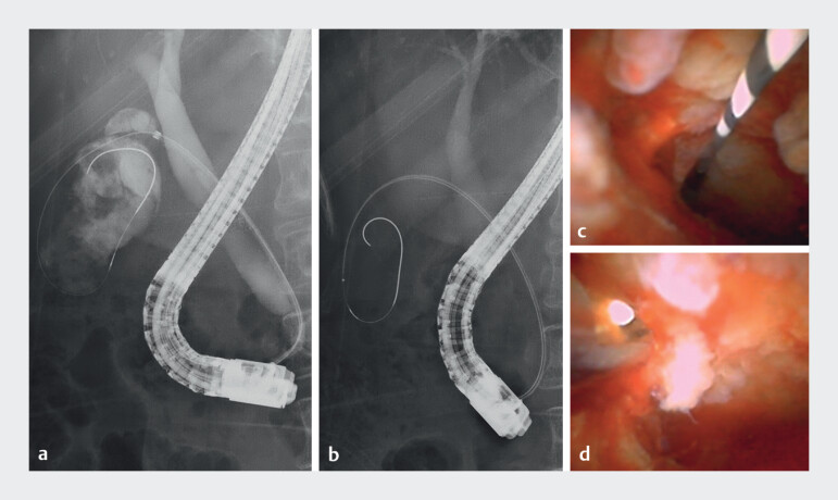 Fig. 3
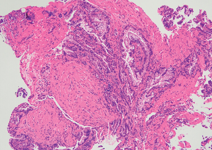 Fig. 4
Peer Reviews
No public reviews on file for this paper yet. If you reviewed it on a platform where reviews are public (OpenReview, ICLR, NeurIPS, ICML), you can paste yours below so the community can read it here.
Videos
No videos yet. Explain this paper in a talk, walkthrough, or lecture? Add one.
Taxonomy
TopicsCholangiocarcinoma and Gallbladder Cancer Studies · Gallbladder and Bile Duct Disorders · Minimally Invasive Surgical Techniques
Endoscopic ultrasound (EUS)-guided tissue acquisition and percutaneous biopsy are not recommended for the preoperative diagnosis of resectable gallbladder cancer because of the risk of dissemination 1 . Definitive diagnosis using imaging tests is difficult, and transpapillary fluoroscopic biopsy and cytology lack high accuracy 2 . Direct observation and biopsy are desirable for a more accurate diagnosis; however, advancing a cholangioscope into the gallbladder through the narrow cystic duct is impractical. We report a case of successful direct observation and biopsy of a gallbladder cancer using a new ultraslim cholangioscope.
An 83-year-old woman was referred for a suspected gallbladder tumor based on a computed tomography scan ( Fig. 1 ). EUS revealed irregular wall thickening of the gallbladder and cystic duct, with continuous mild wall thickening of the common bile duct ( Fig. 2 ). Cholangiography revealed irregular gallbladder tumors, but no irregularities or stenosis in the common bile duct. An ultraslim cholangioscope (DRES Slim Scope and CMOS Camera; Japan Lifeline. Co. Ltd, Tokyo, Japan) was easily inserted into the gallbladder beyond the stenotic cystic duct. A papillary, friable, and irregular mucosa was observed from the gallbladder to the upper part of the cystic duct, whereas the mucosa in the lower part of the cystic duct and common bile duct was intact. Cholangioscopic biopsy of the gallbladder mucosa performed using a tapered sheath device (ERCP Guide Sheath; Olympus, Tokyo, Japan) and slim biopsy forceps (SpyBite Max; Boston Scientific, Tokyo, Japan; Video 1 , Fig. 3 ) revealed adenocarcinoma ( Fig. 4 ). The preoperative diagnosis was gallbladder cancer without bile duct invasion. There were no adverse events associated with the endoscopic procedure. The patient underwent surgical treatment, and the final diagnosis was the gallbladder cancer pT2N0M0.
Computed tomography showing irregular wall thickening with enhancement in the gallbladder. a Axial, plane. b Axial, early phase. c Axial, portal phase. d Axial, delayed phase. e Coronal, early phase.
Endoscopic ultrasound images. a B mode imaging showing irregular wall thickening in the gallbladder. b and c Color doppler and contrast harmonic imaging confirmed blood flow within the lesion. d B mode imaging showing the cystic duct and common bile duct with mild wall thickening.
Cholangioscope showing a papillary, friable, irregular mucosa of the gallbladder. Observation and cholangioscopic biopsy of resectable gallbladder cancer using a new ultra-slim cholangioscope.Video 1
Cholangiography and cholangioscopy. a Cholangiography showing no irregularity or stenosis in the common bile duct and irregular gallbladder tumor. b An ultra-slim cholangoscope was inserted into the gallbladder. c Cholangioscope showing a papillary, friable, irregular mucosa of the gallbladder. d Cholanigoscopic biopsy of the gallbladder mucosa.
Biopsy specimen showing epithelium with marked atypia and diagnosed as adenocarcinoma.
There are few reports on cholangioscopic biopsy for gallbladder cancer; however, it may be a promising new diagnostic option for resectable gallbladder cancer 3 .
Endoscopy_UCTN_Code_TTT_1AR_2AD
The reference list from the paper itself. Each links out to its DOI / PubMed record.
- 1Hijioka S Nagashio Y Ohba A The Role of EUS and EUS-FNA in Differentiating Benign and Malignant Gallbladder Lesions Diagnostics (Basel)20211110.3390/diagnostics 11091586 PMC 846741234573929 · doi ↗ · pubmed ↗
- 2Itsuki H Serikawa M Sasaki T Indication and Usefulness of Bile Juice Cytology for Diagnosis of Gallbladder Cancer Gastroenterol Res Pract 201820185.410349 E 610.1155/2018/5410349 PMC 593244029849591 · doi ↗ · pubmed ↗
- 3Ogura T Bessho K Hattori N Technical aspects of transpapillary biopsy for gallbladder cancer using a novel cholangioscope Endoscopy 202355 E 1085 E 108610.1055/a-2173-789337758212 PMC 10533355 · doi ↗ · pubmed ↗