Ultrasound-guided treatment of giant isolated gastric varices with gastric-renal shunt using coil, cyanoacrylate, and titanium clips
Yuchuan Bai, Zhihong Wang, Yaxian Kuai, Xuecan Mei, Derun Kong

Abstract
Genes, proteins, chemicals, diseases, species, mutations and cell lines named across the full text — each resolved to its canonical identifier and authoritative record.
Click any figure to enlarge with its caption.
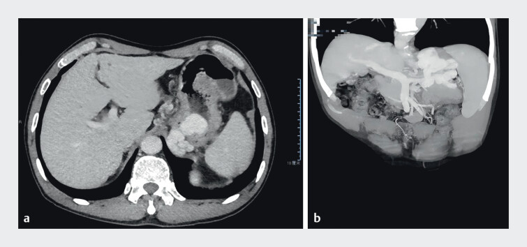 Fig. 1
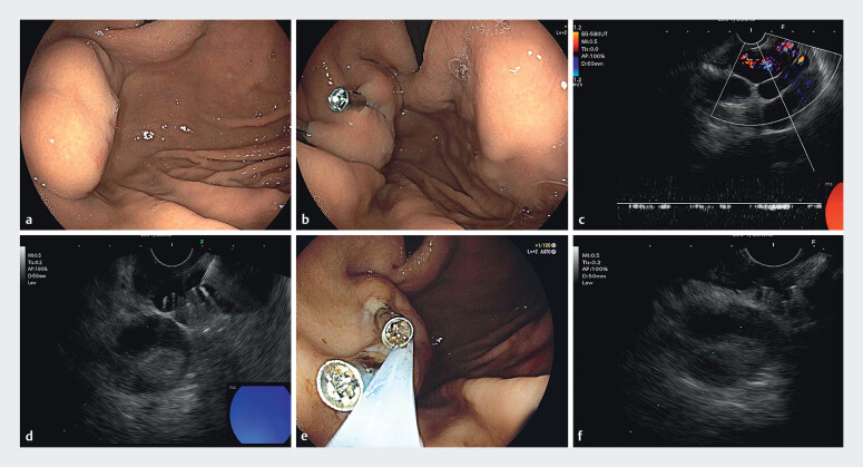 Fig. 2
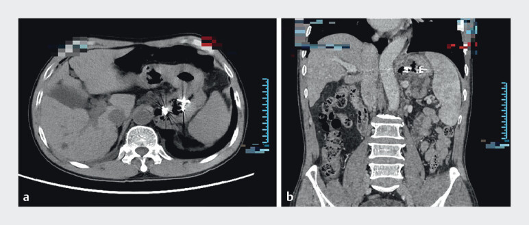 Fig. 3
- —The sixth batch of appropriate technology for health promotion project of Anhui Provincial Health Commission
Peer Reviews
No public reviews on file for this paper yet. If you reviewed it on a platform where reviews are public (OpenReview, ICLR, NeurIPS, ICML), you can paste yours below so the community can read it here.
Videos
No videos yet. Explain this paper in a talk, walkthrough, or lecture? Add one.
Taxonomy
TopicsLiver Disease and Transplantation · Diagnosis and Treatment of Venous Diseases · Abdominal vascular conditions and treatments
In cases where isolated gastric varices (IGV) are complicated by gastrorenal shunt (GRS), endoscopic injection of cyanoacrylate (CYA) carries a risk of ectopic embolism, especially when the GRS diameter is 1 cm or greater 1 . Balloon-occluded retrograde transvenous obliteration (BRTO) is one of the most commonly used methods for reducing ectopic embolism 2 3 . However, after reducing the lumen of IGV, the placement of coils with an appropriate diameter under endoscopic ultrasonography is also a viable option.
A 71-year-old male was initially diagnosed with alcoholic cirrhosis (Child-Pugh class A). Preoperative abdominal CTA revealed a GRS measuring 10.6 mm ( Fig. 1 ). Direct endoscopy revealed a large IGV ( Fig. 2 a ), with a maximum diameter of approximately 3.5 cm measured. Two titanium clips (ROCC-F-26-195, Micro-Tech (Nanjing) Co., Ltd) were applied to clamp the vessel ( Fig. 2 b ). The maximal vascular diameter measured by EUS (EG-580UT; Fujifilm) was 2.1 cm, whereas the diameter at the titanium clip site measured only 1 cm ( Fig. 2 c ). Two coils (M0013120660, Boston Scientific) were placed under EUS guidance ( Fig. 2 d , Video 1 ), followed by CYA injection. Under direct endoscopy, vascular lesions with palpable softness were treated with CYA injection (DEI-CYA) ( Fig. 2 e ). EUS was used again to observe, and the vascular lumen was completely occluded ( Fig. 2 f ). Follow-up CTA showed the disappearance of varices and extramural vessels, with the GRS still present ( Fig. 3 ), and no clinical symptoms of ectopic embolism were observed. The patient did not experience bleeding or other complications 1 month postoperatively.
Preoperative abdominal and pelvic CTA revealed a massive variceal mass in the gastric fundus, which was connected to extramural vessels, indicating the presence of GRS.
a Direct endoscopy revealed a large tumor-like IGV; b Under direct endoscopy, titanium clips were observed blocking the blood vessel; c After titanium clips treatment, EUS measurement showed that the diameter of the blood vessel was approximately 21.56 mm; d EUS revealed a high-echoic shadow in the shape of “∞”; e DEI-CYA; f The varicose veins disappeared after treatment.
Follow-up abdominal and pelvic CTA after treatment showed the disappearance of varices and extramural vessels, with the GRS still present.
A giant IGV is visible under white-light endoscopy. First, some blood vessels are clipped with titanium clips, and then spring coil-tissue adhesive treatment is administered under EUS guidance.Video 1
In this case, we reduced the diameter of IGV using titanium clips and combined coil placement with CYA injection to avoid ectopic embolism. For vessels that were difficult to inject under EUS, DEI-CYA treatment was combined to achieve complete occlusion of the large variceal mass. Postoperative CTA also confirmed the success of this treatment in this case.
Endoscopy_UCTN_Code_TTT_1AS_2AB
The reference list from the paper itself. Each links out to its DOI / PubMed record.
- 1Nardelli S Riggio O Gioia S Spontaneous porto-systemic shunts in liver cirrhosis: Clinical and therapeutical aspects World J Gastroenterol 2020261726173210.3748/wjg.v 26.i 15.172632351289 PMC 7183860 · doi ↗ · pubmed ↗
- 2Xiao Y Huang Z Cao J Balloon-occluded retrograde transvenous obliteration combined with EUS-guided coil embolization and endoscopic cyanoacrylate injection therapy of gastric varices with huge gastrorenal shunt (with videos)Endosc Ultrasound 20231215715936861515 10.4103/EUS-D-21-00157 PMC 10134918 · doi ↗ · pubmed ↗
- 3Garcia-Tsao G Abraldes JG Berzigotti A Portal hypertensive bleeding in cirrhosis: Risk stratification, diagnosis, and management: 2016 practice guidance by the American Association for the study of liver diseases Hepatology 20176531033510.1002/hep.2890627786365 · doi ↗ · pubmed ↗