Accidental chest penetration of a glass foreign body
Simone Duarte Damato, Alessandro Severo Alves de Melo, Edson Marchiori

Abstract
Genes, proteins, chemicals, diseases, species, mutations and cell lines named across the full text — each resolved to its canonical identifier and authoritative record.
Click any figure to enlarge with its caption.
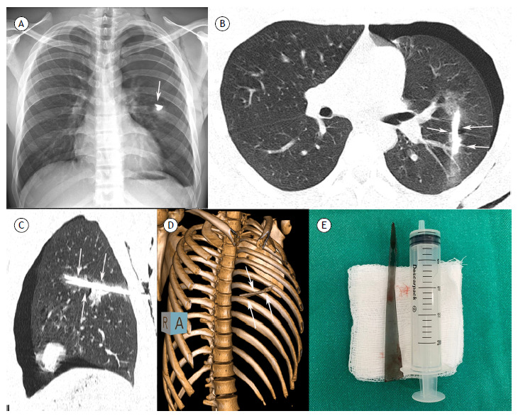 Figure 1
Peer Reviews
No public reviews on file for this paper yet. If you reviewed it on a platform where reviews are public (OpenReview, ICLR, NeurIPS, ICML), you can paste yours below so the community can read it here.
Videos
No videos yet. Explain this paper in a talk, walkthrough, or lecture? Add one.
Taxonomy
TopicsTraumatic Ocular and Foreign Body Injuries · Foreign Body Medical Cases · Restraint-Related Deaths
A 20-year-old previously healthy man reported falling from his own height onto a mirror and suffering a cut in the left infrascapular region. He sought emergency care, where the cut was sutured. He continued to have pain in the area and returned the following day, when a chest X-ray showed a foreign body on the left (Figure 1A). The patient was referred to the hospital, where further imaging showed a high-density linear foreign body in the left hemithorax, in addition to a pneumothorax (Figure 1B-E). He underwent pleural drainage and surgery, during which a glass foreign body (mirror fragment) measuring approximately 30 cm was removed (Figure 1E). The patient progressed well and was discharged four days later in excellent condition.
Figure 1. Chest radiograph (A) showing a dense foreign body in the left hemithorax (arrow). Axial (B) and sagittal (C) chest CT images and 3D reconstruction (D) demonstrating a dense linear foreign body in the left lung and ground-glass opacities (hemorrhage) in the periphery. Note also the pneumothorax on the left. (E) The mirror fragment removed from the patient, measuring about 30 cm.
Intrathoracic foreign bodies include iatrogenic foreign bodies, objects that have migrated through the airways, and traumatic intrathoracic foreign bodies. Glass exhibits high density on chest radiography and CT. CT is the best imaging method for the evaluation of such trauma. Even minor impalement injuries may cause serious complications, including organ damage and life-threatening bleeding. Accurate visual, manual, and instrumental wound exploration is always necessary. Surgical removal of the foreign body is the first-choice treatment.1 ^,^ 2
The reference list from the paper itself. Each links out to its DOI / PubMed record.
- 1Koyama M Matai K Kinoshita A Ishizaki S Okazaki K Inoue M A case of traumatic intrapleural foreign body with progressive supranuclear palsy removed by thoracoscopic surgery Trauma Case Rep 2023434310.1016/j.tcr.2023.100761 PMC 984293336660402 · doi ↗ · pubmed ↗
- 2Luks B Dworzynska A Dobrogowski M Pomorski L Discovery of a Glass Splinter in the Abdominal Cavity After an Old Impalement Injury A Case Report and Literature Review Am J Case Rep 2020212110.12659/AJCR.922599 PMC 727450132457284 · doi ↗ · pubmed ↗