Correction: BDNF augmentation reverses cranial radiation therapy-induced cognitive decline and neurodegenerative consequences
Sanad M. El-Khatib, Arya R. Vagadia, Anh C. D. Le, Janet E. Baulch, Ding Quan Ng, Mingyu Du, Kevin G. Johnston, Zhiqun Tan, Xiangmin Xu, Alexandre Chan, Munjal M. Acharya

Abstract
Genes, proteins, chemicals, diseases, species, mutations and cell lines named across the full text — each resolved to its canonical identifier and authoritative record.
Click any figure to enlarge with its caption.
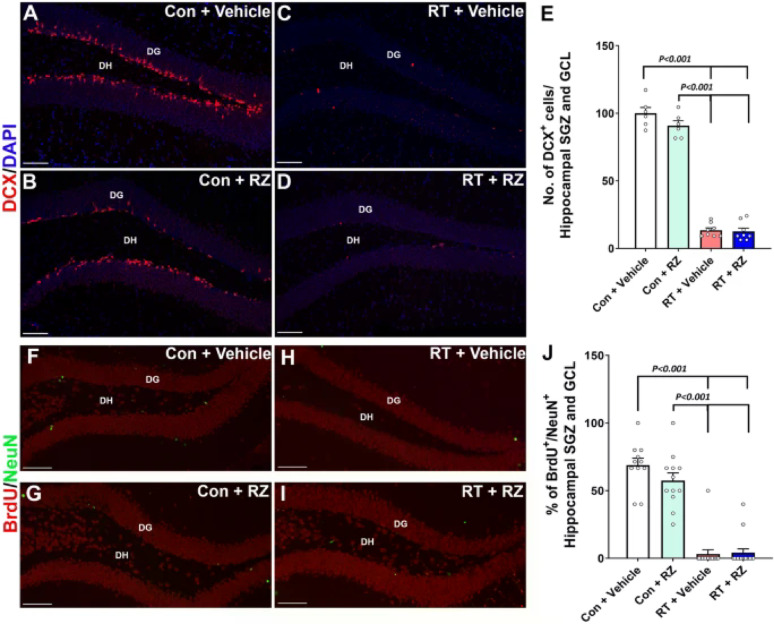 Figure 1
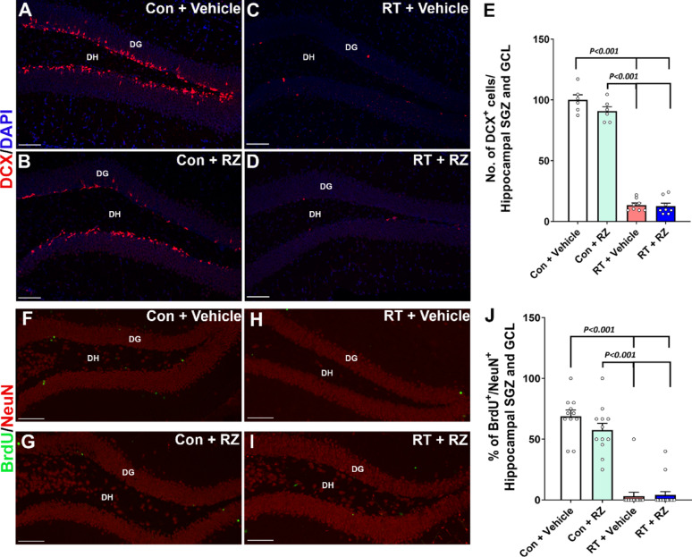 Figure 2
Peer Reviews
No public reviews on file for this paper yet. If you reviewed it on a platform where reviews are public (OpenReview, ICLR, NeurIPS, ICML), you can paste yours below so the community can read it here.
Videos
No videos yet. Explain this paper in a talk, walkthrough, or lecture? Add one.
Taxonomy
TopicsNerve injury and regeneration · Brain Metastases and Treatment · Cancer-related cognitive impairment studies
Correction to: Acta Neuropathologica Communications (2024) 12:190 10.1186/s40478-024-01906-9
In Figure 5F–I, of this article [1], the label color for BrdU-NeuN is swapped from green to red. The incorrect label is . The correct label should be: . For completeness and transparency, both incorrect and correct versions with updated figure legends are displayed below.
Incorrect Fig. 5
Fig. 5 Riluzole treatment did not prevent cranial radiation-induced decline in neurogenesis. WT adult male mice received cranial RT (9 Gy) and treated with riluzole (RZ, 13 mg/kg) 48 h later in their drinking water for 6–7 weeks. Two weeks post-RT, mice were treated with BrdU and hippocampal neurogenesis was quantified using newly born neuron marker, doublecortin (DCX), and BrdU-NeuN dual-immunofluorescence staining in the hippocampal dentate gyrus (DG) sub-granular zone (SGZ) and molecular layer (ML) 6–7 weeks after the BrdU treatment. Cranial RT (RT + Vehicle) significantly reduced the number of DCX^+^ neurons (red, A–D) in the hippocampus compared with either Control + Vehicle or Control + RZ groups E. RT also significantly reduced neurogenesis, as indicated by the reduced percentage of BrdU^+^ cells (green) differentiating into the mature neurons (red, NeuN; F–I) in the RT + Vehicle group compared with either Control + Vehicle or Control + RZ groups (J). Riluzole treatment to the irradiated animals did not prevent the loss of DCX^+^ newly born neurons and the decline in dentate neurogenesis (BrdU^+^-NeuN^+^ dual-fluorescent cells). Data is presented as mean ± SEM (N = 6–16 mice per group). P values were derived from two-way ANOVA and Bonferroni's multiple comparisons test. Scale bars, 50 μm, (A–D) and (F–I)
Correct Fig. 5
Fig. 5 Riluzole treatment did not prevent cranial radiation-induced decline in neurogenesis. WT adult male mice received cranial RT (9 Gy) and treated with riluzole (RZ, 13 mg/kg) 48 h later in their drinking water for 6–7 weeks. Two weeks post-RT, mice were treated with BrdU and hippocampal neurogenesis was quantified using newly born neuron marker, doublecortin (DCX), and BrdU-NeuN dual-immunofluorescence staining in the hippocampal dentate gyrus (DG) sub-granular zone (SGZ) and molecular layer (ML) 6–7 weeks after the BrdU treatment. Cranial RT (RT + Vehicle) significantly reduced the number of DCX^+^ neurons (red, A–D) in the hippocampus compared with either Control + Vehicle or Control + RZ groups E. RT also significantly reduced neurogenesis, as indicated by the reduced percentage of BrdU^+^ cells (green) differentiating into the mature neurons (red, NeuN; F–I) in the RT + Vehicle group compared with either Control + Vehicle or Control + RZ groups (J). Riluzole treatment to the irradiated animals did not prevent the loss of DCX^+^ newly born neurons and the decline in dentate neurogenesis (BrdU^+^-NeuN^+^ dual-fluorescent cells). Data is presented as mean ± SEM (N = 6–16 mice per group). P values were derived from two-way ANOVA and Bonferroni's multiple comparisons test. Scale bars, 50 μm, (A–D) and (F–I)