Chronic Recurrent Multifocal Osteomyelitis Associated in a Child with Inflammatory Bowel Disease and Primary Sclerosing Cholangitis: A Case Report
Yuto Nishiura, Yuta Tsukagoshi, Daisuke Nozawa, Hirota Saito, Kenichi Honma, Toru Hoshi, Masazumi Suzuki, Yasuaki Hosono, Hayato Shimada, Shinsen Nomura, Yoshiyasu Ikezawa

TL;DR
A 14-year-old boy with inflammatory bowel disease and primary sclerosing cholangitis developed chronic recurrent multifocal osteomyelitis, highlighting the need for early diagnosis to avoid unnecessary antibiotics.
Contribution
This case report presents a rare co-occurrence of CRMO with both IBD and PSC in a pediatric patient.
Findings
CRMO was diagnosed after recurrent musculoskeletal symptoms and imaging revealed multifocal bone marrow edema.
The patient was successfully treated with analgesics alone, avoiding long-term antibiotic use.
Early MRI detection of multifocal lesions is critical for diagnosing CRMO in similar cases.
Abstract
Chronic recurrent multifocal osteomyelitis (CRMO) is a rare, non-infectious autoinflammatory bone disorder that predominantly affects children. Some studies have reported cases of CRMO in patients with autoimmune diseases such as inflammatory bowel disease (IBD). We encountered a rare pediatric case of CRMO in a patient with both IBD and primary sclerosing cholangitis (PSC). The patient was a 14-year-old male with a history of ulcerative colitis and PSC, both controlled with vedolizumab, who developed recurrent right wrist pain. Imaging revealed bone marrow edema suggestive of osteomyelitis. Initially suspected to be bacterial osteomyelitis, the patient was treated with antibiotics; however, symptoms recurred, and new lesions appeared on the heel, ultimately leading to a diagnosis of CRMO. The patient was successfully managed with analgesics alone, without antibiotics, and no long-term…
Genes, proteins, chemicals, diseases, species, mutations and cell lines named across the full text — each resolved to its canonical identifier and authoritative record.
Click any figure to enlarge with its caption.
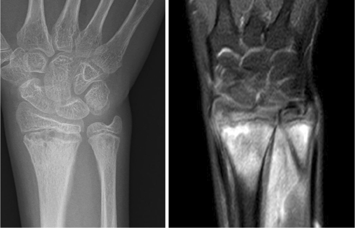 Figure 1
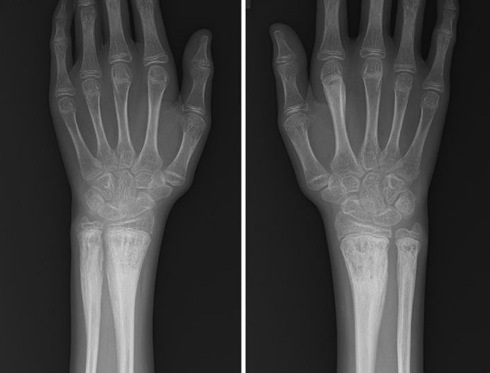 Figure 2
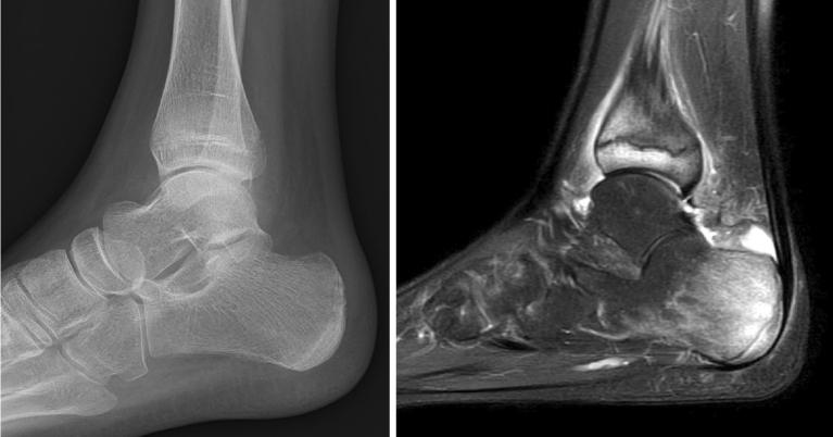 Figure 3
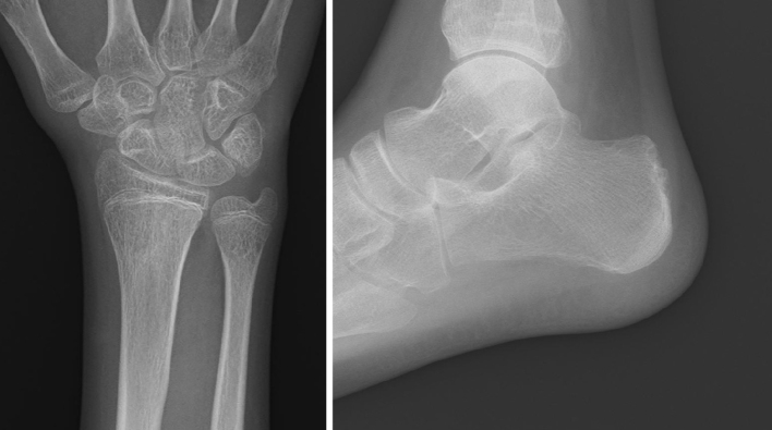 Figure 4
|
|
|
|
| OR |
Peer Reviews
No public reviews on file for this paper yet. If you reviewed it on a platform where reviews are public (OpenReview, ICLR, NeurIPS, ICML), you can paste yours below so the community can read it here.
Videos
No videos yet. Explain this paper in a talk, walkthrough, or lecture? Add one.
Taxonomy
TopicsOsteomyelitis and Bone Disorders Research · Inflammatory Myopathies and Dermatomyositis · Dermatological and Skeletal Disorders
Introduction
Chronic recurrent multifocal osteomyelitis (CRMO) is a rare and poorly understood non-infectious autoinflammatory bone disease characterized by recurrent episodes of bone pain and inflammation ^(1)^. It primarily affects children and adolescents, with a higher prevalence in females ^(2)^. CRMO commonly involves long bones, the spine, and the mandible ^(3)^, presenting as pain, swelling, and redness. The disease is frequently associated with autoimmune conditions such as inflammatory bowel disease (IBD), psoriasis, and palmoplantar pustulosis ^(4)^.
Primary sclerosing cholangitis (PSC) is a chronic cholestatic liver disease characterized by progressive inflammation and fibrosis of the bile ducts, often leading to liver cirrhosis. PSC is strongly associated with IBD, particularly ulcerative colitis (UC), and is considered an immune-mediated disorder. Although the exact relationship between CRMO and PSC remains unclear, both conditions involve dysregulated immune responses and are linked to IBD, suggesting a possible shared pathogenic mechanism ^(5)^.
The pathophysiology of CRMO remains unclear but likely involves innate immune dysregulation influenced by genetic and environmental factors. Unlike bacterial osteomyelitis, CRMO is sterile and unresponsive to antibiotics. Diagnosis relies on magnetic resonance imaging (MRI), which reveals bone marrow edema at multiple sites, with biopsy used to exclude infection or malignancy. In cases with concomitant PSC and IBD, immunological and genetic evaluations may provide further insights ^(6)^.
Case Report
A 14-year-old male with UC, controlled with vedolizumab, presented with right wrist pain persisting for 2 months. He was diagnosed with UC and PSC at age 12 following persistent diarrhea, liver dysfunction, and biopsy.
Physical examination revealed redness and swelling of the right wrist, along with untreated gingivitis. Laboratory findings showed a white blood cell count of 11,700/μL and C-reactive protein 41.3 mg/L; however, blood cultures were negative. Ultrasound examination showed no synovitis, though a small amount of joint fluid was present; puncture fluid culture was also negative. Although MRI showed significant bone marrow edema, the absence of active synovitis on ultrasound made juvenile idiopathic arthritis unlikely. MRI (Short Tau Inversion Recovery) demonstrated bone marrow edema, suggesting osteomyelitis (Figure 1). Due to immunosuppression, hematogenous bacterial osteomyelitis was suspected, and empirical antibiotics were initiated. As C-reactive protein levels gradually decreased and symptoms resolved, antibiotics were discontinued.
Radiograph and MRI of the heel performed on the initial visit.MRI with STIR sequences demonstrated bone marrow edema.MRI: magnetic resonance imaging; STIR: short tau inversion recovery.
Six months later, wrist pain recurred. Radiographs of both wrists revealed chronic osteomyelitis, although the contralateral wrist remained asymptomatic (Figure 2). Given the patient’s immunosuppression state, a bacterial flare-up was suspected, and antibiotics were resumed. Symptoms again resolved. One year and 6 months later, the patient developed right heel pain, and MRI confirmed bone marrow edema (Figure 3).
Radiograph of both wrists performed 6 months after the initial visit. Radiographs showing chronic osteomyelitis, though the contralateral wrist was asymptomatic.
Radiograph and MRI of the heel performed 1 year and 6 months after the initial visit.MRI revealed bone marrow edema, despite no abnormalities being observed on plain radiography.MRI: magnetic resonance imaging
CRMO was suspected, and treatment was limited to analgesics. Symptoms gradually improved with acetaminophen, and no skeletal deformities or long-term complications were observed (Figure 4).
Radiographs obtained at the final follow-up, 3 years after the initial visit. No significant skeletal deformities or long-term complications were observed.
Bone biopsy was not performed. However, due to a lack of improvement with antibiotics, bacterial osteomyelitis was ruled out. Because the conditions were not progressive, malignancy and Langerhans cell histiocytosis were also excluded. Based on the Bristol diagnostic criteria for CRMO ^(7)^ (Table 1), a diagnosis of CRMO was made.
Discussion
Previous studies have reported an association between CRMO and IBD, with CRMO preceding IBD in 53% of cases, while IBD appears first in 35% ^(6)^. This highlights the need for clinicians to consider CRMO when patients with IBD present with unexplained musculoskeletal pain. Diagnosis is challenging due to overlapping features with bacterial osteomyelitis, particularly in immunocompromised patients. MRI has a high sensitivity in detecting CRMO and can identify lesions even in asymptomatic cases, making it useful for both initial diagnosis and follow-up. In the present case, performing MRI of both wrist joints at disease onset might have enabled an earlier diagnosis ^(8)^.
Although the relationship between IBD and CRMO, or IBD and PSC, has been recognized for some time, each condition is rare, and the molecular biological relationship remains unclear. Only one prior case involving the combination of IBD, CRMO, and PSC―similar to this case―has been reported ^(5)^. In that case, CRMO was diagnosed based on nonbacterial osteomyelitis in both femurs and was followed by a diagnosis of PSC due to persistent elevation of liver enzymes. Further investigation into the association between PSC and UC in that case revealed the presence of asymptomatic UC. In contrast, in the present case, UC and PSC were already being managed with strict immunosuppressive therapy when osteomyelitis of the right distal radius developed. A key difference from the previous case is that all three conditions were symptomatic, with CRMO being the last to develop. The combination of CRMO and UC is frequently reported, and the coexistence of all three conditions may be underpinned by genetic and immunological factors that remain unclear. Vedolizumab has been shown to have an effect on UC, but there are no reports mentioning its effect on CRMO. Accumulating case reports of complicated cases like this one will help clarify the immunological and genetic differences between CRMO and UC. Unfortunately, these investigations were not performed in this case because the patient and family declined them. This is a limitation of this case.
Nonsteroidal anti-inflammatory drugs are typically the first-line therapy for CRMO. Biologic agents have shown efficacy in treating both CRMO and IBD. Alternative therapies, such as bisphosphonates or interleukin-1 inhibitors, may be considered in refractory cases ^(4)^.
In conclusion, an important point from this case report is that nonbacterial osteomyelitis occurred in a patient with IBD and PSC. This case report supports the avoidance of unnecessary antibiotic use for osteomyelitis in patients with IBD and PSC, with consideration of the possibility of CRMO.
Article Information
Author Contributions
Study design, conceptualization, and proof outline: Yuta Tsukagoshi,. Data collection: Yuto Nishiura and Yuta Tsukagoshi. Interpretation of results and manuscript preparation: Yuto Nishiura and Yuta Tsukagoshi. Project supervision: Yuta Tsukagoshi. Manuscript writing: Yuto Nishiura and Yuta Tsukagoshi. All authors discussed the results and commented on the manuscript.
Conflicts of Interest
None
IRB Approval Code and Name of the Institution
No, Ethical approval was not required for this case report, as it does not involve interventional research. Parental consent was obtained for the use of radiographs and clinical photographs included in this study.
The reference list from the paper itself. Each links out to its DOI / PubMed record.
- 1Jurik AG, Møller BN. Chronic sclerosing osteomyelitis of the clavicle. A manifestation of chronic recurrent multifocal osteomyelitis. Arch Orthop Trauma Surg (1978). 1987;106(3):144-51.3606353 10.1007/BF 00452200 · doi ↗ · pubmed ↗
- 2Girschick H, Finetti M, Orlando F, et al. The multifaceted presentation of chronic recurrent multifocal osteomyelitis: a series of 486 cases from the Eurofever international registry. Rheumatology (Oxford). 2018;57(7):1203-11.29596638 10.1093/rheumatology/key 058 · doi ↗ · pubmed ↗
- 3Falip C, Alison M, Boutry N, et al. Chronic recurrent multifocal osteomyelitis (CRMO): a longitudinal case series review. Pediatr Radiol. 2013;43(3):355-75.23263195 10.1007/s 00247-012-2544-6 · doi ↗ · pubmed ↗
- 4Rao AP, Mallya PP, Ranjani S, et al. Chronic recurrent multifocal osteomyelitis - a case series from India. Indian J Orthop. 2018;52(6):672-7.30532310 10.4103/ortho.IJ Ortho_464_17PMC 6241066 · doi ↗ · pubmed ↗
- 5Ng HY, Guttman OR, Tucker LB. Chronic recurrent multifocal osteomyelitis and primary sclerosing cholangitis with Type 1 autoimmune hepatitis in a child with ulcerative colitis: a case report. BMC Rheumatol. 2021;5(1):16.34078478 10.1186/s 41927-021-00186-3PMC 8173920 · doi ↗ · pubmed ↗
- 6Tzaneti A, Athanasopoulou E, Fessatou S, et al. Chronic nonbacterial osteomyelitis in inflammatory bowel disease. Life (Basel). 2023;13(12):2347.38137947 10.3390/life 13122347 PMC 10745028 · doi ↗ · pubmed ↗
- 7Roderick MR, Shah R, Rogers V, et al. Chronic recurrent multifocal osteomyelitis (CRMO) - advancing the diagnosis. Pediatr Rheumatol Online J. 2016;14(1):47.27576444 10.1186/s 12969-016-0109-1PMC 5006369 · doi ↗ · pubmed ↗
- 8Beck C, Morbach H, Beer M, et al. Chronic nonbacterial osteomyelitis in childhood: prospective follow-up during the first year of anti-inflammatory treatment. Arthritis Res Ther. 2010;12(2):R 74.20433730 10.1186/ar 2992 PMC 2888230 · doi ↗ · pubmed ↗