In Vivo Wound Healing and Antimicrobial Activities of the Hydroalcoholic Extract and Solvent Fractions of Arisaema schimperianum Schott. (Araceae) Tuber in Mice
Betelhem Awoke, Daniel Bisrat, Betelhem Gebreamlak, Abiy Abebe, Sileshi Degu, Kaleab Asres

TL;DR
This study shows that a plant extract from Arisaema schimperianum helps heal wounds and fight bacteria in mice, supporting its traditional use in Ethiopian medicine.
Contribution
The study provides first-time scientific validation of the wound healing and antimicrobial properties of Arisaema schimperianum tuber extracts in an animal model.
Findings
The aqueous fraction of the extract showed the strongest wound healing effect, improving contraction, epithelialization, and tensile strength.
The extract inhibited bacterial growth, with stronger activity against Gram-positive bacteria.
Histopathological analysis confirmed the extract's beneficial effects on wound healing.
Abstract
Globally, wounds have become a growing health concern due to the increasing prevalence of vascular diseases. In Ethiopian traditional medicine, plants belonging to the genus Arisaema have long been used to treat wounds and infections. This study aimed at evaluating the wound healing and antimicrobial activities of 80% methanol extract and its solvent fractions from the tubers of Arisaema schimperianum Schott (Araceae). Wound healing activity was assessed in mice using excision and incision wound models, while skin irritation tests were conducted in rats. The evaluation included measurements of wound contraction rate, epithelialization period, tensile strength, hydroxyproline content, and histological analysis. Antibacterial and antifungal activities were assessed in vitro using the broth dilution method. Ointments formulated at 5% and 10% (w/w) concentrations were tested for wound…
Genes, proteins, chemicals, diseases, species, mutations and cell lines named across the full text — each resolved to its canonical identifier and authoritative record.
Click any figure to enlarge with its caption.
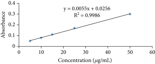 Figure 1
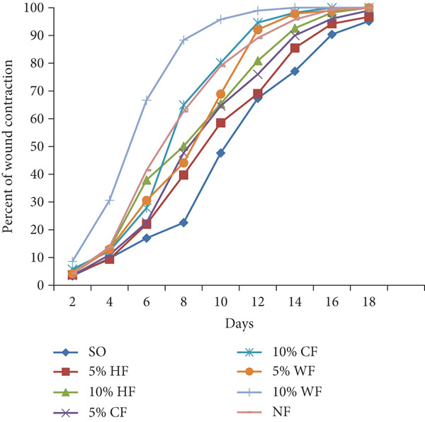 Figure 2
|
|
|
|
|
|---|---|---|---|
| Erythema | Edema | ||
| No erythema | 0 | No edema | 0 |
| Very slight erythema | 1 | Very slight edema | 1 |
| Well‐defined erythema | 2 | Well‐defined edema (edge by define raising) | 2 |
| Moderate to severe erythema | 3 | Moderate edema (raising ~1 mm) | 3 |
| Severe erythema (beet redness) to scar formation | 4 | Severe edema (raising more than 1 mm and extended beyond the area of exposure) | 4 |
|
|
|
|
|
|
|
|
|
|
|
|---|---|---|---|---|---|---|---|---|---|
| SO | 3.33 ± 2.96 (3.39%) | 283.50 ± 2.62 (9.71%) | 260.66 ± 2.77 (16.98%) | 224.33 ± 4.08 (28.55%) | 164.50 ± 7.10 (47.61%) | 102.50 ± 4.34 (67.35%) | 71.83 ± 4.49 (77.12%) | 30.16 ± 2.70 (90.39%) | 15.16 ± 2.02 (95.17%) |
| 5% HAE | 301.00 ± 4.56 (4.14%) | 275.50 ± 2.27 (12.26%) | 236.66 ± 2.21 (24.63%)d2 | 192.50 ± 6.61 (38.69%)a2,d1 | 120.00 ± 2.22 (61.78%)a1 | 90.50 ± 8.06 (71.17%)d2 | 33.50 ± 1.76 (89.33)a1 | 12.66 ± 1.22 (95.94)a1 | 5.00 ± 1.06 (98.39)a1 |
| 10% HAE | 297.16 ± 4.57 (5.36%) | 262.66 ± 2.29 (16.35%)a1,b3 | 176.33 ± 12.79 (43.83%)a1,d1,e1 | 90.5 ± 5.92 (70.99%)a1,d1,e1 | 50.83 ± 4.36 (83.70%)a1,d1,e1 | 18.33 ± 1.47 (94.12%)a1,d1,e1 | 5.33 ± 0.49 (98.29%)a1,e1 | 0 (100%)a1,e1 | 0 (100)a1 |
| 5% HF | 302.16 ± 4.36 (3.77%) | 284.50 ± 2.78 (9.39%) | 244.83 ± 2.40 (22.02%)c1,d1 | 189.16 ± 3.15 (39.75%)a1,c1,d1 | 130.16 ± 1.35 (58.54%) | 97.16 ± 4.48 (69.05%)c1,d1 | 46.50 ± 3.38 (85.50%)a1,c3,d1 | 18.16 ± 2.02 (94.21%)a1,c3,d1 | 6.16 ± 0.90 (96.71%)a1,c3,d1 |
| 10% HF | 298.33 ± 3.79 (4.99%) | 272.66 ± 2.47 (13.16%) | 194.83 ± 2.18 (37.95%)a1,b1 | 156.66 ± 2.92 (50.10%)a1,c1,d1 | 108.66 ± 2.89 (65.39%)a1,b3,c1 | 60.00 ± 2.43 (80.89%)a1,c1,d1 | 23.16 ± 11.00 (92.62%)a1,b1 | 5.50 ± 0.42 (98.24%)a1,b1 | 0 (100%)a1,b1 |
| 5% CF | 302.83 ± 2.21 (3.55%) | 279.66 ± 1.22 (10.93%) | 243.00 ± 2.08 (22.61%)a2,c1,d1 | 170.50 ± 1.38 (47.70%)a1,b1,c1 | 111.00 ± 3.00 (64.64%)a1,c1 | 75.33 ± 5.05 (76.00%)a1,b1,c1 | 30.00 ± 2.81 (90.00%)a1,b1,c1 | 12.33 ± 1.54 (96.04%)a1,c1,d3 | 3.16 ± 0.47 (98.98%)a1 |
| 10% CF | 295.83 ± 3.52 (5.78%) | 275.33 ± 1.83 (12.33%) | 226.50 ± 2.66 (27.86%)a1,b2,c1 | 110.00 ± 0.68 (64.96%)a1,b1,d1 | 62.33 ± 6.69 (80.14%)a1,b1,d1 | 16.83 ± 0.79 (94.60%)a1,b1,d1 | 5.50 ± 1.20 (98.23%)a1,b1,d1 | 0 (100%)a1,b | 0 (100%)a1,b1 |
| 5% WF | 301.00 ± 4.56 (4.14%) | 273.16 ± 2.97 (13.00%) | 218.00 ± 2.22 (30.57%)a1,b1,c1,d1 | 175.50 ± 3.90 (44.10%)a1,c1,d1 | 97 ± 3.47 (68.91%)a1,a2,c1 | 24.67 ± 2.78 (92.09%)a1,b1,d1 | 6.67 ± 0.84 (97.86%)a1,b1,d1 | 0 (100%)a1,b1 | 0 (100%)a1,b1 |
| 10% WF | 285.16 ± 4.57 (8.60%) | 216.50 ± 8.92 (30.60%)a1,b1,c1 | 103.83 ± 6.60 (66.72%)a1,b1,c1,d1 | 36.33 ± 1.52 (88.35%)a1,b1,c1,d1, | 13.16 ± 1.35 (95.78%)a1,b1,c1,d1 | 3.20 ± 0.50 (98.97%)a1,b1,c1,d1 | 0 (100%)a1,b1,d1 | 0 (100%)a1,b1 | 0 (100%)a1,b1 |
| NF | 300.50 ± 3.30 (4.29%) | 270.66 ± 1.76 (13.80%)a3 | 183.83 ± 3.32 (41.45%)a1,d1 | 116.50 ± 2.39 (62.66%)a1,b1,d1,e1 | 65.33 ± 6.60 (79.06%)a1,b1,d1,e1 | 25.00 ± 1.91 (91.98%)a1,b1,d1,e1 | 13.16 ± 1.42 (95.78%)a1,b1 | 2.16 ± 0.47 (98.80%)a1,b1 | 0 (100%)a1,b1 |
|
|
|
|---|---|
| SO | 18.67 ± 0.51 |
| 5% HAE | 16.00 ± 0.44a2 |
| 10% HAE | 13.00 ± 0.67a1 |
| 5% HF | 17.33 ± 0.42 |
| 10% HF | 16.00 ± 0.73a1 |
| 5% CF | 16.00 ± 0.51c1 |
| 10% CF | 15.83 ± 0.54a1 |
| 5% WF | 15.67 ± 0.61b1 |
| 10% WF | 13.67 ± 0.61a1,b1,c2 |
| NF | 14 ± 0.73a1,b1,c2 |
|
|
|
|
|
|
|
|
|---|---|---|---|---|---|---|
| SO | + | + | + | ++ | ++ | + |
| 5% HAE | ++ | ++ | ++ | ++ | ++ | ++ |
| 10% HAE | +++ | +++ | +++ | ++ | + | ++ |
| 5% HF | ++ | ++ | ++ | ++ | +++ | + |
| 10% HF | ++ | ++ | ++ | +++ | ++ | +++ |
| 5% CF | ++ | ++ | ++ | +++ | +++ | + |
| 10% CF | +++ | +++ | ++ | +++ | +++ | ++ |
| 5% WF | ++ | ++ | ++ | ++ | ++ | ++ |
| 10% WF | +++ | +++ | ++ | +++ | +++ | ++ |
| NF | ++ | ++ | +++ | ++ | ++ | ++ |
|
|
|
|---|---|
| SO | 10.00 ± 0.57 |
| 5% HAE | 41.00 ± 2.23a1,d1 |
| 10% HAE | 58.67 ± 1.15a1,e1 |
| 5% HF | 12.33 ± 1.20c1,d1 |
| 10% HF | 31.33 ± 1.29a1,b1,d1 |
| 5% CF | 28.00 ± 1.57a1,b1,d1 |
| 10% CF | 34.33 ± 0.881a1,b1,d1 |
| 5% WF | 43.67 ± 1.20a1,b1,c1,d1 |
| 10% WF | 61.00 ± 2.30a1,b1,c1,d1 |
| NF | 58.02 ± 1.11a1,b1,c1,e1 |
|
|
|
|
|---|---|---|
| Untreated control | 187.50 ± 7.04 | — |
| SO | 230.83 ± 19.07 | 18.86 |
| 5% HAE | 304.50 ± 15.01a1,d2,e1 | 31.91 |
| 10% HAE | 425.00 ± 18.02a1,d1,e1 | 84.11 |
| 5% HF | 276.66 ± 18.95c2,d1 | 19.85 |
| 10% HF | 333.33 ± 17.44a2,b1 | 44.40 |
| 5% CF | 340.00 ± 6.83a2,b1 | 47.29 |
| 10% CF | 378.33 ± 18.87a1,b1 | 63.89 |
| 5% WF | 359.16 ± 9.69a1,b1 | 55.59 |
| 10% WF | 440.00 ± 32.04a1,b1,c1 | 90.61 |
| NF | 406.67 ± 16.67a1,b1,c1,e1 | 76.32 |
|
|
| |||||||
|---|---|---|---|---|---|---|---|---|
|
|
|
|
|
|
|
|
| |
| HAE | 8 | 16 | 8 | 2 | 2 | 2 | NA | NA |
| HF | 8 | 16 | 8 | 8 | 8 | 0.5 | NA | NA |
| CF | 8 | 16 | 8 | 8 | 8 | 2 | NA | NA |
| WF | 8 | 16 | 4 | 1 | 0.5 | 1 | NA | NA |
| Negative control | NA | NA | NA | NA | NA | NA | NA | NA |
- —Addis Ababa University10.13039/501100007941
Peer Reviews
No public reviews on file for this paper yet. If you reviewed it on a platform where reviews are public (OpenReview, ICLR, NeurIPS, ICML), you can paste yours below so the community can read it here.
Videos
No videos yet. Explain this paper in a talk, walkthrough, or lecture? Add one.
Taxonomy
TopicsWound Healing and Treatments · Burn Injury Management and Outcomes · Ethnobotanical and Medicinal Plants Studies
1. Introduction
Wounds are disruptions in the skin that impair its normal function [1]. The global incidence and healthcare costs of chronic wounds have increased significantly, largely due to the rising prevalence of vascular diseases. Approximately 6 million individuals are estimated to develop chronic wounds each year, placing a financial burden equivalent to 2%–4% of healthcare budgets [2]. In developing regions, chronic wounds affect around 1%–2% of the population over a lifetime [3]. Wounds can result from mechanical injury, chemical exposure, thermal damage, or microbial infections [4]. Although various treatment options exist, many conventional therapies are associated with undesirable side effects [5]. In recent years, herbal remedies have gained attention as potentially safer and more effective alternatives [6].
In Ethiopia, Arisaema schimperianum Schott is known by various vernacular names, including Amoch in Amharic, Qolxo in Sidama, and Cherana in Afaan Oromo. It is a herbaceous tuberous plant in the Araceae family and is widely distributed across Ethiopia, the Imatong Hills (on the Uganda–Sudan border), the Democratic Republic of Congo, Kenya, and southern Arabia [7]. It is a seasonal plant that flourishes during the rainy season and remains dormant during the dry months, with its main flowering season occurring in June and July [8].
In traditional Ethiopian medicine, the dried tuber of A. schimperianum, often mixed with petroleum jelly, is used to treat wounds associated with glandular tuberculosis, ringworm, scabies, and other skin conditions [9, 10]. In addition to its medicinal uses, the boiled tubers are consumed as food, particularly in southern Ethiopia during periods of drought [11]. The high oxalate content in fresh tubers was identified as a major obstacle to safe consumption. However, Hazo et al. [12] reported that boiling A. schimperianum tubers significantly reduces oxalate levels and the associated irritation, thereby allowing occasional consumption.
Despite its widespread traditional use, no prior studies have specifically examined the wound healing and antimicrobial properties of this plant. This study is aimed at filling that gap by investigating the therapeutic potential of A. schimperianum tubers. We believe that scientific validation of traditionally used wound healing plants not only advances biomedical research and drug discovery but also reinforces the credibility of traditional medical practices, supporting their integration into modern healthcare.
2. Materials and Methods
2.1. Plant Material
Fresh tubers of A. schimperianum were collected in July 2023 from the Sedie Muja district in the South Gondar Zone, Amhara Region, about 774 km north of Addis Ababa. The plant material was authenticated by the National Herbarium, Department of Plant Biology and Biodiversity Management, College of Natural and Computational Sciences, Addis Ababa University (AAU), where a botanical specimen was deposited (Voucher Number BA‐001) for future reference.
2.2. Experimental Animals
Healthy adult Swiss albino mice of either sex (25–30 g, 8–10 weeks old) and female rats (150–250 g) were used for wound healing activity and dermal toxicity studies, respectively. The animals were procured from the Animal House of the School of Pharmacy, AAU, housed under standard laboratory conditions (22°C, 12 h light/dark cycle) with food and water provided ad libitum. All experiments followed internationally accepted guidelines, and animals were acclimatized for 1 week before the study and housed individually [13].
2.3. Test Bacteria and Fungi
In vitro antibacterial and antifungal activity tests were performed using clinical or drug‐sensitive standard strains obtained from the Ethiopian Public Health Institute (EPHI) Microbiology Laboratories, Addis Ababa. The Gram‐positive bacteria used were Staphylococcus aureus (ATCC 25923), Staphylococcus epidermidis (ATCC 12228) and methicillin‐resistant Staphylococcus aureus (MRSA) (clinical isolate). Escherichia coli (ATCC 25922), Proteus mirabilis (ATCC 35659), and Pseudomonas aeruginosa (ATCC 27853) were the Gram‐negative bacteria employed. Candida albicans (clinical isolate) and Aspergillus niger (ATCC 10535) were used as test fungal strains.
2.4. Preparation of Hydroalcoholic Extract
Fresh tubers of A. schimperianum were cleaned and dried under shade at room temperature. They were then powdered using a mortar and pestle. The powdered tuber (800 g) was soaked in 4 L of 80% methanol for 72 h. The mixture was initially filtered using a nylon cloth, and the marc was remacerated three times (each time for 24 h) to maximize the yield. The combined filtrate was then allowed to pass through Whatman No. 1 filter paper using a pressurized suction system, concentrated under reduced pressure using a rotary evaporator set at 121 mbar, 45 rpm, and a temperature of 40°C. The remaining aqueous solution was dried in an oven at a temperature not exceeding 40°C. The resulting dried extract was weighed, transferred into an amber‐colored vial, and stored in a refrigerator at 4°C until use.
2.5. Preparation of Solvent Fractions
Fractionation of the hydroalcoholic extract was performed using solvent–solvent partitioning. The process involved successive solvent–solvent fractionation using n‐hexane, chloroform, and water. Initially, the hydroalcoholic extract (30 g) was suspended in 100 mL of distilled water to which an equal volume of n‐hexane was added and vigorously shaken in a separatory funnel. After allowing the mixture to stand until clear separation occurred, the process was repeated three times, and the n‐hexane layer was collected. This was followed by the addition of an equal volume of chloroform to the aqueous layer, and the lower chloroform layer was carefully collected in a beaker, repeating the process three times to obtain a chloroform fraction. The remaining upper layer was taken as the water fraction. The organic solvent fractions were concentrated in a rotatory evaporator, while the water fraction was dried in an oven at a temperature not exceeding 40°C. The dried fractions were then packed in airtight containers and labeled and stored in a refrigerator until used for further experiments.
2.6. Ointment Formulation
A simple ointment was prepared using the hydroalcoholic extract and solvent fractions following the British Pharmacopoeia formula [14]. Thus, three formulations were prepared: control (simple ointment) and medicated ointments with 5% and 10% (w/w) extract. Ingredients were melted, blended, cooled, and levigated for uniform consistency. The 5% ointment contained 1 g of test substance in 19 g base, while the 10% ointment had 2 g in 18 g base.
2.7. Grouping and Dosing of Experimental Animals
Ten groups of mice, each containing six animals, were used for the excision model. One group served as a negative control, eight groups as test groups, and one group as a positive control. Group I received the simple ointment base (negative control). Groups II and III received 5% and 10% (w/w) hydroalcoholic extract, respectively. Groups IV and V were treated with 5% and 10% (w/w) n‐hexane fraction, Groups VI and VII with 5% and 10% (w/w) chloroform fraction, and Groups VIII and IX with 5% and 10% (w/w) water fraction. Group X received 0.2% w/v nitrofurazone (positive control). For the incision model, animals were assigned similarly, with the addition of Group XI, which served as an untreated negative control.
2.8. Toxicity Studies
2.8.1. Acute Oral Toxicity Study
This study followed the Organization for Economic Cooperation and Development (OECD) guideline 425 [15], using five female mice. In the initial phase, one mouse received 2000 mg/kg of 80% methanol A. schimperianum tuber extract, dissolved in 5% Tween 80, via oral gavage after an 8‐h fasting period. Observations were made for 30 min and continued over 24 h, with close attention during the first 4 h. Since no mortality was observed, the same dose was administered to four additional mice the following day. All animals were monitored for 14 days for behavioral changes, including alterations in skin, fur, and eye condition, appetite loss, piloerection, lacrimation, tremors, convulsions, and mortality.
2.8.2. Acute Dermal Toxicity Study
A skin irritation test was performed on three groups of Wistar rats, each comprising three males and three females, to evaluate the 80% methanol extract of A. schimperianum tubers. Following a 7‐day acclimatization period, the dorsal skin of each rat (approximately 500 mm^2^) was shaved. A simple ointment was applied to the shaved area of the first group, while 5% and 10% extract ointments were applied thinly and uniformly to the shaved areas of the second and third groups, respectively. The sites were covered with gauze and nonocclusive bandages. After 24 h, the bandages were removed, and the sites rinsed with distilled water [16]. Skin reactions were observed at 1, 24, 48, and 72 h using a standardized scoring system (Table 1).
The score of primary irritation (SPI) was calculated for each rat as follows:
The primary irritation index (PII) was determined as the arithmetic mean of the SPI values from all six rats. The degree of irritation was categorized as follows: negligible (0–0.4), slight (0.5–1.9), moderate (2–4.9), or severe (5–8).
2.9. Wound Healing Studies
2.9.1. Excision Wound Model
On the day of wounding, animals were anesthetized with ketamine (50 mg/kg) and diazepam (5 mg/kg) administered intraperitoneally (IP) [4]. The dorsothoracic fur was shaved, and a circular area (~314 mm^2^) was marked and excised using sterilized scissors. After recovery, animals were returned to individual cages, with the day of surgery designated as Day 0. From Day 1, wounds were treated daily with topical ointment applications [1, 17].
2.9.1.1. Measurement of Wound Contraction
Wound closure was assessed every other day by tracing the wound area onto transparent paper and transferring it to graph paper for calculation. The percentage of wound contraction was determined using the initial wound size (314 mm^2^) as 100%:
where n is the number of days, that is, 2nd, 4th, 6th, etc., until complete wound healing.
2.9.1.2. Estimation of Hydroxyproline Content
After 10 days of treatment, granulation tissue was collected on Day 11, cleaned, weighed, and dried at 60°C for 12 h. Dried tissue was hydrolyzed in 6N HCl at 110°C for 24 h [18]. Hydrolysates (1 mL) were analyzed alongside standard hydroxyproline solutions, with absorbance measured at 572 nm using a UV‐Vis spectrophotometer (Jenway 6500, England). Hydroxyproline content was calculated using a calibration curve [17].
2.9.1.3. Preparation of Calibration Curve
Standard L‐hydroxyproline (0.05 g) was dissolved in 20 mL of HCl and water to prepare a 100 μg/mL stock solution. From this stock, graded concentrations of 5, 10, 15, 25, and 50 μg/mL were prepared for subsequent experiments. Each solution was treated with 1 mL of 0.05 M CuSO₄ and 1 mL of 2.5 N NaOH, incubated at 40°C for 3–5 min, followed by 1 mL of 6% H₂O₂. Then, 4 mL of 3N H₂SO₄ and 2 mL of 5% p‐dimethylaminobenzaldehyde were added sequentially, with swirling after each addition. Tubes were capped and heated in a water bath at 70°C for 16 min. After cooling and mixing, absorbance was measured at 572 nm. Unknown hydroxyproline concentrations were determined from the calibration curve (Figure 1) [17].
Standard curve for determination of hydroxyproline concentration.
2.9.2. Incision Wound Model
On the day of incision, animals were anesthetized as previously described. The dorsal fur was shaved, and a 3 cm longitudinal paravertebral incision was made. The wound was sutured using surgical silk (No. 000) and a curved needle (No. 9), with stitches spaced 1 cm apart. From Day 1 postwounding, animals received topical ointment treatment as per the grouping and dosing section (excluding the untreated group). On Day 8, sutures were removed, and treatment continued. Tensile strength was measured on Day 10 using the continuous water flow method, and percent tensile strength was calculated accordingly.
2.9.3. Histopathological Analysis
On Day 12 postwounding, the mice were euthanized using a high dose of IP ketamine and diazepam. Skin samples were collected and fixed in 10% buffered formalin and then processed for paraffin embedding. Sections of 5 μm thickness were prepared and stained with hematoxylin and eosin (H&E) [19]. A light microscope equipped with a digital image analysis system was used to evaluate the stages of wound healing. Parameters such as re‐epithelialization, fibroblast proliferation, collagen deposition, polymorphonuclear cell infiltration, mononuclear cell infiltration, and neovascularization were assessed. Each parameter was graded as mild (+), moderate (++), or marked (+++) to evaluate the extent of epidermal and dermal remodeling [20].
2.10. Antimicrobial Activity Test
Mueller–Hinton agar (MHA) and broth (MHB) were used for bacterial cultures, while Sabouraud dextrose agar (SDA) and broth (SDB) were used for fungal cultures. Nutrient broth (NB) served as a general control. Minimum inhibitory concentration (MIC) was determined using the microbroth dilution method in 96‐well plates according to CLSI guidelines [21]. Test samples and controls (ciprofloxacin for bacteria and ketoconazole for fungi) underwent serial dilution. Bacterial cultures were incubated at 37°C for 24 h and fungal cultures at 25°C for 7 days. Growth was assessed using 2,3,5‐triphenyltetrazolium chloride, with MIC defined as the lowest concentration without color change. All tests were conducted in triplicate under sterile conditions [22, 23].
2.11. Statistical Analysis
All experimental data are presented as m e a n ± s t a n d a r d e r r o r of the m e a n (SEM). Statistical comparisons were performed using one‐way analysis of variance (ANOVA), followed by Tukey’s post hoc test. A p v a l u e < 0.05 was considered statistically significant. Data analysis was conducted using SPSS Version 23.
3. Results
3.1. Yields of Extract and Fractions
The tuber of A. schimperianum yielded a 4.75% (w/w) viscous dark brown hydroalcoholic extract, which was subsequently fractionated into 33.33% n‐hexane, 16.66% chloroform, and 50.00% aqueous fractions.
3.2. Acute Oral Toxicity Test
A single oral dose of 2000 mg/kg of the hydroalcoholic extract of A. schimperianum tuber was found to be safe, with no mortality or observable signs of gross or behavioral toxicity over a 14‐day observation period. The LD₅₀ of the extract is therefore estimated to be greater than 2000 mg/kg.
3.3. Acute Dermal Toxicity Test
In the skin irritation study, topical application of a 5% (w/w) ointment prepared from the 80% methanol extract of A. schimperianum tuber did not cause any irritation or visible inflammation. The 10% (w/w) ointment caused slight redness 24 h post‐application, but no edema or inflammation was noted at 72 h. No signs of toxicity or mortality were observed during the 14‐day monitoring period. Based on the scoring system for classifying skin reactions, their PII values ranged from 0 to 0.4, indicating that the test samples were non‐irritant.
3.4. Wound Healing Effects on the Excision Wound
3.4.1. Hydroalcoholic Extract and Solvent Fractions
3.4.1.1. Effects on Wound Contraction
As shown in Table 2, the 10% ointment formulation of the 80% methanol extract and the standard drug induced significant wound contraction (p < 0.001 and p < 0.05, respectively) from the fourth day post‐wounding compared to the negative control. Among the solvent fractions, the 10% (w/w) aqueous fraction exhibited a highly significant (p < 0.001) wound contraction effect starting from Day 4. All fractions, except the 5% (w/w) n‐hexane fraction, demonstrated significant effects from the sixth day post‐wounding (p < 0.01–p < 0.001) (Figure 2). The 10% formulations outperformed their 5% counterparts. Complete wound closure was achieved on Day 14 by the 10% aqueous fraction, on Day 16 by the 10% methanol extract, and on Day 18 by the standard drug (Figure 3). In contrast, the simple ointment achieved complete closure only after 20 days.
Effect of solvent fractions of Arisaema schimperianum tuber on the percentage wound contraction of excision wound model in mice. SO (negative control), simple ointment, HF, n‐hexane fraction; CF, chloroform fraction; WF, water fraction; NF (positive control), 0.2% (w/v) nitrofurazone ointment.
Figure 3. Photograph of skin after topical application of (a) 10% (w/w) ointment prepared from 80% methanol extract of the tuber of Arisaema schimperianum, (b) 10% (w/w) ointment prepared from the water fraction of the tuber of Arisaema schimperianum, and (c) 0.2% (w/v) nitrofurazone ointment for wound contraction test.(a)(b)(c)
3.4.1.2. Effect on Epithelialization Period
Table 3 shows that the time required for complete epithelialization was significantly shorter in the extract‐ and standard drug–treated groups compared to the control. Treatment with 10% and 5% (w/w) aqueous fractions and 10% chloroform fraction significantly reduced the epithelialization period (p < 0.01). Similarly, 5% (w/w) chloroform and 10% (w/w) n‐hexane fractions also showed a significant reduction (p < 0.05). Although differences existed between the 5% and 10% preparations, significance was only observed in the water fraction group (p < 0.05).
3.4.1.3. Histological Analysis
As detailed in Table 4, histological examination revealed enhanced healing in animals treated with 10% (w/w) aqueous and chloroform fractions, as well as 10% hydroalcoholic extract (Figures 4a, 4b, and 4c), which exhibited increased fibroblast activity, moderate collagen deposition, neovascularization, and reduced inflammatory cell infiltration compared to controls. Standard drug–treated tissues (Figure 4d) showed high collagen content, moderate fibroblast presence, and improved vascularization with fewer inflammatory cells. Treatments with 5% (w/w) hydroalcoholic extract and aqueous fraction also demonstrated moderate levels of healing markers (Figure 4e,f), while tissues from the simple ointment group displayed limited fibroblast and collagen presence, increased inflammation, and reduced vascularization (Figure 4j).
Figure 4. Photomicrograph of histopathological section of wound tissue (stained with H&E, 100× magnifications) obtained from mice treated with (a) 10% (w/w) water fraction of Arisaema schimperianum tuber, (b) 10% (w/w) chloroform fraction of Arisaema schimperianum tuber, (c) 10% (w/w) 80% methanol extract of Arisaema schimperianum tuber, (d) nitrofurazone 0.2% (w/v) ointment, (e) 5% (w/w) water fraction of Arisaema schimperianum tuber, (f) 5% (w/w) 80% methanol extract of Arisaema schimperianum tuber, (g) 10% (w/w) n‐hexane fraction of Arisaema schimperianum tuber, (h) 5% (w/w) chloroform fraction of Arisaema schimperianum tuber, (i) 5% (w/w) n‐hexane fraction of Arisaema schimperianum tuber, and (j) simple ointment. C, collagen fiber; F, fibroblast; M, macrophage.(a)(b)(c)(d)(e)(f)(g)(h)(i)(j)
3.4.1.4. Effects on Tissue Hydroxyproline Content
As shown in Table 5, hydroxyproline levels in granulation tissue were significantly higher (p < 0.01) in animals treated with the 10% hydroalcoholic extract and all solvent fractions than in the negative control group. The highest hydroxyproline concentrations were recorded for the 10% aqueous fraction (61.00 mg/g tissue) and the standard drug nitrofurazone (58.02 mg/g tissue). The 10% hydroalcoholic extract and the standard drug showed comparable results.
3.5. Wound Healing Effects on Incision Wound
3.5.1. Effects on Tensile Strength
As shown in Table 6, the 5% and 10% ointments of the 80% methanol extract and the standard drug significantly increased (p < 0.001) the tensile strength of incision wounds compared to the negative control and untreated groups. All solvent fractions, except for the 5% n‐hexane fraction, also significantly increased tensile strength (p < 0.001) relative to the simple ointment. The 5% n‐hexane fraction‐treated group showed a moderate but significant increase (p < 0.05). No significant difference was observed between the 10% hydroalcoholic extract and the standard drug.
3.6. Antibacterial and Antifungal Activities
The lowest concentration of the extract that prevents the visible growth of each of the test microorganisms was determined as the MIC. As shown in Table 7, the 80% methanol extract of A. schimperianum tuber inhibited the growth of all six bacterial strains tested. However, Gram‐positive bacteria appeared to be more susceptible to the extract than the Gram‐negative ones. The hydroalcoholic extract showed an MIC value of 2 mg/mL against MRSA, while the highest MIC value (16 mg/mL) was obtained against P. mirabilis. The Gram‐positive bacteria tested were also found to be the most inhibited bacterial pathogens by the solvent fractions. The water fraction showed the strongest activity against MRSA (MIC = 0.5 mg/mL), while S. epidemidis was most susceptible to the n‐hexane fraction (0.5 mg/mL). However, neither the hydroalcoholic extract nor the solvent fractions of A. schimperianum displayed activity against the fungal strains tested at the tested doses.
4. Discussion
Wound infection remains a major healthcare challenge, particularly in developing countries like Ethiopia, where comorbidities complicate healing [19]. Herbal ointments are considered safe, effective, and affordable for wound treatment [24]. In Ethiopia, the tuber of A. schimperianum is traditionally used for treating wounds, glandular tuberculosis, ringworm, scabies, and skin lesions [9, 10]. This study evaluated the wound healing activity of the 80% methanolic extract of A. schimperianum. The hydroalcoholic extract was subsequently fractionated with solvents of varying polarity to identify the fraction with the strongest activity and facilitate the isolation of the active compound. Acute oral and dermal toxicity studies showed that the hydroalcoholic extract was nontoxic to mice at an oral dose of 2000 mg/kg. Topical application of 5% and 10% (w/w) ointments was also safe, with mild irritation observed at the higher concentration, likely due to a stronger dose‐dependent effect [25]. Similar studies have also reported that at a higher concentration, topical application of the tuber extract of Arisaema leschenaultii causes skin irritation [26].
For a plant‐based treatment to be scientifically supported as a wound healing agent, it should influence at least two different wound healing processes [27]. Ointments formulated with 5% and 10% (w/w) of 80% methanolic extract of A. schimperianum tuber enhanced wound contraction starting from the 10th and 4th postwounding day, respectively, and increased wound breaking strength, indicating a genuine wound healing effect. In addition, the 10% formulation promoted faster formation of new epithelial tissue and required greater force to break compared to the 5% formulation. This effect is likely due to the higher concentration of bioactive constituents in the 10% ointment, which provided greater local availability of active compounds at the wound site and thereby enhanced tissue repair in a dose‐dependent manner [28, 29].
Further fractionation of the hydroalcoholic extract of A. schimperianum tuber revealed that the water fraction was more effective in promoting wound contraction, reducing epithelialization time, and increasing tensile strength compared to the organic solvent fractions. Histological studies confirmed that the water fraction enhanced healing by reducing inflammation, boosting collagen and fibroblast activity, and promoting tissue maturation. These findings align with previous studies, which demonstrated that polar compounds with anti‐inflammatory, antioxidant, and antimicrobial properties support wound healing [30]. Moreover, water‐soluble compounds are generally more easily incorporated into aqueous biological environments, which may aid their availability at the wound site [31]. Hydroxyproline is a unique amino acid predominantly found in collagen. Therefore, it is a common biochemical approach to estimate collagen content by determining hydroxyproline concentration [32]. The ointment prepared from 10% (w/w) water fraction increased hydroxyproline content more than that of the positive control, suggesting a marked enhancement in collagen synthesis and turnover, which are crucial for wound healing [33].
It is well known that wound infection is detrimental to wound healing and is the most common cause of impairing the wound healing process. In this study, the hydroalcoholic extract and solvent fractions of A. schimperianum displayed antibacterial activity, showing a stronger inhibitory effect against Gram‐positive bacteria compared to Gram‐negative bacteria. This is probably due to the difference in cell wall structure between Gram‐positive and Gram‐negative bacteria. Gram‐negative bacteria have a thin peptidoglycan layer and an outer membrane composed of lipopolysaccharides, which can make it more difficult for antimicrobial compounds to penetrate compared to Gram‐positive bacteria [34]. The water fraction of A. schimperianum displayed a strong effect against S. aureus (MIC = 1 mg/mL), the most prevalent causative organisms associated with infected wounds [35, 36]. Interestingly, the water fraction was most active against MRSA (MIC = 0.5 mg/mL), which is among the most dangerous resistant microorganisms responsible for skin infections [36]. The weaker effect of this polar fraction against the Gram‐negative bacteria implied that the active hydrophilic compound(s) present in it struggled to cross the outer lipophilic membrane. Previous studies carried out on Arisaema jacquemontii showed that the methanol extracts of the leaves and tubers possess activity (MIC = 0.37 mg/mL) against S. aureus [35]. As plant extracts with MIC values between 100 and 625 μg/mL are regarded as moderately active antibacterial agents [37], the tuber extracts of A. schimperianum have the potential to be used as sources of antibacterial compounds. Previous studies by Bhat et al. [38] demonstrated that the methanol extract of Arisaema utile tuber inhibits the growth of both bacteria and fungi. However, in the present study, both the hydroalcoholic extract and solvent fractions of A. schimperianum tuber failed to show any antifungal activity at the tested doses. Further phytochemical studies are in progress to isolate, characterize, and identify the specific active compounds in this plant responsible for wound healing and antimicrobial activity.
5. Conclusion
In conclusion, the present findings disclosed that the tubers of A. schimperianum possess wound healing properties, lending scientific support to folkloric or anecdotal use of the plant for the treatment of wounds. The results further confirmed that the aqueous fraction is the most active, suggesting that the active components are polar compounds. It can also be concluded that the antibacterial activity of the plant might be among the contributing factors involved in the enhancement of the wound healing process. In addition, the safety of the hydroalcoholic extract at a high dose and minimal topical irritation in the acute dermal toxicity study ensures its potential as a natural wound therapy.
Ethics Statement
The animal study protocol was approved by the Institutional Review Board of the School of Pharmacy, College of Health Sciences, Addis Ababa University (Approval Code ERB/SOP/620/16/2024).
Disclosure
All authors have read and agreed to the published version of the manuscript.
Conflicts of Interest
The authors declare no conflicts of interest.
Author Contributions
B.A. and K.A. designed the research studies. B.A. carried out the experiments and drafted the original manuscript. K.A. and D.B. supervised the research work. A.A. and S.D. carried out the antimicrobial tests. B.G. carried out the histopathological analysis. B.A., D.B., and K.A. analyzed the data. D.B. reviewed the statistical analysis. K.A. and D.B. reviewed and edited the manuscript.
Funding
The research was partially funded by the School of Graduate Studies, Addis Ababa University.
The reference list from the paper itself. Each links out to its DOI / PubMed record.
- 1Velnar T. , Baliley T. , and Smrkolj V. , The Wound Healing Process: An Overview of the Cellular and Molecular Mechanisms, Journal of International Medical Research. (2009) 37, no. 5, 1528–1542, 10.1177/147323000903700531, 2-s 2.0-70449659580.19930861 · doi ↗ · pubmed ↗
- 2Järbrink K. , Sönnergren H. , Schmidtchen H. , Pang C. , Bajpai R. , and Car J. , Prevalence and Incidence of Chronic Wounds and Related Complications: A Protocol for a Systematic Review, Systematic Reviews. (2016) 5, no. 1, 10.1186/s 13643-016-0329-y, 2-s 2.0-84986275965.PMC 501704227609108 · doi ↗ · pubmed ↗
- 3Nussbaum S. R. , Carter M. J. , Fife C. E. , Da Vanzo J. , Haught R. , Nusgart M. , and Cartwright D. , An Economic Evaluation of the Impact, Cost and Medicare Policy Implications of Chronic Nonhealing Wounds, Value in Health. (2018) 21, no. 1, 27–32, 10.1016/j.jval.2017.07.007, 2-s 2.0-85029572227, 29304937.29304937 · doi ↗ · pubmed ↗
- 4Thakur R. , Jain N. , Pathak R. , and Sandhu S. S. , Practices in Wound Healing Studies of Plants, Evidence-Based Complementary and Alternative Medicine. (2011) 2011, 438056, 10.1155/2011/438056, 2-s 2.0-80052776616.21716711 PMC 3118986 · doi ↗ · pubmed ↗
- 5Gupta A. , Upadhyay N. K. , Sawhney R. C. , and Kumar R. , A Polyherbal Formulation Accelerates Normal and Impaired Diabetic Wound Healing, Wound Repair and Regeneration. (2008) 16, no. 6, 784–790, 10.1111/j.1524-475X.2008.00431.x, 2-s 2.0-54949132506, 19128249.19128249 · doi ↗ · pubmed ↗
- 6Khan M. S. A. and Ahmad I. , Khan M. S. A. , Ahmad I. , and Chattopadhyay D. , Chapter 1 - Herbal Medicine: Current Trends and Future Prospects, New Look to Phytomedicine, 2019, Academic Press, 3–13, 10.1016/B 978-0-12-814619-4.00001-X. · doi ↗
- 7Mayo S. J. and Gilbert M. G. , A Preliminary Revision of Arisaema (Araceae) in Tropical Africa and Arabia, Kew Bulletin. (1986) 41, no. 2, 261–278, 10.2307/4102929. · doi ↗
- 8Hazo H. and Yirgalem A. , Proximate Composition of Qolxo (Arisaema Schimperianum Schott) and Different Processing Effect on its Composition, Journal of Food Processing and Technology. (2021) 12, no. 8.