LeGO-3D: 3D imaging of lung metastases and vascularisation using light sheet fluorescence microscopy
Sabrina M. Lewis, Jean Berthelet, Lachlan W. Whitehead, Pradeep Rajasekhar, Farrah El-Saafin, Caroline Bell, Shalin Naik, Delphine Merino, Verena C. Wimmer, Kelly L. Rogers

TL;DR
This paper introduces LeGO, a method using light sheet microscopy to study how breast cancer cells spread to the lungs and interact with blood vessels in 3D.
Contribution
The novel contribution is the use of lentiviral-based optical barcoding and light sheet microscopy to analyze metastatic cancer cell dynamics in 3D lung tissue.
Findings
Polychromatic metastases are less common and closer to blood vessels than monochromatic metastases in mouse models.
The 3D imaging pipeline provides insights into metastatic heterogeneity and cancer cell-vascular interactions.
The method enables large-volume analysis of lung metastases and therapeutic response.
Abstract
Cancer metastasis involves a complex cascade of events, where cancer cells migrate from their site of origin to secondary sites via the lymphatic and circulatory system. During this process, some cancer subclones will successfully ‘seed’ at distant organs to generate lethal metastases. Here, we optimised a method for tracking cancer cells in metastatic breast cancer tumours and investigated their complex interplay with the lung vasculature using lentiviral-based optical barcoding (LeGO). Given the regional heterogeneity in lung tissue microenvironments as well as lobar asymmetry, we used light sheet microscopy to perform three-dimensional (3D) imaging of wholemount lung lobes. The results revealed that polychromatic metastases occurred less frequently than monochromatic metastases and were more likely to be located nearer to blood vessels in both spontaneous (i.e. mammary fat pad…
Genes, proteins, chemicals, diseases, species, mutations and cell lines named across the full text — each resolved to its canonical identifier and authoritative record.
Click any figure to enlarge with its caption.
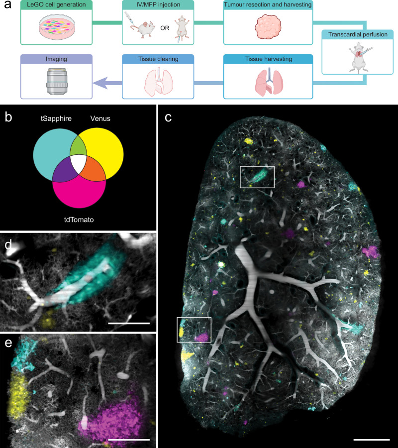 Figure 1
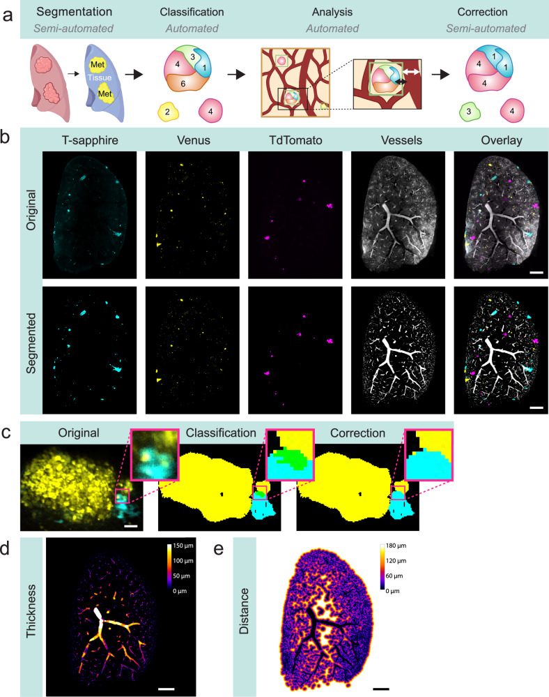 Figure 2
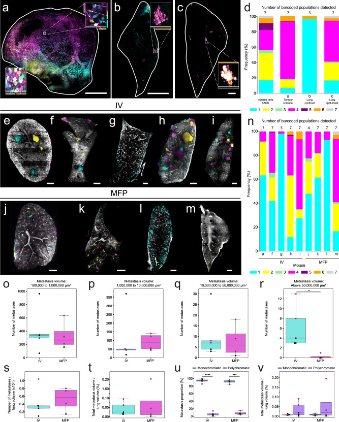 Figure 3
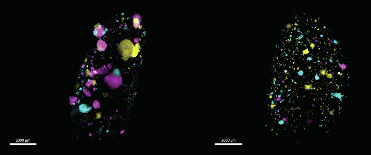 Figure 4
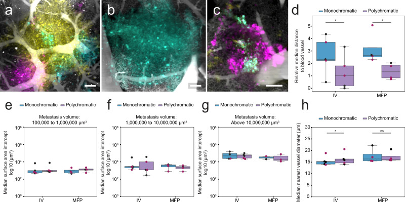 Figure 5
Peer Reviews
No public reviews on file for this paper yet. If you reviewed it on a platform where reviews are public (OpenReview, ICLR, NeurIPS, ICML), you can paste yours below so the community can read it here.
Videos
No videos yet. Explain this paper in a talk, walkthrough, or lecture? Add one.
Taxonomy
TopicsAdvanced Fluorescence Microscopy Techniques · Digital Holography and Microscopy · Optical Coherence Tomography Applications