Nonnegative matrix factorization incorporating domain specific constraints for four dimensional scanning transmission electron microscopy
Koji Kimoto, Fumihiko Uesugi, Koji Harano, Jun Kikkawa, Ovidiu Cretu, Yuki Shibazaki, Motoki Shiga, Atsushi Togo

TL;DR
This paper introduces a new nonnegative matrix factorization method for electron microscopy data that incorporates domain-specific constraints to improve material analysis.
Contribution
A novel constrained NMF technique for 4D STEM that integrates domain-specific knowledge to enhance decomposition and classification.
Findings
The constrained NMF successfully decomposed simulated and experimental 4D STEM data into interpretable diffractions and maps.
Nanometer-sized crystalline precipitates in ZrCuAl were detected and classified using the proposed method.
The method outperforms PCA and primitive NMF by incorporating domain-specific constraints and reducing artifacts.
Abstract
Modern electron microscopy enables the acquisition of extremely large datasets, necessitating optimized machine learning techniques, such as dimensionality reduction and clustering, to extract material insights. We propose a novel nonnegative matrix factorization (NMF) technique that integrates domain-specific constraints inherent to electron microscopy, including spatial resolution and continuous intensity features without downward-convex peaks. This constrained NMF was applied to four-dimensional (4D) scanning transmission electron microscopy (STEM). Using the constrained NMF, both simulated and actual experimental data were successfully decomposed into interpretable diffractions and maps that cannot be achieved using principal component analysis (PCA) and primitive NMF methods. Additionally, hierarchical clustering was optimized based on diffraction similarity, which is a combination…
Genes, proteins, chemicals, diseases, species, mutations and cell lines named across the full text — each resolved to its canonical identifier and authoritative record.
Click any figure to enlarge with its caption.
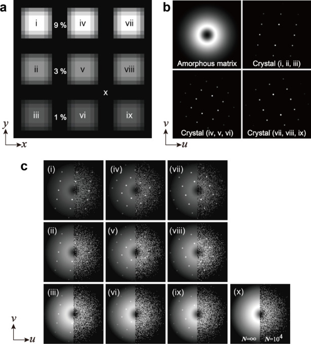 Figure 10
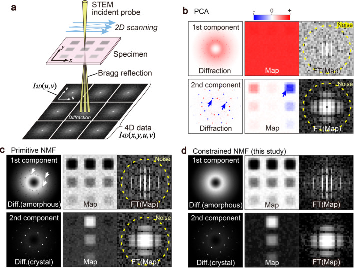 Figure 1
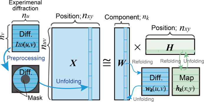 Figure 2
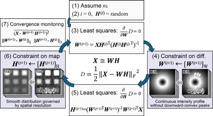 Figure 3
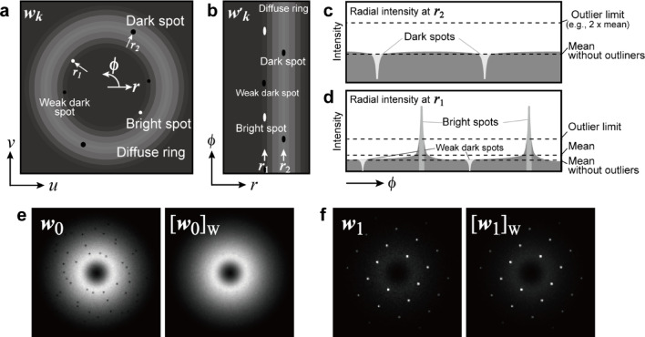 Figure 4
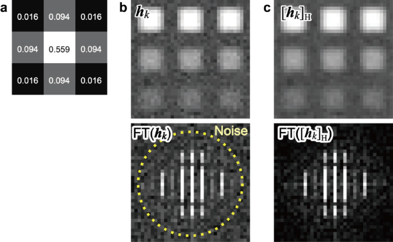 Figure 5
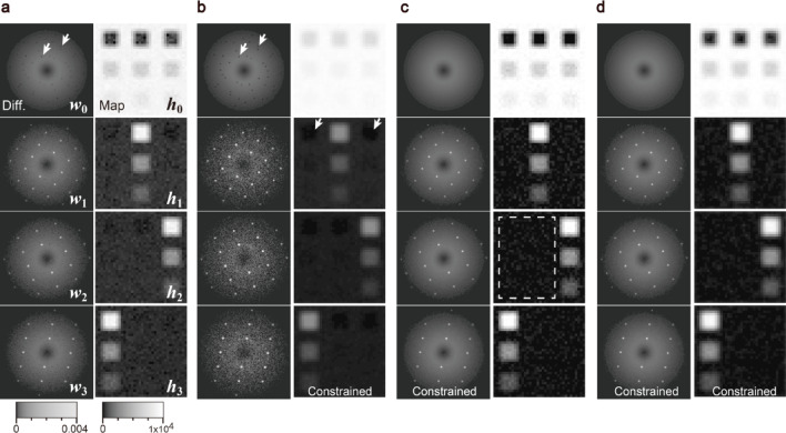 Figure 6
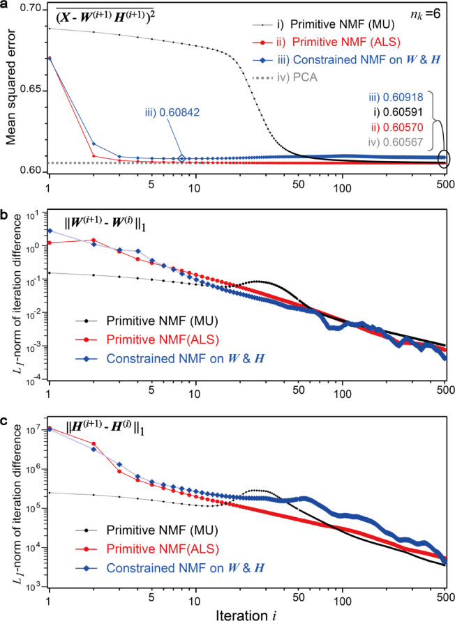 Figure 7
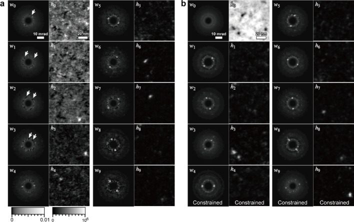 Figure 8
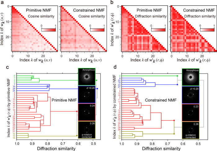 Figure 9
Peer Reviews
No public reviews on file for this paper yet. If you reviewed it on a platform where reviews are public (OpenReview, ICLR, NeurIPS, ICML), you can paste yours below so the community can read it here.
Videos
No videos yet. Explain this paper in a talk, walkthrough, or lecture? Add one.
Taxonomy
TopicsAdvanced Electron Microscopy Techniques and Applications · Quasicrystal Structures and Properties · Machine Learning in Materials Science