Phagophores originate from endoplasmic reticulum membranes in vasopressin neurons in a mouse model of familial neurohypophysial diabetes insipidus
Takashi Miyata, Daisuke Hagiwara, Ryosei Ashida, Satoshi Naito, Yohei Kawaguchi, Tomoko Handa, Tomoko Kobayashi, Mariko Sugiyama, Takeshi Onoue, Shintaro Iwama, Hidetaka Suga, Ryoichi Banno, Mami Matsumoto, Hidetoshi Urakubo, Nobuhiko Ohno, Hiroshi Arima

TL;DR
This study shows that phagophores, which help clear cellular waste, originate from endoplasmic reticulum membranes in neurons affected by a genetic disorder in mice.
Contribution
The first in vivo evidence of structural continuity between phagophores and ER membranes in AVP neurons in a mouse model of FNDI.
Findings
Phagophores envelop aggregates within dilated ER in AVP neurons of FNDI mice.
Serial imaging reveals physical connections between phagophores and intact ER membranes.
ER-phagy is induced in AVP neurons under stress conditions like water deprivation.
Abstract
Familial neurohypophysial diabetes insipidus (FNDI) is an autosomal dominant disorder caused by mutations in the arginine vasopressin (AVP) gene. In AVP neurons in a mouse model of FNDI, aggregates of mutant AVP precursors accumulate within a specific compartment of the endoplasmic reticulum (ER). However, as FNDI mice aged, or were exposed to repeated water deprivation, the ER lumen dilated and mutant aggregates dispersed throughout the ER. Meanwhile, autophagic isolation membranes, known as phagophores, emerged to envelop ER containing these aggregates, indicating induction of ER-phagy. Previous in vitro studies showed that phagophores originate from ER membranes, but the structural relationship between phagophores and the ER membrane in vivo remains unknown. In this study, we used serial block-face scanning electron microscopy to investigate the structural relationship between…
Genes, proteins, chemicals, diseases, species, mutations and cell lines named across the full text — each resolved to its canonical identifier and authoritative record.
Click any figure to enlarge with its caption.
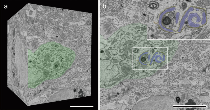 Figure 1
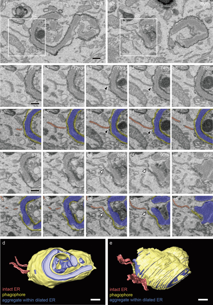 Figure 2
Peer Reviews
No public reviews on file for this paper yet. If you reviewed it on a platform where reviews are public (OpenReview, ICLR, NeurIPS, ICML), you can paste yours below so the community can read it here.
Videos
No videos yet. Explain this paper in a talk, walkthrough, or lecture? Add one.
Taxonomy
TopicsPancreatic function and diabetes · Ion Transport and Channel Regulation · Erythrocyte Function and Pathophysiology