Experimental Evidence for the Solid-State Nitrite-Ligand Photoisomerization Mechanism in Nickel(II) Square-Planar Complexes
Krystyna A. Deresz, Artem Mikhailov, Joanna Jankowska, Lorenzo Donà, Bartolomeo Civalleri, Adam Krówczyński, Radosław Kamiński, Dominik Schaniel, Katarzyna N. Jarzembska

TL;DR
This paper shows how light can switch the structure of nickel complexes in solid form, confirming a previously predicted mechanism.
Contribution
First experimental evidence of a nitro-to-nitrito isomerization mechanism in nickel(II) complexes via exo-nitrito form.
Findings
Nitro form dominates in ground-state crystals of Ni-4d and Ni-4d′.
Endo-nitrito isomer achieves nearly 100% conversion at 100 K using 530 nm LED light.
Exo-nitrito isomer is generated up to 25% using 660 nm LED light at 90 K for Ni-4d.
Abstract
Two square-planar nitrite nickel(II) photoswitches were designed and synthesized, referred to as Ni-4d and the related oxime Ni-4d′. In the ground-state single crystals of both compounds, the nitro form (Ni–N(O)2) is the dominant one. For the two systems, it was possible to generate and detect both endo-nitrito and exo-nitrito linkage isomers using 530 nm light-emitting diode (LED) light at 100 K. Over time, the endo-nitrito form takes over, and almost 100% conversion can be achieved, as confirmed by photocrystallographic and infrared (IR) spectroscopic experiments. The stability of this isomer is similar for both systems, which is confirmed by the experimentally determined decay temperature (T d) values. Furthermore, when 660 nm LED light is applied at 90 K, the exo-nitrito form can be generated up to about 25% population with no admixture of the endo-nitrito isomer for Ni-4d. Based…
Genes, proteins, chemicals, diseases, species, mutations and cell lines named across the full text — each resolved to its canonical identifier and authoritative record.
Click any figure to enlarge with its caption.
 1
1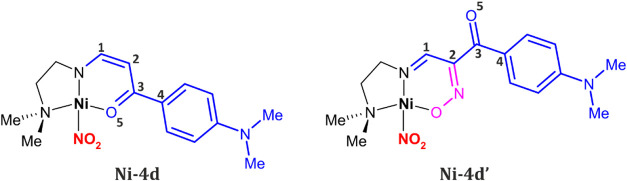 2
2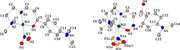 1
1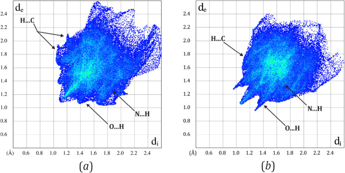 2
2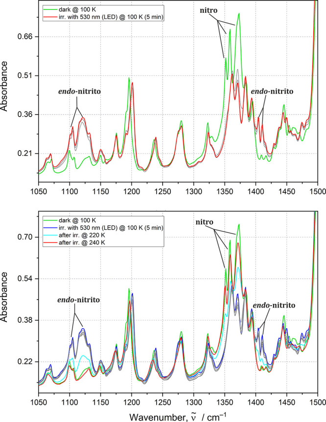 3
3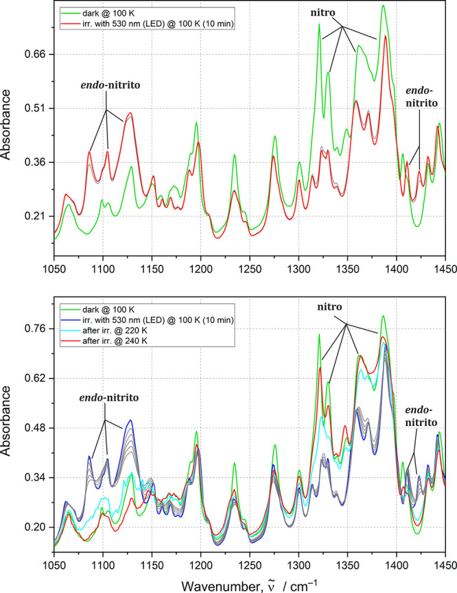 4
4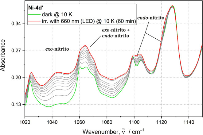 5
5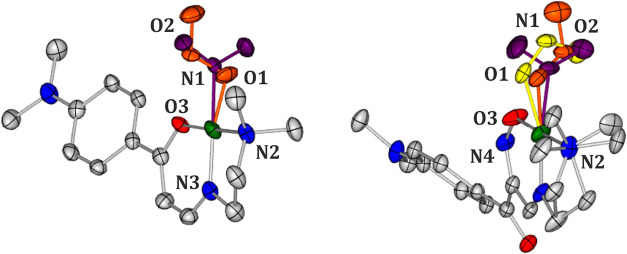 6
6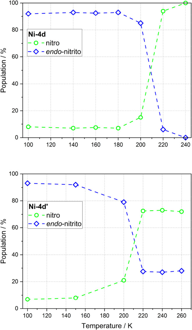 7
7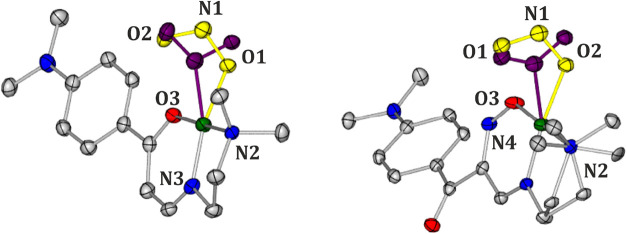 8
8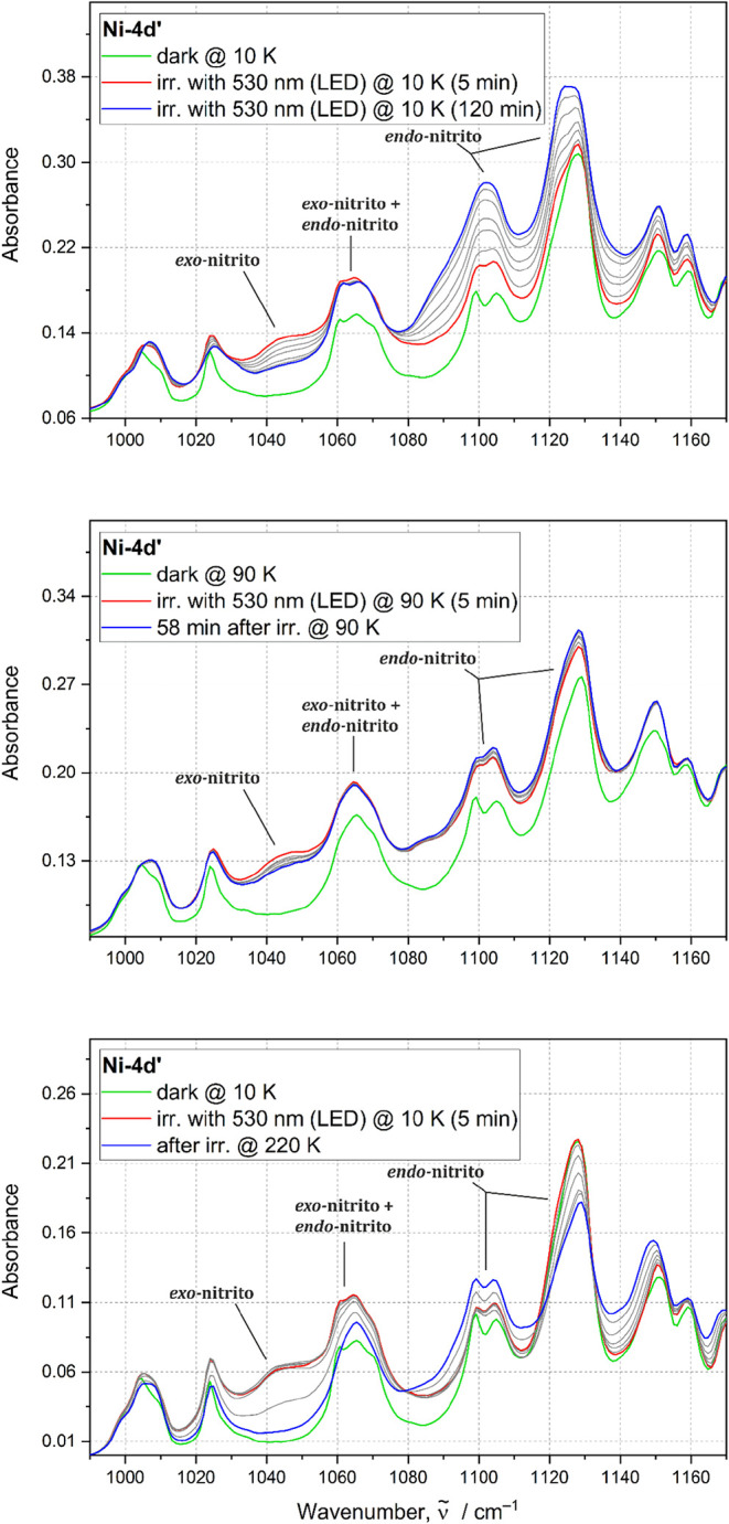 9
9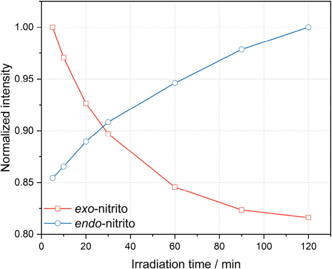 10
10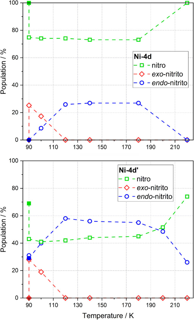 11
11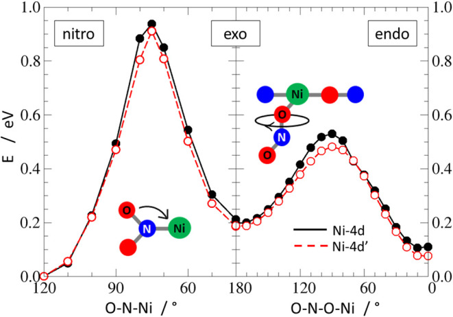 12
12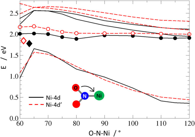 13
13| structure/compound |
|
|
|---|---|---|
| data set |
|
|
| moiety formula | C15H22N4Ni1O3 | C15H21N5Ni1O4 |
| moiety formula mass, | 365.06 | 394.06 |
| crystal system | monoclinic | triclinic |
| space group |
|
|
|
| 4 | 2 |
|
| 768 | 412 |
| crystal color and shape | orange block | orange block |
|
| 100 | 100 |
|
| 11.0495(5) | 7.3612(2) |
|
| 10.4673(4) | 10.0034(3) |
|
| 14.7449(6) | 11.8479(4) |
| α/° | 90 | 85.320(3) |
| β/° | 107.461(5) | 89.590(2) |
| γ/° | 90 | 72.842(3) |
|
| 1626.79(12) | 830.71(5) |
|
| 1.490 | 1.575 |
|
| 4.45% | 3.47% |
|
| 11.8% | 9.03% |
| ϱres min/max/e·Å–3 | –0.41/+0.59 | –0.69/+0.47 |
| CCDC code | 2433376 | 2433395 |
| Ni···H | N···O | N···C | N···H | O···O | O···C | O···H | C···C | C···H | H···H | |
|---|---|---|---|---|---|---|---|---|---|---|
|
| 2.1% | 0.2% | 0.2% | 2.9% | 0.1% | 23.2% | 19.7% | 51.5% | ||
|
| 1.2% | 0.7% | 0.3% | 10.3% | 0.4% | 1.2% | 30.1% | 0.7% | 17% | 38% |
| motif |
| selected interactions |
|
| θD‑H···A/Å |
|---|---|---|---|---|---|
|
| –33.82 | O2···H7b–C7#1 | 2.50 | 3.113(4) | 121.74 |
|
| –107.05 | O1···H1a–C1#1 | 2.53 | 3.293(4) | 136.61 |
| C3···H6b–C6#2 | 2.86 | 3.693(5) | 145.13 | ||
| C4···H6b–C6#2 | 2.80 | 3.500(5) | 130.74 | ||
|
| –59.86 | O1···H1b–C1#3 | 2.78 | 3.460(4) | 128.83 |
| C10–H10···C3#3 | 2.92 | 3.693(5) | 138.51 |
| motif |
| selected interactions |
|
| θD‑H···A/Å |
|---|---|---|---|---|---|
|
| –26.08 | O2···H15c–C15#1 | 2.63 | 3.494(13) | 150.42 |
|
| –25.79 | O2···H15c–C15#1 | 2.51 | 3.379(11) | 150.54 |
|
| –49.66 | C6–H6c···O2#2 | 3.84 | 3.740(16) | 76.68 |
|
| –37.29 | N1···H5a–C5#2 | 3.84 | 3.813(15) | 81.45 |
| O2···H6c–C6#2 | 2.39 | 2.640(11) | 94.10 |
|
|
| |||
|---|---|---|---|---|
|
|
|
|
| |
|
| 66(2) | 77 (8) | 30(3) | 11.4(8) |
| log( | 13.5(5) | 15.8(2) | 12.77(13) | 3.4(5) |
|
| 211(7) | 214 (2) | 100(9) | 93(7) |
|
|
| |||||
|---|---|---|---|---|---|---|
| irradiation time | nitro |
|
| nitro |
|
|
| before irradiation | 100% | 0% | 0% | 69% | 31% | 0% |
| after 30 min irradiation | 75% | 0% | 25% | 43% | 29% | 28% |
|
|
| |||||
|---|---|---|---|---|---|---|
| irradiation time | nitro |
|
| nitro |
|
|
| before irradiation | 100% | 0% | 0% | 70% | 30% | 0% |
| after 1 h irradiation | 57% | 35% | 8% |
|
|
|
| after 2 h irradiation | 26% | 74% | 0% | 19% | 82% | 0% |
| after 4 h irradiation | 8% | 92% | 0% | 7% | 93% | 0% |
|
| ||||
|---|---|---|---|---|
|
|
| |||
| isomer | isolated mol. |
| isolated mol. |
|
| nitro | 0.00 | 0.00 | 0.00 | 0.00 |
|
| 8.16 | 16.79 | 5.78 | 5.66 |
|
| 16.44 | 30.93 | 17.69 | 17.50 |
|
| ||
|---|---|---|
| isomer |
|
|
| nitro | 21.12 | 28.74 |
|
| 17.06 | 29.22 |
|
| 22.54 | 30.44 |
| motif |
| selected interactions |
|
| θD‑H···A/Å |
|---|---|---|---|---|---|
|
| –30.69 | O2···H7b–C7#1 | 2.50 | 3.113 | 121.74 |
|
| –92.91 | O1···H1a–C1#2 | 2.53 | 3.293 | 136.61 |
| C3···H6b–C6#2 | 2.86 | 3.693 | 145.13 | ||
| C4···H6b–C6#2 | 2.80 | 3.500 | 130.74 | ||
|
| –91.90 | O1···H1b–C1#2 | 2.46 | 3.193 | 132.74 |
| C3···H6c–C6#2 | 2.87 | 3.717 | 147.95 | ||
| C4···H6c–C6#2 | 2.82 | 3.497 | 128.44 | ||
|
| –112.83 | N1···H1a–C1#2 | 2.52 | 3.36 | 146.72 |
| C4···H6b–C6#2 | 2.79 | 3.507 | 131.86 | ||
| C5···H6b–C6#2 | 2.73 | 3.454 | 132.87 | ||
|
| –37.30 | C11···H6a–C6#3 | 3.00 | 3.638 | 125.18 |
| N4···H6a–C6#3 | 2.85 | 3.729 | 152.03 | ||
|
| –36.76 | C11···H6a–C6#3 | 2.96 | 3.604 | 125.44 |
| N4···H6a–C6#3 | 2.78 | 3.663 | 152.59 | ||
|
| –35.25 | C14–H14b···O1#3 | 3.17 | 3.772 | 122.03 |
| C11···H6a–C6#3 | 3.00 | 3.638 | 125.18 | ||
| C12···H6a–C6#3 | 3.57 | 4.049 | 113.08 | ||
|
| –60.96 | O1···H1b–C1#4 | 2.78 | 3.460 | 128.83 |
| C10–H10···C3#4 | 2.92 | 3.693 | 138.51 | ||
|
| –55.27 | N1···H1a–C1#4 | 2.63 | 3.325 | 129.25 |
| N1···H2b–C2#4 | 2.70 | 3.191 | 112.55 | ||
| C10–H10···C3#4 | 2.92 | 3.772 | 147.92 | ||
|
| –48.73 | O2···H1b–C1#4 | 2.20 | 3.014 | 141.56 |
| C10–H10···C3#4 | 2.92 | 3.715 | 141.26 |
| motif |
| selected interactions |
|
| θD‑H···A/Å |
|---|---|---|---|---|---|
|
| –25.67 | O2···H15c–C15#1 | 2.63 | 3.494 | 150.42 |
|
| –25.87 | O2···H15c–C15#1 | 2.50 | 3.38 | 151.2 |
|
| –24.79 | N1···H15c–C15#1 | 2.63 | 3.494 | 150.42 |
|
| –86.16 | O3···H1a–C1#2 | 2.62 | 3.191 | 118.39 |
| N4···H2b–C2#2 | 2.67 | 3.187 | 113.93 | ||
| C9···H2b-C2#2 | 2.81 | 3.670 | 148.95 | ||
| C14–H14c···O4#2 | 2.32 | 3.136 | 142.89 | ||
|
| –84.53 | O3···H1a–C1#2 | 2.62 | 3.191 | 118.39 |
| N4···H2b–C2#2 | 2.67 | 3.187 | 113.93 | ||
| C9···H2b-C2#2 | 2.81 | 3.670 | 148.95 | ||
| C14–H14c···O4#2 | 2.32 | 3.136 | 142.89 | ||
|
| –87.34 | O2···H6a–C6#2 | 2.50 | 3.415 | 160.37 |
| O3···H1a–C1#2 | 2.62 | 3.191 | 118.39 | ||
| N4···H2b–C2#2 | 2.67 | 3.187 | 113.93 | ||
| C9···H2b-C2#2 | 2.81 | 3.670 | 148.95 | ||
| C14–H14c···O4#2 | 2.32 | 3.136 | 142.89 | ||
|
| –47.27 | C6–H6c···O2#3 | 3.84 | 3.740 | 76.68 |
|
| –46.77 | N1···H5a–C5#3 | 3.84 | 3.813 | 81.45 |
| O2···H6c–C6#3 | 2.39 | 2.640 | 94.10 | ||
|
| –51.00 | O2···H5a–C5#3 | 2.88 | 2.920 | 82.94 |
| O2···H6c–C6#3 | 2.71 | 3.481 | 91.35 |
- —Agence Nationale de la Recherche10.13039/501100001665
- —Agence Nationale de la Recherche10.13039/501100001665
- —Narodowe Centrum Nauki10.13039/501100004281
- —Akademia G??rniczo-Hutnicza im. Stanislawa Staszica10.13039/501100007751
- —European Commission10.13039/501100008530
- —Wroclawskie Centrum Sieciowo-Superkomputerowe, Politechnika Wroclawska10.13039/501100011009
- —Narodowa Agencja Wymiany Akademickiej10.13039/501100014434
Peer Reviews
No public reviews on file for this paper yet. If you reviewed it on a platform where reviews are public (OpenReview, ICLR, NeurIPS, ICML), you can paste yours below so the community can read it here.
Videos
No videos yet. Explain this paper in a talk, walkthrough, or lecture? Add one.
Taxonomy
TopicsPolyoxometalates: Synthesis and Applications · Nanocluster Synthesis and Applications · Inorganic Chemistry and Materials
Introduction
1
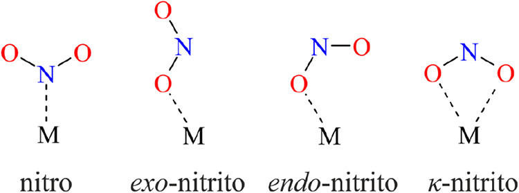
The term photoswitchable compounds refers to materials that undergo various types of transformations under light irradiation, such as cyclization, cis-trans isomerization, or linkage isomerization.? Thanks to the ability to transform from one form to another upon exposure to light, photoswitches may find wide applications in materials science,? optoelectronics,? or medicine.? Transition-metal complexes containing ambidentate ligands, e.g., SO_2_, NO, and NO_2_, ?−? ? ? ? constitute an interesting and readily modifiable group of molecular photoswitches. The ambidentate ligands are key fragments in such molecules, as they may bind to a metal center in more than one way, depending on the conditions. For instance, the nitrite group may adopt four different binding modes in mononuclear complexes (Scheme): nitro-(η^1^-N(O)2) (further referred to as nitro), exo-nitrito-(η^1^-ONO) (further referred to as exo-nitrito), endo-nitrito-(η^1^-ONO) (further referred to as endo-nitrito), and κ-nitrito-(η^2^-O_2_N),? which, under some circumstances, may be interchanged.
Selected Possible Coordination Modes of the Nitrite Group in Mononuclear Metal Complexes
Understanding of the influence of supporting ligands, metal centers, or crystal packing on the photoswitchable properties, as well as the isomerization reaction mechanism itself, is crucial in the context of designing new efficient photoswitches. To the best of our knowledge, to date, there is no direct experimental confirmation of the nitro-to-endo-nitrito reaction mechanism in square-planar nickel complexes. However, quite recently, Warren et al. proposed a mechanism according to which the nitro isomer transforms to the exo-nitrito form * first *, and subsequently converts to the final photoproduct, the endo-nitrito isomer.? More recently, a possible isomerization reaction pathway for square-planar complexes was discussed in two other articles. The isomerization reaction mechanism fully consistent with the one described above was reported by us? for crystals of the nickel nitro complex with the 1-phenyl-2-hydroxyimino-3-[(2′-dimethylamino)ethyl]imino-1-propanone moiety as an (N,N,O)-donor supporting ligand (Ni-1d′). The computationally evaluated mechanism (nitro → exo-nitrito → endo-nitrito) sheds light on the experimental observations. In contrast, Mikhailov et al.? presented two possible reaction pathways based on the experiments performed for a series of complex salts of the [Pd(NH_3_)4] [Pd(NH_3_)3_NO_2] [M(Ox)3]·y H_2_O type (M = Cr, Rh, Co; Ox = oxalate). In the first considered mechanism, the endo-nitrito isomer served as an intermediate product, while in the other potential pathway, the transformation went through the exo-nitrito isomer, similarly as suggested by the above-discussed papers.
Hence, in this paper, two related photoswitchable square-planar Ni^II^ nitrite complexes, namely Ni-4d and Ni-4d′, are introduced (Scheme). These compounds were designed based on the nickel nitro complexes with (N,N,O)-donor supporting ligands previously studied by some of us. ?,?,? The two compounds were crystallized and completely characterized, both experimentally and theoretically, in order to shed more light on the nitro-to-endo-nitrito photoreaction mechanism.
Schematic Representation of the Studied Systems: Ni-4d and Ni-4d′
Experimental Section
2
Synthesis
2.1
The compounds Ni-4d and Ni-4d′ were synthesized in the same manner as in literature procedures ?,? using the following compounds to create a chelating ligand: 4-dimethyloaminoacetophenone, ethyl formate, and N,N-dimethyloethylenediamine. It is worth stressing that the Ni-4d compound is obtained when using a deficiency of LiNO_2_ in the reaction, and when an excess of LiNO_2_ is used, Ni-4d′ is derived. The reaction steps are schematically presented in Figure S1 (Supporting Information). The yields of the reaction for Ni-4d and Ni-4d′ were 55 and 49%, respectively.
X-ray Diffraction
2.2
All X-ray diffraction experiments (including the preliminary ones) were carried out on a Rigaku Oxford Diffraction SuperNova single-crystal diffractometer equipped with an Atlas CCD detector, a copper microfocus X-ray source (Cu–K_α_ radiation, λ = 1.54184 Å) coupled with the multilayer mirror optics, a low-temperature nitrogen gas flow Oxford Cryosystems device, and our homemade light-delivery device,? which allows in situ photocrystallographic experiments. Single crystals were irradiated with 530 and 660 nm LED light (LED = light-emitting diode). The optimal data-collection strategy took into account the mounted light-delivery device and was automatically prepared by using the native diffractometer software. In the case of photocrystallographic experiments, the same strategy was used for all data collected for a given crystal (only the exposure time was adjusted for various temperature points). All data collections were carried out in complete darkness (the sample mounting and centering were done at room temperature prior to any further data collection, and all experiments were performed with all of the diffractometer lights permanently switched off). For details concerning photocrystallographic data-collection codes and the measurement sequence for each sample, see the Supporting Information (Tables S5 and S6). Further data processing (i.e., unit-cell determination, raw diffraction-frame integration, absorption correction, and scaling) was the same for all data sets collected. All structures were solved using an intrinsic phasing method as implemented in the SHELXT program? and refined with the SHELXL program? in the OLEX2 package? within the independent atom model (IAM) approximation.
Spectroscopy
2.3
All infrared (IR) measurements were performed using a Nicolet 5700 FTIR spectrometer (spectral resolution of 2 cm^–1^ in the range of 360–4000 cm^–1^) equipped with a closed-cycle cryostat (Oxford Optistat V01). The sample was ground, mixed with spectroscopic grade KBr, pressed into pellets (referred to as thin-film samples) via a mechanical press, and glued to the coldfinger of the cryostat using a silver-paste thermal adhesive. During measurements, the sample was kept in a vacuum inside the cryostat. Irradiation of the sample was achieved through the cryostat window using various LEDs (Thorlabs L and LP series), the central wavelengths of which covered the range from violet to red (from 385 to 735 nm). LED powers are presented in the Supporting Information (Table S2).
Theoretical Calculations
2.4
Isolated molecule, dimer interaction energies, and normal-mode frequencies were calculated using the density functional theory (DFT) at the DFT(B3LYP)/6–311++G** level of theory ?−? ? ? using the GAUSSIAN package (ver. 16, rev. C.01).? For harmonic-mode computations, no imaginary frequencies were found. All optimized molecular energies were corrected for the zero-point energy (ZPE). For all of the calculations performed with the GAUSSIAN package, the Grimme empirical dispersion correction ?,? modified by the Becke-Johnson damping function was applied; ?,? in the case of the interaction energy calculations, a correction for basis set superposition error ?,? (BSSE) was also included. The semiautomatic generation of input files was accomplished with the CLUSTERGEN program.? Additionally, unit-cell parameters and atomic positions were optimized using the periodic approach implemented in the CRYSTAL program (ver. 17)? applying the PBESOL0–3C functional and the sol-def2-mSVP basis set for all atoms.?
Potential-energy profiles (PEPs) calculations in the ground (S_0_) and in the lowest excited (S_1_) singlet states, as well as the approximate minimum-energy conical intersection (MECI) optimizations were performed at the time-dependent density functional theory level with the Tamm-Dancoff approximation (TDA-TDDFT).? In these calculations, the B3LYP functional with the three-body Grimme dispersion correction was employed, with the 6–311++G** basis set applied for all atoms but the nickel, for which the 6–31G** basis set? was used. The PEP scans have been performed with the TURBOMOLE suite of programs (ver. 7.1),? while the CIOPT code? interfaced with TURBOMOLE was used for the MECI optimization calculations.
Results and Discussion
3
The examined Ni-4d and Ni-4d′ were synthesized using the same synthetic protocol as in our previous articles. ?,?,? It appears that selection of an (N,N,O)-donor ligand, thus the amine and ketone used in the synthesis, may affect the synthetic route, leading to an expected complex described in a series of our other articles, ?,?,? or to a rarer oxime-type product, as, e.g., the reported Ni-1d′.? In the latter case, it was the use of a more basic aliphatic amine that resulted in a nontypical synthesis product. The obtained compound appeared to be an efficient photoswitch in the solid state and can be successfully isomerized from the ground-state nitro form both to the endo- and exo-nitrito isomers upon light irradiation. The direct experimental confirmation of the proposed reaction pathway was, however, challenging in this case since the endo- and exo-nitrito isomers had a very similar stability, and there was a rather low-energy barrier for transformation in both ways. Hence, for the purpose of the current study, we have decided to use similar ligands in the synthesis so as to possibly obtain both kinds of compounds, a typical product and the oxime-type one, in order to further investigate experimentally the isomerization mechanism and compare the behavior of both systems in the solid state.
Consequently, when compared to the synthesis of the previous Ni-1d′ compound, the same aliphatic amine substrate was used, but the ketone substrate was changed from acetophenone to a more basic N,N-dimethyl acetophenone. In such a case, depending on the amount of LiNO_2_ added during the reaction, different products are obtained. These are the two desired coordination compounds, Ni-4d and an oxime-type Ni-4d′, which differ in the way the carbonyl fragment is attached to the metal center as shown in Scheme. In the case of Ni-4d′, an additional NO fragment is incorporated in the coordination sphere of Ni which is the result of the excess of nitrite salt in the reaction environment along with the basicity of the amine used. Molecular structures of Ni-4d and Ni-4d′ are presented in Figure. Similarly to the photoswitching nickel nitro complexes previously studied by us, the metal center is coordinated by the (N,N,O)-donor supporting ligand and the ambidentate nitrite group, while the metal center coordination geometry is close to square-planar. ?,?
Molecular structures of Ni-4d (left) and Ni-4d′ (right) derived from the X-ray diffraction data sets collected in complete darkness at 100 K (atomic thermal motion is represented as ellipsoids at the 50% probability level; hydrogen atoms are omitted for clarity; note the disorder, shown in gold, at the NO2 group in Ni-4d′).
Crystal Structures
3.1
Ni-4d crystallizes in the monoclinic P2_1_/c space group, with one molecule in the asymmetric unit (ASU) (Table). The nitrite group in the Ni-4d crystal structure adopts the nitro binding mode in the ground state (GS) (Figure).
In turn, Ni-4d′ crystallizes in the triclinic P1̅ space group with one molecule in ASU. In the Ni-4d′ crystal structure, the nitrite group exists in two isomeric forms, i.e., nitro and endo-nitrito in the GS with the nitro binding mode predominating in the crystal structure (population of ca. 70%). Multitemperature measurements of Ni-4d′ crystal structures were performed in the 280–100 K range, showing that the endo-nitrito population remains generally stable (Table S7, Supporting Information). The small deviations are within experimental error.
Crystal packings of Ni-4d and Ni-4d′ are different. Molecules in the crystal structure of Ni-4d are arranged in a herringbone-manner along the [010] direction (Figure S4, Supporting Information), which is mainly supported by C–H···π interactions and hydrogen bonds involving nitrite ligands. Furthermore, the molecules are arranged into a ribbon-like structure along the [001] direction (Figure S5, Supporting Information) which is formed mainly by C–H···π interactions and hydrogen bonds involving the nitrite ligand as in the former case. Main motif in the Ni-4d′ structure is a dimeric plane along the [100] direction (Figure S6, Supporting Information), majorly supported by hydrogen bonds involving nitrite group as well as O3, O4, and N4 atoms from the supporting ligand along with C–H···π interactions.
1: Selected Crystal-Structure Parameters of the Studied Complexes at 100 K Prior to Any Light Exposure (“Dark” Structures)
Differences and similarities in the crystal packing of the examined compounds can be well illustrated and analyzed using the concept of Hirshfeld surfaces and further – fingerprint plots (Figure and Table). ?−? ? ? ? The calculated interatomic contact contributions to the Hirshfeld surfaces generated for the Ni-4d and Ni-4d′ crystal structures show that both of them are dominated by H···H interactions, which is typical for molecular crystals of organic compounds. Interestingly, the percentage contribution of these contacts is significantly smaller for Ni-4d′. In this case, this is balanced by notably greater contributions of the N···H and O···H type contacts. The increased impact of these interactions results from the presence of the oxime moiety (N4–O3) and the exposed carbonyl oxygen engaged in hydrogen-bond-type contacts in the crystal structure of Ni-4d′. This kind of interactions are naturally also formed by the nitrite group, involving its exposed oxygen atoms which contribute the most to the total number of hydrogen-bond-like contacts in both systems. From the respective fingerprint plots, it can be concluded that hydrogen bonds are stronger and more distinct in the Ni-4d′ structure compared to Ni-4d. However, theoretical calculations of dimeric motifs (Tables and ? and Figures S14 and S15, Supporting Information) reveal that the nitrite group in Ni-4d is better stabilized by these interactions. This suggests that the strong hydrogen bonding interactions observed in the fingerprint plot for Ni-4d′ involve other parts of the molecule rather than the nitrite group. In turn, the structure of Ni-4d exhibits more effective C–H···π edge-to-face interactions between the phenyl ring, the nickel-containing aromatic ring, and hydrogen atoms from methyl groups, the phenyl ring, and the nonaromatic ring as well. Similar interactions occur in the Ni-4d′ structure; however, there is fewer of them, and the molecules adopt less favorable orientations. Based on the obtained fingerprint plots, it can also be concluded that packing efficiency is higher in the case of the Ni-4d′ crystal structure.
2: Interatomic Contact Contributions to the Hirshfeld Surface Generated for the Ni-4d and Ni-4d′ Molecules in the GS Crystal Structures, Computed with the CRYSTALEXPLORER Program,
Hirshfeld fingerprint plots generated for (a) Ni-4d and (b) Ni-4d′, based on the 100 K ground-state (GS) crystal structures. The intensity of the blue color reflects the contribution of specific intermolecular interactions: the brighter the blue, the greater the contribution. The presence of diffuse regions at d i and d e values (normalized “internal” and “external” surface–nearest-atom distances) around 2.0 Å and higher indicates less efficient crystal packing. The more defined and sharp features are in the plots, the stronger and more directional the corresponding interactions.
3: Dimeric Motifs Engaging the NO2 Group and Respective Interaction Energies Based on the Experimental Ground-State Crystal Structure of the Ni-4d Compound (Figure S14, Supporting Information),
4: Dimeric Motifs Engaging the NO2 Group and Respective Interaction Energies Based on the Experimental Ground-State Crystal Structure of Ni-4d′ Structure (Figure S15, Supporting Information),
Since the nitrite ligand in Ni-4d′ exists in two isomeric forms in the ground state, namely the nitro and endo-nitrito binding modes, intermolecular interactions formed by the nitro and endo-nitrito isomers can be compared. Analyzing the fingerprint plot (Figure S7, Supporting Information), it can be clearly seen that the endo-nitrito isomer is engaged in shorter and better-defined hydrogen bonds. However, theoretical calculations of dimeric motifs (Table) indicate that this does not improve the stability of this isomer, which might be due to the noticeably fewer interactions formed with oxygen in favor of interactions involving nitrogen. Still, part of the intermolecular interactions are similar for both endo-nitrito and nitro form. The packing for Ni-4d′ in endo-nitrito form is visibly less efficient than in the case of the nitro form (Figure S7, Supporting Information).
A very useful tool in the analysis of light-induced transformations in crystals is the concept of the reaction cavity. The term and first computations were introduced by Ohashi and co-workers? with a great deal of success in explaining many subtle phenomena occurring when molecules change in crystals. The method is relatively simple. It requires only “cutting out” the photoactive part from the crystal structure (in our case this would be the NO_2_ group) and analyzing the shape and volume of the artificially created “void.” Nowadays several programs can compute the crystal voids and related properties. The most well-known are PLATON ? (grid search with the van der Waals radii criteria?), MERCURY ? (“rolling-ball” method?), or CRYSTALEXPLORER (promolecule electron density cutoff criterion?). Here, we used MERCURY, with the settings identical to those in our previous work for consistency.
The reaction-cavity volumes calculated for both compounds amount to 26.3 Å^3^ for Ni-4d and 33.9 Å^3^ for Ni-4d′. Although both values are within the volume range reported for compounds exhibiting photoswitching behavior of the nitro group, the cavity in the Ni-4d crystal structure is at the lower limit. ?,?,? In the context of isomerization reaction, the cavity shape in the Ni-4d′ crystal structure, which already hosts two linkage isomers, appears to be more favorable as it is rather evenly distributed in all directions (Figures S8 and S9, Supporting Information) providing enough space for rearrangement of the ambidentate ligand.
In view of the above, both compounds have an isomerization potential. The nitro group in both structures mainly forms hydrogen-bond-like interactions with the neighboring molecules, which, unlike strong hydrogen-bond interactions involving more electronegative atoms, may promote the linkage isomerism reaction by stabilizing the resulting isomers while also being easy to break when exposed to visible light. ?,? Furthermore, the reaction cavities in both crystal structures are sufficient to accommodate the ambidextrinated ligand transformation.
Formation and Decay of Photoinduced Linkage
Isomers
3.2
Infrared Spectroscopy
3.2.1
A series of solid-state infrared spectroscopic measurements were conducted for samples just after being exposed to different LEDs’ light for varying periods of time at selected temperatures. Such an approach enables characterization of the photoswitching behavior of the examined material and defines optimal conditions of the analyzed photoinduced transformation, based on intensity changes of bands assigned to the vibrations of the nitrite group. The bands’ intensity changes with irradiation time and during sample relaxation provide us also with some information about the kinetics of the process. Thermal behavior of nonirradiated samples was also investigated.
For both compounds, the respective vibration ranges for each linkage isomer under consideration are comparable. In all cases, the bands were assigned to specific vibrations based on the calculated frequencies (Table S3, Supporting Information) and available literature. ?−? ? For the nitro and endo-nitrito binding modes, these are bands located from 1300 to 1400 cm^–1^ and from 1070 to around 1140 cm^–1^, respectively. Upon 530 nm LED irradiation, in the IR spectrum of Ni-4d (Figure), the intensity of the 1068, 1105, and 1121 cm^–1^ bands associated with the symmetrical stretching vibrations of the endo-nitrito group are increasing, which is accompanied by lowering of the 1352, 1358, and 1372 cm^–1^ bands corresponding to symmetrical stretching (1352 cm^–1^) and asymmetrical stretching (1358 and 1372 cm^–1^) vibrations of the nitro isomer, respectively. The presence of the endo-nitrito binding mode is also evidenced by the appearance of the 1404 and 1409 cm^–1^ bands, which can be attributed to their symmetrical stretching. For Ni-4d′, the nitro-to-nitrito isomerization process is manifested by the intensity rise of the following bands upon 530 nm LED irradiation: 1065, 1085, 1104, and 1128 cm^–1^ related to symmetrical vibrations of the endo-nitrito binding mode (Figure). Furthermore, spectral changes accompanying this transformation are also visible in the higher vibrational frequency regions, i.e., around 1410 and 1426 cm^–1^. In turn, the nitro isomer gradual depopulation can be monitored by observation of the following bands 1321, 1330, 1361, and 1386 cm^–1^, which are assigned to asymmetrical stretching vibrations.
IR spectra were collected for the Ni-4d sample. Upper panel: before (green line) and after (red line) optimal irradiation time with 530 nm LED at 100 K for generating endo-nitrito form; gray lines correspond to subsequent irradiation points (1–5 min); bottom panel: before (green line) and after irradiation (blue line) with 530 nm LED at 100 K and during temperature relaxation (cyan and red line) of endo-nitrito form; gray lines correspond to subsequent temperature points during sample heating.
IR spectra collected for the Ni-4d′ sample. Upper panel: before (green line) and after (red line) optimal irradiation time with 530 nm LED light at 100 K for generating the endo-nitrito form; gray lines correspond to subsequent irradiation point (5 min), bottom panel: before (green line), after irradiation (blue line) at 100 K, and during temperature relaxation (cyan and red line) of the endo-nitrito form; gray lines correspond to subsequent temperature points during sample heating.
In order to investigate the temperature effect on the samples, multitemperature measurements were conducted in the RT → 10 K range. The temperature was gradually reduced, and an IR spectrum was collected every 20 K. For both compounds, no spectral changes were observed during these experiments. However, it can be noticed that in the Ni-4d′ sample, bands associated with the endo-nitrito binding mode are already visible without any irradiation, which is in agreement with the single-crystal structure of this compound.
The most significant spectral changes associated with the nitro-to-endo-nitrito isomerization for the examined compounds were observed after sample irradiation with a 530 nm LED light. The optimal irradiation time was determined at 100 K. It appears that maximal population of the endo-nitrito form is reached faster for Ni-4d, for which 5 min of LED irradiation, equal to 21.3 J·cm^–2^ fluence (or energy density; Table S2, Supporting Information), of thin-film sample is sufficient. In turn, the Ni-4d′ sample requires 10 min of irradiation (42.5 J·cm^–2^ fluence) to reach the maximum conversion level. Photoinduced linkage isomers in both cases are stable up to 220 K at which their populations visibly decrease with time. At 240 K, in both cases, full relaxation to the initial state is noted.
Throughout the IR experiments, the generation of the exo-nitrito isomer under 530 and 660 nm LED light irradiation was detected for both Ni-4d and Ni-4d′. For Ni-4d′, the existence of this metastable form was clearly evidenced by the distinct appearance of 1044, 1065, and 1460 cm^–1^ bands after irradiation. These spectral changes were particularly well-defined, allowing for definitive identification of the formation of the exo-nitrito form. For the Ni-4d complex, the transition to the exo-nitrito form was confirmed by an increase of the 1068 and 1436 cm^–1^ bands. However, the 1068 cm^–1^ band substantially overlaps with that from the endo-nitrito binding mode vibrations, complicating the precise identification of the exo-nitrito isomer. This contrasts with the Ni-4d′ complex, where the spectral changes allow for more reliable characterization of the isomeric transition.
The optimal irradiation wavelengths for inducing the formation of the exo-nitrito isomer were determined at 10 K, where this isomer can be more effectively generated. For Ni-4d′, short irradiation with 530 nm LED (5 min, 4.3 J·cm^–2^ fluence) produced excellent results, with clearly visible spectral bands corresponding to the exo-nitrito form. Notably, Ni-4d′ requires significantly lower LED power compared to Ni-4d to achieve the exo-nitrito form with minimal admixture of the endo-nitrito isomer. This difference is attributed to the rapid formation of the endo-nitrito binding mode when Ni-4d is exposed to 530 nm LED light (Figures S12 and S13, Supporting Information).
Further experiments revealed that 660 nm LED light could generate even higher populations of the exo-nitrito form in both compounds with lower conversion to the endo-nitrito isomer (Figure). The optimal irradiation time for Ni-4d′ was longer (1 h, 451.7 J·cm^–2^ fluence) compared to that of Ni-4d (Figure S12, Supporting Information). While 530 nm proved to be the energy-optimal wavelength for generating the exo-nitrito isomer in both complexes, the 660 nm LED provided better conditions for monitoring the isomerization process due to the slower course of the photoinduced transformation in this case.
IR spectra collected before (green line) and after (red line) optimal irradiation time at 10 K for generating the exo-nitrito form in the Ni-4d′ sample with 660 nm LED; gray lines correspond to subsequent irradiation points (1–60 min).
Relaxation Kinetics of the Photoinduced
Linkage Isomers
3.2.2
The kinetics of the relaxation of the metastable endo-nitrito and exo-nitrito isomers in the solid state was examined for both compounds (Table). For this purpose, a series of isothermal IR spectroscopic measurements during the relaxation process were conducted. In order to determine the decay constants (k) at each temperature, the first-order kinetic equation was employed. The activation energy (E a) and frequency factor (k 0) were calculated based on the Arrhenius equation. The decay temperature (T d), which corresponds to the point at which k = 10^–3^ s^–1^, was calculated based on the obtained kinetic parameters. Despite the fact that the activation energy and frequency factor differ between the two compounds, the resulting decay temperatures, derived for both the exo-nitrito and endo-nitrito metastable forms, are comparable. For the endo-nitrito isomers, T d is almost equal; hence, it can be concluded that their stability in the Ni-4d and Ni-4d′ thin films is very similar. In turn, T d determined for the exo-nitrito forms differs more significantly between the studied compounds, which indicates slightly lower stability of this linkage isomer in the Ni-4d′ solid-state sample.
5: Kinetic Parameters of the Endo-Nitrito and Exo-Nitrito Form Relaxation Were Determined by IR Spectroscopy
Photocrystallography
3.2.3
Having the basic knowledge about the Ni-4d and Ni-4d′ crystal structures along with the results of IR spectroscopic experiments, photocrystallographic studies on single crystals were conducted. In accordance with the spectroscopic findings, single crystals were irradiated with 530 and 660 nm LED light and examined toward formation of the nitrito linkage isomers. Such experiments allow us to evaluate the actual structures of the photoinduced linkage isomers, to relatively accurately estimate their populations, and to contrast the outcomes with those deduced from the spectroscopic studies for thin-film samples. The metastable state population is determined from structural refinement of the disordered nitrite group. Based on our rough analysis (Supporting Information), the estimated standard deviations do not exceed 3%. Thus, in the following discussion, the populations are rounded and analyzed, focusing mainly on the observed trends and/or changes, taking into account possible slight deviations from the given values.
Photocrystallography at 90 K
3.2.3.1
Based on the above-described kinetic measurements for the exo-nitrito form in the Ni-4d compound, it is evident that achieving a substantial population of this isomer would be a challenging task at 100 K and above. This is because the estimated lifetime of this isomer at 100 K is about 16 min, as calculated applying the Arrhenius equation with the determined and above-described kinetic parameters. Thus, during a crystallographic measurement, a significant portion of this form may undergo thermal relaxation. In turn, at 90 K, the exo-nitrito form’s lifetime extends to approximately 10 h, which facilitates obtaining more reliable results. The highest conversion to the exo-nitrito form, 25%, was reached after irradiation for 30 min with a 660 nm LED (1.86 J·cm^–2^ fluence) (Table). The same experiment was conducted for **Ni-4d′**in this case, the predicted lifetime of the exo-nitrito form at 90 K reaches about 27 min. Nevertheless, the reaction was stopped after 30 min irradiation with a 660 nm LED (2.59 J·cm^–2^ fluence); it was possible to generate the exo-nitrito isomer and estimate its population to 28%, while the endo-nitrito population remained unchanged within the estimation error. Crystal structures obtained after irradiation are listed in Figure.
Molecular structures of Ni-4d (left) and Ni-4d′ (right) derived from the X-ray diffraction data sets collected after 30 min of irradiation with 660 nm LED at 90 K. Note the disorder on the NO2 site (nitro isomer - violet color, exo-nitrito - orange, endo-nitrito - yellow, atomic thermal motion is represented as ellipsoids at the 50% probability level, hydrogen atoms are omitted for clarity, only selected atom labels are shown - disorder labels, A/B/C are not shown for clarity, atoms N1, O1, N2 are approximately shown).
6: Populations of Linkage Isomers before and after Irradiation with a 660 nm LED for 30 Min at 90 K for Ni-4d and Ni-4d′
Photocrystallography at 100 K
3.2.3.2
The ground-state structure of Ni-4d at 100 K contained exclusively the nitro form. In order to determine the optimal irradiation time to generate the metastable states, the sample was irradiated within three time steps with 530 nm LED (Figure and Table). After 1 h (1.05 J·cm^–2^ fluence), a population of 35% of the endo-nitrito and 8% of the exo-nitrito linkage isomer was generated. During the subsequent 1 h of light exposure (2.10 J·cm^–2^ fluence), the exo-nitrito isomer completely vanished, while the endo-nitrito binding mode’s population increased to 74%. The maximal conversion to this latter form was achieved after 2 additional hours of irradiation (4.21 J·cm^–2^ fluence) and reached about 92%. Regarding the relaxation of the endo-nitrito linkage isomer in Ni-4d crystals, it appears that up to 180 K, no population changes are observed. At 200 K, there is a small reduction of the endo-nitrito (by 7%) population, which sharply decreases to 6% at 220 K. At 240 K, complete relaxation to the ground-state structure was noted.
Average populations estimated for each isomer (green line - nitro, blue line - endo-nitrito) after irradiation with 530 nm LED at 100 K and during temperature relaxation. Upper panel presents results obtained for Ni-4d while bottom panel presents results obtained for Ni-4d′. These population values are averaged, accounting for the concurrent decay occurring during the data-collection period at each temperature point.
7: Populations of Different Linkage Isomers before and during Subsequent Steps of Irradiation with 530 nm LED at 100 K for Ni-4d and Ni-4d′
Similar investigations were conducted for the Ni-4d′ crystal structure (Figure and Table). As already mentioned, in the ground-state structure at 100 K, it consists of ca. 30% of the endo-nitrito form and ca. 70% of the nitro form. The crystal was irradiated with a 530 nm LED in two time steps, each lasting for 2 h. The initial 2 h light exposure (16.94 J·cm^–2^ fluence) led to an increase of the endo-nitrito population to 82%, while during the subsequent 2 h of irradiation (33.88 J·cm^–2^ fluence), the population of the endo-nitrito isomer further increased reaching 93%. Due to high population of the endo-nitrito isomer in the ground state, the relaxation of the photoinduced endo-nitrito form is particularly interesting. The population of endo-nitrito isomer remains stable up to 150 K and decreases to 79% at 200 K. As the temperature rises to 220 K, the population further diminishes to 28%, reaching a thermally invariant state at this pointthe population of the endo-nitrito isomer remains constant at higher temperatures. Crystal structures obtained after irradiation are listed in Figure.
Molecular structures of Ni-4d (left) and Ni-4d′ (right) derived from the X-ray diffraction data sets collected after 4 h of irradiation with 530 nm LED at 100 K. Note the disorder on NO2 group (nitro isomer - violet color, exo-nitrito - orange, endo-nitrito - yellow, atomic thermal motion is represented as ellipsoids at the 50% probability level, hydrogen atoms are omitted for clarity, only selected atom labels are shown - disorder labels, A/B/C are not shown for clarity, atoms N1, O1, N2 are approximately shown).
Mechanism of the Nitro-to-Endo-Nitrito Transformation
3.3
As previously described, irradiation of the samples with a 530 nm LED can result in the generation of both endo- and exo-nitrito forms. For both compounds, the isomerization reaction is notably slower at 10 than at 100 K. Thus, IR experiments with gradual irradiation with 530 nm LED at 10 K were conducted in order to shed light on the photoisomerization reaction mechanism.
Formation of both photogenerated binding modes is easier to trace for Ni-4d′; since, in this case, it is possible to differentiate bands related to both metastable forms. It can be clearly seen that after the first cycle of irradiation for 5 min, the bands associated with the exo-nitrito and endo-nitrito form appear (Figure). After the second irradiation step (additional 10 min of irradiation), there is a noticeable decrease of the exo-nitrito population with a concurrent increase in the population of the endo-nitrito linkage isomer. During further light exposure (for 20, 30, 60, 90, and 120 min), the exo-nitrito isomers’ population gradually diminishes until it completely disappears, while the population of the endo-nitrito binding mode continuously increases (Figure).
IR spectra collected for the Ni-4d′ sample during: (i) top panel: subsequent cycles of irradiation with 530 nm LED at 10 K, (ii) middle panel: exo-nitrito isomer relaxation over time at 90 K, (iii) bottom panel: thermal relaxation of the exo-nitrito isomer in the 10–220 K temperature range. Green line indicates the initial state, red line represents spectra collected after irradiation, and blue line signifies the final state. Gray lines represent spectra collected between irradiation and final state.
Intensity evolution of the characteristic bands for the exo- and endo-nitrito binding modes during irradiation of Ni-4d′ with 530 nm LED at 10 K.
The reaction mechanism can also be investigated via tracing of the exo-nitrito state thermal relaxation (Figure). Thus, after reaching maximal intensity of the band associated with the exo-nitrito form at 10 K with 530 nm, the sample was heated and IR spectra were collected with a 20 K step until the complete disappearance of the endo-nitrito isomer at 220 K. Additionally, it was observed that at 90 K, the exo-nitrito binding mode, once generated, transforms into the endo-nitrito isomer in time when the irradiation is stopped. This clearly suggests that the exo-nitrito isomer converts directly to the endo-nitrito binding mode by thermal relaxation.
Populations estimated for each isomer (green line - nitro, red line - exo-nitrito, blue line - endo-nitrito) before irradiation at 90 K (filled points), after irradiation with 660 nm LED for 30 min at 90 K, and during temperature relaxation. Upper panel presents results obtained for Ni-4d compound while bottom panel presents results obtained for the Ni-4d′ compound.
In the IR experiments for Ni-4d, an identical pattern was observed under irradiation with a 530 nm LED at 10 K and during thermal and time relaxation of the exo-nitrito form.
Finally, related photocrystallographic experiments were conducted in order to verify the applicability of spectroscopic observations to single-crystal samples. Experimental conditions were selected based on kinetic considerations and the available lowest temperature for the X-ray diffraction studies at our home laboratory. Hence, single crystals of Ni-4d and Ni-4d′ were irradiated with a 660 nm LED light for 30 min at 90 K so as to generate the exo-nitrito binding mode. 25% of this isomer was generated in the case of the Ni-4d system, whereas for Ni-4d′, such population reached 29% (the admixture of around 30% of the endo-nitrito form remained the same as for the ground-state structure). In order to verify the isomerization mechanism, i.e., the endo-nitrito isomer formation through the exo-nitrito form as an intermediate photoproduct, the X-ray diffraction measurements were conducted during heating at the following temperatures: 100, 120, 140, 180, and 220 K. This way the relaxation channel of the process was probed. Based on these experiments, it is clearly visible that during relaxation, the exo-nitrito form fully transforms to the endo-nitrito linkage isomer. Starting at 100 K, partial conversion of the exo-nitrito form to the endo-nitrito binding mode was observed. In the Ni-4d crystal, population of the exo-nitrito species decreased to about 16% concurrently generating 9% of the endo-nitrito form. Similarly, in Ni-4d′, the relaxation of the exo-nitrito isomer to 19% resulted in 12% increase of the endo-nitrito form’s population. Full conversion to the endo-nitrito isomer occurred at 120 K in both systems and its population remained stable up to 220 K, at which the endo-nitrito isomer fully relaxed to the nitro form in Ni-4d, while in Ni-4d′, the population of this isomer decreased to 26%which is comparable to its initial value.
The theoretical modeling of the mechanism coincides with the results obtained in our previous paper concerning the Ni-1d′ compound in the solid state. First, to get a notion of a possible source of differences in the interisomer transformation dynamics observed in the electronic ground state for Ni-4d and Ni-4d′, we had a look at the S_0_ potential-energy profiles obtained for respective isolated molecules. The S_0_ profiles between the nitro, exo-nitrito, and endo-nitrito forms, calculated along the O–N–Ni angle (nitro → exo-nitrito), and O–N–O–Ni torsion angle (exo-nitrito → endo-nitrito) coordinates, are shown in Figure. One can notice that, in both cases, the barriers for the nitro → exo-nitrito transformation, estimated to ca. 0.936 (90.3 kJ·mol^–1^) and 0.910 eV (87.8 kJ·mol^–1^) for Ni-4d and Ni-4d′, respectively, significantly exceed the corresponding exo-nitrito → endo-nitrito reaction barriers of 0.321 (31.0 kJ·mol^–1^) and 0.293 eV (28.3 kJ·mol^–1^). Moreover, the slightly smaller values obtained for the Ni-4d′ system stay in line with the observed lower thermal stability of its nitrito forms, on the one hand, and the faster exo-nitrito to endo-nitrito transformation, on the other.
Potential-energy profiles (PEPs) calculated for the S0 state along the O–N–Ni valence angle (nitro → exo-nitrito, left panel), and the O–N–O–Ni torsion angle (exo-nitrito → endo-nitrito, right panel), respectively (black curve - Ni-4d, red curve - Ni-4d′).
In the next step, to investigate the possible mechanism of the photoinduced nitro → exo-nitrito transformation expected to be the primary photoswitching channel in the studied compounds, we calculated the S_1_-optimized potential-energy profiles (Figure) along the O–N–Ni angle: a reaction coordinate used also in the S_0_ PEP calculations. Both for Ni-4d and Ni-4d′, it can be observed that after the S_0_ → S_1_ excitation, initially the system ends up in a rather flat S_1_ potential-energy surface area, protected from the reactive transformation by a small energy barrier of 0.061 (5.9 kJ·mol^–1^) and 0.054 eV (5.2 kJ·mol^–1^) for Ni-4d and Ni-4d′, respectively. At the same time, the expected kinetic energy excess gained at the Franck–Condon vicinity due to the initial relaxation from the respective vertically excited GS structures amounts for Ni-4d and Ni-4d′ to 0.392 (37.8 kJ·mol^–1^) and 0.403 eV (38.9 kJ·mol^–1^). Thus, it can be expected that after some time spent in the Franck–Condon area, the excited nitro isomer may easily reach a potential-energy surface region characterized with a very small S_1_–S_0_ energy gap and, eventually, relax through a nearby, energy-accessible conical intersection. Such minimum-energy conical intersection points have been identified for both studied systems; their geometrical structures can be found in the Supporting Information (Figure S18). Energy-wise, both found MECIs lie below the scanned S_1_ profiles, characterized by relative energies of 1.780 (171.7 kJ·mol^–1^) and 1.848 eV (178.3 kJ·mol^–1^) for Ni-4d and Ni-4d′, respectively.
Potential-energy profiles along the O–N–Ni reaction coordinate optimized in the S1 state for the Ni-4d (black lines) and the Ni-4d′ (red lines) systems. The optimized state in each set has been marked with circles: full for Ni-4d and empty for Ni-4d′, while the other states, calculated vertically on the S1 geometries, are marked with solid/dashed lines for Ni-4d/Ni-4d′, respectively. The black-full/red-empty diamonds mark positions of the optimized MECI points of Ni-4d and Ni-4d′.
At the same time, we note that no direct photoreaction path between the nitro and endo-nitrito isomers was established in the course of the present study, which is in agreement with our previous study on a closely related system.?
Molecular Stability and Intermolecular Interactions
Analysis
4
To support the experimental findings and possibly gain deeper insights into the examined processes, structure optimizations and interaction energy computations were executed. Isolated-molecule geometry optimizations were performed in the GAUSSIAN program, as well as crystal-structure optimizations in CRYSTAL.? The obtained results are summarized in Tables and ?. Based on the calculations and experimental outcomes, it is evident that the nitro form constitutes the most stable linkage isomer in the case of both compounds, whereas the exo-nitrito binding mode itself is the least energetically favored one. The stability of the exo- and endo-nitrito isomers is comparable for the Ni-4d and Ni-4d′ compounds as far as the isolated molecules are concerned. However, after optimizing the whole crystal structure, it becomes clear that packing and intermolecular interactions play an important role in the stability of different forms of the nitrite ligand, especially in the case of the Ni-4d crystal structure. The calculated energy differences between the considered linkage isomers become more significant in this case, which is in contrast to Ni-4d′ for which they remain similar to the ones calculated for the isolated molecules. The minor role of intermolecular interactions for Ni-4d′, thus very similarly energetically stabilized nitro and endo-nitrito isomers in the crystal environment (<6 kJ·mol^–1^ energy difference), supports their coexistence in the crystal structure. In turn, the deformation energies provided in Table appear significantly larger for **Ni-4d′**this might, to some extent, explain a greater difficulty in forming crystals which was noted during crystallization in the experimental process.
8: Energy Differences (E diff) between the Ground (Nitro) and Metastable (Exo- and Endo-Nitrito) Linkage Isomers Computed for the Optimized Isolated-Molecule Geometries and for the Molecular Geometries from the CRYSTAL-Optimized Ni-4d and Ni-4d′ Crystal Structures
9: Deformation Energy (E def) between Optimized Isolated Molecule and the Molecule Optimized in the Crystal Environment (E def = E cry – E isol) Presented for the Nitro, Endo-Nitrito, and Exo-Nitrito Isomers Both for Ni-4d and Ni-4d′
Additionally, interaction energies between dimeric motifs’ components involving different ambidentate ligand’s binding modes were analyzed (Tables and ?, and Figures S14 and S15, Supporting Information). Generally, the nitrite ligand is engaged mostly in weak, hydrogen-bond-type interaction, such as O···H–C and N···H–C. The dimeric motifs in the Ni-4d structure are overall better energetically stabilized than those in Ni-4d′. The energies and interactions of the corresponding dimeric motifs of Ni-4d noticeably vary between isomeric forms in some cases. Primarily, the nitro isomer in the Ni-4d structure creates an additional interaction relative to the exo- and endo-nitrito isomers, which is present in the D4 motif. The exo-nitrito form is best stabilized in dimer D6, where its interaction energy is lower by ca. 20 kJ·mol^–1^ when compared to other isomers, which results from the formation of a hydrogen bond with the nitrogen atom from the NO_2_ group. All of the isomers are similarly stabilized in motif D7. In motif D8, each isomer is engaged in different intermolecular interactions, which results in noticeable differences in their energies with the nitro group being the most stabilized and the exo-nitrito form the least. In turn, the stabilization energies for the corresponding dimeric motifs formed by various isomers in Ni-4d′ are quite similar. In motif D1, the exo-nitrito form is slightly less stabilized due to the formation of hydrogen bonds with the nitrogen instead of the oxygen atom in the case of the remaining isomers. However, D2 and D8 engaging the exo-nitrito form are characterized by the lowest energy. The overall stabilization energies of the endo-nitrito and nitro forms are very similar, which, combined with the small energy gap between the optimized molecules of these isomers, calculated both for the isolated molecule and within the crystal environment (Table), could explain the initial high population of the endo-nitrito isomer in the ground state of the Ni-4d′ crystal.
10: Dimeric Motifs Engaging the NO2 Group and the Respective Interaction Energies Based on the CRYSTAL-Optimized Crystal Structures for Each Linkage Isomer in the Ni-4d Structure (Figure S14, Supporting Information),
11: Dimeric Motifs Engaging the NO2 Group and the Respective Interaction Energies Based on the CRYSTAL-Optimized Crystal Structures for Each Linkage Isomer in the Ni-4d′ Structure (Figure S15, Supporting Information),
Conclusions
5
Two related square-planar nickel(II) complexesNi-4d and **Ni-4d′**were designed in order to investigate the mechanism of photoinduced nitro-to-endo-nitrito transformation. For both Ni-4d and Ni-4d′, we were able to generate and experimentally observe all three forms of the nitrite ligandnitro, endo-nitrito, and exo-nitrito isomerswhich made these complexes promising for tracking the photoisomerization pathway. For both systems, the endo-nitrito isomer can be generated most efficiently using 530 nm LED light with nearly 100% conversion at 100 K. This isomer showed comparable stability for both compounds (T d ∼ 210–215 K) in the solid state. In turn, the exo-nitrito isomer is formed under 530 or 660 nm LED light irradiation. However, its observation is facilitated at temperatures below 100 K, due to its lower thermal stability (T d amounts to 100 K for Ni-4d and 93 K for Ni-4d′). The maximal obtained populations of this form based on the photo-XRD experiments amounted to ca. 25% at 90 K in single crystals of both compounds when irradiated with 660 nm LED light. The lower stability of the exo-nitrito isomer compared to the endo-nitrito form in these compounds and slower kinetics at lower temperatures enabled experimental tracking of the nitro-to-endo-nitrito isomerization pathway.
We report the first experimental evidence of the previously predicted nitro-to-nitrito isomerization mechanism for square-planar nickel(II) nitrite coordination compounds in the solid state. The formation of the exo-nitrito linkage isomer in the first stage of the photoreaction has been confirmed spectroscopically and structurally during extended irradiation with 530 nm LED, as well as via monitoring of both thermal and time relaxation processes. The experimental findings are supported by theoretical calculations performed for both compounds. The barriers for the nitro → exo-nitrito transformation (ca. 90.3 and 87.8 kJ·mol^–1^ for Ni-4d and Ni-4d′, respectively) significantly exceed the corresponding exo-nitrito → endo-nitrito reaction barriers (31.0 for Ni-4d and 28.3 kJ·mol^–1^ for Ni-4d′). Finally, the photoswitching reactivity of the studied systems is confirmed by determination of low-energy S_1_–S_0_ conical intersection points, easily accessible after the respective nitro-form excitation. Importantly, no direct photoreaction path between the nitro and endo-nitrito isomers was established, which indicates the sequential mechanism via the exo-nitrito intermediate.
In summary, the presented experimental evidence clearly shows that the nitrite group photoisomerization in square-planar Ni^II^ nitro complexes can be described as the nitro → exo-nitrito → endo-nitrito transformation in the solid state. This is in agreement with theoretical modeling and indicates that the mechanism * differs
- from that proposed for the Ni nitro * octahedral
- complexes. Nevertheless, there is still an open question whether an analogous mechanism of the nitrite group isomerization applies for other transition-metal square-planar nitro complexes. This will be further investigated.
Supplementary Material
The reference list from the paper itself. Each links out to its DOI / PubMed record.
- 1Hatcher L. E.Skelton J. M.Warren M. R.Raithby P. R.Photocrystallographic studies on transition metal nitrito metastable linkage isomers: manipulating the metastable state Acc. Chem. Res.2019521079108810.1021/acs.accounts.9b 0001830916544 PMC 7005940 · doi ↗ · pubmed ↗
- 2Dong H.Zhu H.Meng Q.Gong X.Hu W.Organic photoresponse materials and devices Chem. Soc. Rev.20124151754180810.1039/C 1CS 15205 J 22158983 · doi ↗ · pubmed ↗
- 3Kamtekar K. T.Monkman A. P.Bryce M. R.Recent advances in white organic light-emitting materials and devices (WOLE Ds)Adv. Mater.201022557258210.1002/adma.20090214820217752 · doi ↗ · pubmed ↗
- 4Holick M. F.Mac Laughlin J.Clark M.Holick S.Potts J.Anderson R.Blank I.Parrish J.Elias P.Photosynthesis of previtamin D 3 in human skin and the physiologic consequences Science 1980210446620320510.1126/science.62515516251551 · doi ↗ · pubmed ↗
- 5Schaniel D.Casaretto N.Bendeif E.-E.Woike T.Gallien A. K. E.Klüfers P.Kutniewska S. E.Kamiński R.Bouchez G.Boukheddaden K.Pillet S.Evidence for a photoinduced isonitrosyl isomer in ruthenium dinitrosyl compounds Cryst Eng Comm 201921385804581010.1039/C 9CE 01119 F · doi ↗
- 6Schaniel D.Bendeif E.-E.Woike T.Böttcher H.-C.Pillet S.Wavelength-selective photoisomerisation of nitric oxide and nitrite in a rhodium complex Cryst Eng Comm 2018207100710810.1039/C 8CE 01345 D · doi ↗
- 7Hatcher L. E.Skelton J. M.Warren M. R.Stubbs C.Silvaa E. L. d.Raithby P. R.Monitoring photo-induced population dynamics in metastable linkage isomer crystals: a crystallographic kinetic study of [Pd(Bu 4dien)NO 2]B Ph 4 Phys. Chem. Chem. Phys.20182085874588610.1039/C 7CP 05422 J 29417100 · doi ↗ · pubmed ↗
- 8Warren M. R.Brayshaw S. K.Hatcher L. E.Johnson A. L.Schiffers S.Warren A. J.Teat S. J.Warren J. E.Woodall C. H.Raithby P. R.Photoactivated linkage isomerism in single crystals of nickel, palladium and platinum di-nitro complexes – a photocrystallographic investigation Dalton Trans.201241131731317910.1039/c 2dt 30314 k 22996434 · doi ↗ · pubmed ↗