Incidental Splenic Leiomyoma in a Child Uncovered During Workup for Eosinophilic Gastroenteritis
Reza Khorvash, Maryam Monajemzadeh

TL;DR
A 9-year-old boy with eosinophilic gastroenteritis was found to have a rare splenic tumor, expanding the known clinical spectrum of this condition.
Contribution
First reported case of EGE associated with splenic leiomyoma in an immunocompetent child.
Findings
The patient had eosinophilic infiltration consistent with EGE and a splenic mass confirmed as leiomyoma.
The splenic tumor was benign and showed specific immunohistochemical markers (desmin, smooth muscle actin).
The case suggests broader clinical contexts for splenic leiomyoma beyond immunodeficiency.
Abstract
Eosinophilic gastroenteritis (EGE) is a rare, heterogeneous inflammatory disorder characterized by eosinophilic infiltration of the gastrointestinal tract in the absence of secondary causes. Clinical presentation is variable and often mimics other gastrointestinal conditions, making diagnosis challenging. Splenic leiomyoma, in contrast, is a rare benign smooth muscle tumor, typically reported in immunocompromised patients. Pediatric cases are exceedingly uncommon. We describe a 9-year-old boy presenting with persistent diarrhea and peripheral blood eosinophilia. Endoscopy showed esophageal furrowing, gastric and colonic erythema, and biopsies demonstrated marked eosinophilic infiltration in multiple gastrointestinal sites, consistent with EGE. During work-up, an abdominal ultrasound identified a well-defined splenic mass. Splenectomy revealed a solitary spindle cell tumor composed of…
Genes, proteins, chemicals, diseases, species, mutations and cell lines named across the full text — each resolved to its canonical identifier and authoritative record.
Click any figure to enlarge with its caption.
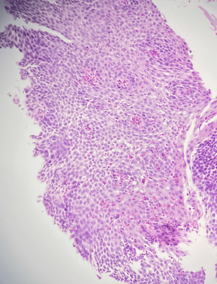 Figure 1
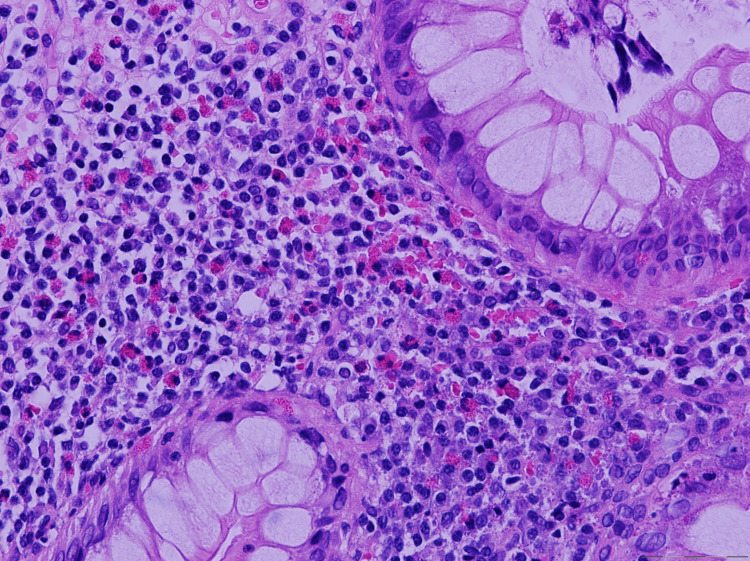 Figure 2
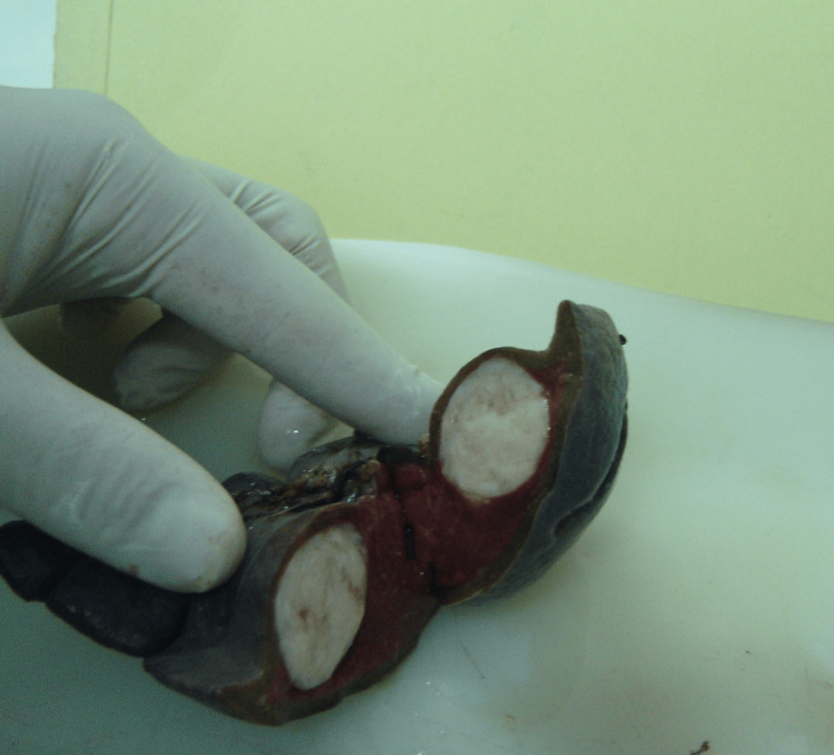 Figure 3
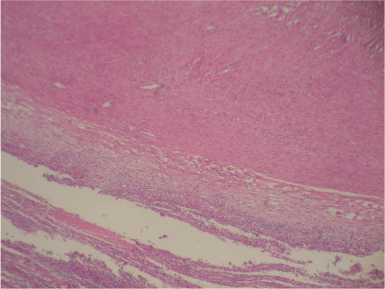 Figure 4
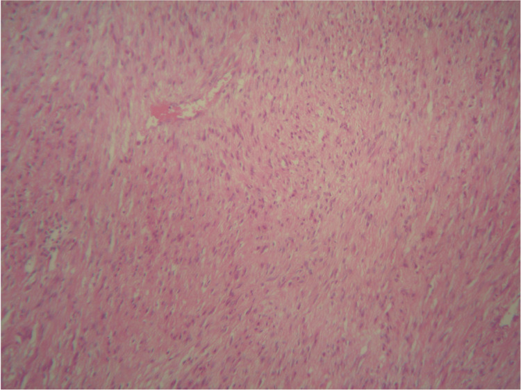 Figure 5
Peer Reviews
No public reviews on file for this paper yet. If you reviewed it on a platform where reviews are public (OpenReview, ICLR, NeurIPS, ICML), you can paste yours below so the community can read it here.
Videos
No videos yet. Explain this paper in a talk, walkthrough, or lecture? Add one.
Taxonomy
TopicsEosinophilic Esophagitis · Eosinophilic Disorders and Syndromes · Gastrointestinal disorders and treatments
Introduction
Eosinophilic gastroenteritis (EGE) is an uncommon, heterogeneous inflammatory disorder of the gastrointestinal tract characterized by eosinophilic infiltration in the absence of causes such as parasitic infections, drug reactions, or systemic diseases [1,2]. The clinical manifestations are diverse and largely depend on the distribution of eosinophilic infiltration across the gastrointestinal tract. Patients may present with nonspecific symptoms, including abdominal pain, diarrhea, vomiting, nausea, gastrointestinal bleeding, protein-losing enteropathy, and, in severe cases, malnutrition [3,4]. Because eosinophils are generally present in the gastrointestinal tract, the diagnosis requires clinicopathologic correlation, recognition of abnormal eosinophil density, and exclusion of secondary causes [5]. Diagnosis of EGE is particularly challenging, often mimicking inflammatory bowel disease, infectious enterocolitis, or food allergy [6]. Its pathogenesis is incompletely understood but appears to be multifactorial. Immune dysregulation with a Th2-mediated response, hypersensitivity to allergens, and genetic predisposition have all been implicated [7,8]. Increased levels of cytokines, such as IL-5, and chemokines, such as eotaxin, recruit and activate eosinophils, leading to mucosal injury and chronic inflammation [4]. Endoscopic findings range from normal mucosa to nonspecific erythema, friability, or ulcers [9]. Treatment usually involves corticosteroids, dietary modification, and, in refractory cases, biologic agents [10].
Splenic leiomyoma, on the other hand, is a rare benign smooth muscle tumor, usually arising from the splenic capsule, trabeculae, or vascular structures. Few cases have been reported in the literature, and most were identified incidentally or in patients with underlying immunodeficiency such as HIV/AIDS, post-transplant immunosuppression, or ataxia-telangiectasia [11-13]. In the pediatric population, benign splenic tumors are themselves rare, with cysts, hamartomas, and hemangiomas being more common, while leiomyomas are extraordinarily unusual [14]. Susmitha et al. also highlighted that splenic leiomyomas may present asymptomatically, often discovered during evaluation for unrelated conditions [12].
We report the case of a 9-year-old boy with EGE who presented with diarrhea. During the investigation, an incidental solitary splenic mass was identified and histologically confirmed as a leiomyoma. The first published pediatric case of splenic leiomyoma involved a boy with ataxia-telangiectasia, further emphasizing its exceptional rarity [15]. In a retrospective review of 30 pediatric cases with benign splenic lesions, leiomyoma was identified in only one patient, underscoring its rarity [16].
Case presentation
A 9-year-old boy was referred for evaluation of persistent diarrhea. His past medical history was unremarkable, with no evidence of chronic illness, acute infections, or prior gastrointestinal disorders. Routine laboratory investigations, including complete blood count and biochemical profile, were primarily within normal limits, except for peripheral blood eosinophilia. Stool cultures and parasitological examinations showed no significant findings. Given the persistence of symptoms, endoscopic evaluation and abdominal ultrasound were performed.
Upper gastrointestinal endoscopy and colonoscopy revealed esophageal furrowing and trachealization, as well as gastric and colonic erythema, without evidence of ulcers, polyps, or other discrete lesions. Multiple biopsies were obtained from the esophagus, stomach, duodenum, and colon. Histopathologic examination demonstrated marked eosinophilic predominant infiltration in all samples, including dense mucosal eosinophilia, more than 50 high-power fields or HPF, epithelial injury as eosinophilic pititis and cryptitis in the gastric and colon, respectively, and degranulation, suggestive of EGE. Figures 1, 2 show esophageal and duodenal biopsies infiltrated with eosinophils, respectively.
Eosinophils infiltrating the esophageal squamous mucosa
Large intestinal mucosa with eosinophil infiltration
Abdominal ultrasonography revealed a well-defined mass in the spleen. The differential diagnosis for this mass included abscess, hamartoma, and neoplastic processes, such as lymphoma. Due to the uncertain nature of the lesion, a surgical excision was performed. Gross examination of the splenectomy specimen showed a solitary, well-circumscribed intraparenchymal mass (Figure 3). Histopathological evaluation showed a spindle cell neoplasm composed of bland-looking smooth muscle fibers arranged in intersecting fascicles, without atypia, necrosis, or significant mitotic activity. Immunohistochemistry confirmed the smooth muscle phenotype, with tumor cells diffusely positive for desmin and smooth muscle actin (Figures 4, 5) and negative for C-Kit, CD34, and S100. Congo red staining demonstrated no amyloid deposition. These findings established the diagnosis of a benign smooth muscle tumor consistent with splenic leiomyoma.
Well-demarcated splenic mass
Spleen with the leiomyoma
High-power view shows bland-looking spindle cell tumor cells
Follow-up after 6 months showed significant symptomatic improvement with dietary modification and proton pump inhibitor therapy. Repeat endoscopy demonstrated mild eosinophilic infiltration within the esophagus, duodenum, and colon.
Discussion
Due to its heterogeneous presentation and overlap with other gastrointestinal conditions, EGE is a diagnostic challenge. In this patient, the presence of diffuse eosinophilic infiltration within the esophagus, stomach, duodenum, and colon highlighted the widespread nature of the disease. The finding of massive esophageal and eosinophilic cryptitis and pititis underscores the mucosal injury, which is consistent with EGE [1,17,18]. Notably, there were no other causes, such as parasitic infections, drug reactions, or systemic disorders.
The accidental finding of a splenic leiomyoma during the examination is significant. Splenic leiomyoma is a rare benign smooth muscle tumor of the spleen, most commonly reported in immunocompromised patients, including those with post-renal transplantation status, AIDS, or genetic disorders such as ataxia-telangiectasia [19,20]. The pathogenesis in these cases is thought to be associated with Epstein-Barr virus-related smooth muscle proliferation occurring in the context of an impaired immune system [19]. Most patients present with nonspecific abdominal pain, splenomegaly, cytopenias, or the tumor is found as an incidental lesion [19, 20]. Histologically, splenic leiomyomas resemble leiomyomas found elsewhere in the body, consisting of spindle-shaped smooth muscle cells arranged in interlacing fascicles. Immunohistochemistry shows positivity for smooth muscle actin and desmin, which helps distinguish them from other spindle cell neoplasms. Pediatric reports are exceedingly scarce. In a review of 30 pediatric cases with benign splenic lesions, leiomyoma was identified in only a single patient [16]. The first pediatric case described in the literature involved an 8-year-old boy with ataxia-telangiectasia [15]. Additionally, a case was reported involving an 18-year-old girl with an incidentally discovered splenic leiomyoma, which occurred alongside a duodenal ulcer, without any signs of immune deficiency [12]. Such observations indicate that splenic leiomyomas can also occur sporadically in patients with intact immune systems.
Our case, therefore, expands the spectrum of clinical contexts in which splenic leiomyoma may occur by documenting its presence in an immunocompetent child with EGE, although the association between EGE and splenic leiomyoma appears to be incidental and has not been mechanistically explored.
Conclusions
Our case is unique because it occurred in a child with EGE, an inflammatory disorder not typically linked to smooth muscle tumors, and it has not been shown to increase the risk of smooth muscle tumors. Although the presentation may represent a coincidental finding, it raises the possibility that splenic leiomyoma could develop in immunocompetent patients with underlying inflammatory disorders. Reporting such cases is important, as it expands the clinical contexts in which splenic leiomyomas may arise and contributes to a better understanding of their pathogenesis and natural history. Further accumulation of cases is needed to clarify any potential associations between inflammatory diseases and rare smooth muscle tumors of the spleen.
The reference list from the paper itself. Each links out to its DOI / PubMed record.
- 1Eosinophilic gastroenteritis: a clinicopathological study of patients with disease of the mucosa, muscle layer, and subserosal tissues Gut Talley NJ Shorter RG Phillips SF Zinsmeister AR 5458311990231843210.1136/gut.31.1.54PMC 1378340 · doi ↗ · pubmed ↗
- 2Eosinophilic gastroenteritis Best Pract Res Clin Gastroenterol Khan S 1771981920051583368710.1016/j.bpg.2005.01.009 · doi ↗ · pubmed ↗
- 3Eosinophilic gastroenteritis: a state-of-the-art review J Gastroenterol Hepatol Zhang M Li Y 647232201710.1111/jgh.1346327253425 · doi ↗ · pubmed ↗
- 4Eosinophilic colitis: epidemiology, clinical features, and current management Therap Adv Gastroenterol Alfadda AA Storr MA Shaffer EA 3013094201110.1177/1756283 X 10392443 PMC 316520521922029 · doi ↗ · pubmed ↗
- 5Clinical characteristics, treatment outcomes, and resource utilization in children and adults with eosinophilic gastroenteritis Dig Liver Dis Reed C Woosley JT Dellon ES 1972014720152554719810.1016/j.dld.2014.11.009PMC 4339627 · doi ↗ · pubmed ↗
- 6Natural history of eosinophilic gastroenteritis Clin Gastroenterol Hepatol Pineton de Chambrun G Gonzalez F Canva JY 950956920112180695210.1016/j.cgh.2011.07.017 · doi ↗ · pubmed ↗
- 7Eosinophilic gastrointestinal disorders (EGID)J Allergy Clin Immunol Rothenberg ME 112811320041471390210.1016/j.jaci.2003.10.047 · doi ↗ · pubmed ↗
- 8Eosinophilic gastrointestinal diseases - pathogenesis, diagnosis, and treatment Allergol Int Kinoshita Y Oouchi S Fujisawa T 4204296820193100044510.1016/j.alit.2019.03.003 · doi ↗ · pubmed ↗