Tapering Levothyroxine Dose for Intra-Amniotic Infusion in the Antenatal Treatment of Fetal Goiter: A Case Report
Wendy Lin, Omar Abuzeid

TL;DR
A case report shows that intra-amniotic levothyroxine may help treat fetal goiter and improve outcomes for mother and baby.
Contribution
This is the first reported case of successful intra-amniotic levothyroxine treatment for fetal goitrous hypothyroidism.
Findings
Weekly intra-amniotic levothyroxine resolved fetal goiter by 30 weeks gestation.
The infant showed no signs of goiter at birth and had no neurodevelopmental impairments.
Both mother and infant had favorable outcomes following the treatment.
Abstract
Fetal goitrous hypothyroidism is associated with important obstetrical complications including preterm birth, polyhydramnios, respiratory disorders, and neurodevelopmental impairments. There are currently no standard treatment guidelines for fetal goitrous hypothyroidism, and further studies are needed to help establish treatment guidelines. We report a case of a healthy 41-year-old female whose fetus was diagnosed with fetal goiter at 20 weeks gestation. The patient underwent weekly intra-amniotic infusions of levothyroxine, and the fetal goiter resolved by 30 weeks gestation. The infant was delivered vaginally at 36 weeks with no evidence of goiter on physical exam and diagnosed with congenital hypothyroidism upon follow-up with pediatric endocrinology. Both mother and infant are doing well today with the infant showing no signs of neurodevelopmental impairment. This case demonstrates…
Genes, proteins, chemicals, diseases, species, mutations and cell lines named across the full text — each resolved to its canonical identifier and authoritative record.
Click any figure to enlarge with its caption.
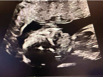 Figure 1
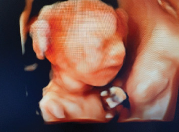 Figure 2
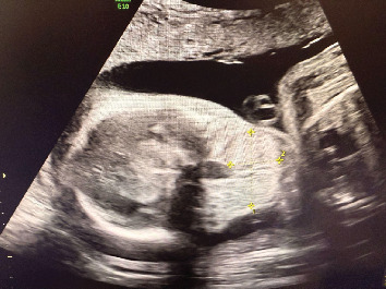 Figure 3
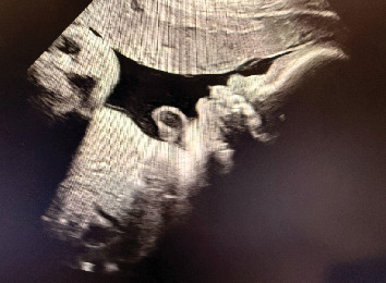 Figure 4
Peer Reviews
No public reviews on file for this paper yet. If you reviewed it on a platform where reviews are public (OpenReview, ICLR, NeurIPS, ICML), you can paste yours below so the community can read it here.
Videos
No videos yet. Explain this paper in a talk, walkthrough, or lecture? Add one.
Taxonomy
TopicsThyroid Disorders and Treatments · Thyroid and Parathyroid Surgery · Thyroid Cancer Diagnosis and Treatment
1. Introduction
Fetal goiter is characterized by enlarged fetal thyroid gland size due to defects in thyroid hormone synthesis or transportation [1]. It can occur in hypothyroid, hyperthyroid, or euthyroid states [2]. In the case of hypothyroid states, it is termed fetal goitrous hypothyroidism [2]. Causes of fetal goiter include genetic abnormalities, maternal thyroid conditions, exposure to maternal antithyroid medications, and maternal iodine deficiencies [3]. Fetal goitrous hypothyroidism is associated with important obstetrical complications including preterm birth, polyhydramnios, respiratory disorders, and neurodevelopmental impairments [4]. A rare condition, fetal goitrous hypothyroidism, affects between 1:30,000 and 1:50,000 newborns in North America and Europe [5]. It is also important to consider differential diagnoses of fetal goiter, including fetal hyperthyroidism, teratoma, cystic hygroma/lymphangioma, hemangioma, and epignathus [6]. There is currently no standard treatment method for fetal goitrous hypothyroidism, and prenatal treatment remains controversial [6]. While there have been a few reported cases that show successful reduction of fetal goiter with intra-amniotic infusions of levothyroxine, more literature is needed to help establish guidelines for standard treatment [3]. Herein, we report a case of fetal goitrous hypothyroidism that was successfully treated with intra-amniotic infusion of levothyroxine.
2. Case Presentation
2.1. Patient History
The patient is a 41-year-old female G5P3 (gravida 5, para 3) at 20 weeks gestation who was referred to maternal fetal medicine (MFM) by her obstetrician–gynecologist for advanced maternal age. She has a history of three prior uncomplicated vaginal deliveries and one miscarriage. She has no significant past medical history. Her only medication is aspirin 81 mg and prenatal vitamins taken daily. Her body mass index was 46.
2.2. Obstetrical Course
At her 20-week Level 2 ultrasound with MFM, the fetus was incidentally found to have an anterior homogeneous neck mass that was suspected to be fetal goiter (Figure 1). No other anomalies were seen. Thyroid studies were recommended for the patient, which showed normal levels of TSH, free T4, free T3, and thyroid stimulating immunoglobulin (TSI). The patient denies a history of thyroid abnormalities in herself and her three prior children. The patient was instructed to follow up to reassess the neck mass and rule out hydrops fetalis.
Patient returned for her follow-up visit at 24 weeks for a repeat ultrasound, which showed the fetal neck mass to still be present and measuring 3.3 × 2.1 × 1.4 cm (Figures 2 and 3). Other findings included polyhydramnios and hyperextension of the fetal neck. The patient was counseled on the risks and benefits and offered options of cordocentesis, amniocentesis, or expectant management. She declined a cordocentesis to evaluate fetal TSH levels due to the potential risks of the procedure. She ultimately decided and consented to an amniocentesis with installation of levothyroxine. Her first amniocentesis for genetic screening was performed at 25 weeks gestation which showed normal results. The patient was recommended to have a fetal MRI for further evaluation.
At 26 weeks gestation, the patient underwent a fetal MRI which confirmed findings of fetal goiter with no other fetal anomalies. The mass measured 3.68 × 1.64 × 1.08 cm. The same week, the patient underwent amniocentesis with instilled infusion of 200 mcg of levothyroxine into the amniotic cavity. The patient subsequently returned weekly for amnioinfusion of levothyroxine. The dose of levothyroxine was increased to 400 mcg at 27 weeks gestation due to the goiter not shrinking in size. The assumed diagnosis at this point was congenital hypothyroidism causing goiter, or fetal goitrous hypothyroidism. Other differentials included (1) fetal hyperthyroidism, which was less likely due to the patient being euthyroid; (2) teratoma; (3) cystic hygroma/lymphangioma; (4) hemangioma; and (5) epignathus, which were ruled out with fetal MRI.
At each weekly visit from 26 weeks and beyond, the patient received amnioinfusion of levothyroxine into the amniotic cavity. Additionally, TSH and gram stains were collected during amniocentesis procedures. The patient was counseled that amniocentesis is not a perfect diagnostic tool for TSH as there are no standard reference ranges. The reference ranges are from case series of similar cases. Despite this, of note, the TSH in the amniotic fluid for this patient showed a downward trend, beginning at 0.71 IU/mL and stabilizing at around 0.2 IU/mL at the final amniocentesis.
The fetal goiter also shrunk with each subsequent visit and amnioinfusion of levothyroxine. At 28 weeks gestation, the goiter had started to shrink in size to 3.0 × 1.7 × 1.5 cm. During this week, the polyhydramnios had also resolved. At 29 weeks gestation, the goiter measured even smaller at 2.7 × 1.7 × 1.7 cm. By 30 weeks gestation, the goiter was no longer appreciated and the fetal neck was no longer hyperextended (Figure 4). The patient continued to receive 300 mcg of levothyroxine via amnioinfusion until 34 weeks gestation.
2.3. Delivery and Postnatal Course
Around 34 weeks gestation, the patient developed gestational hypertension and then preeclampsia. She had planned to have an induction of labor at 37 weeks, but her amniotic membrane ruptured spontaneously at 36 weeks, and she went into labor. She had an uncomplicated vaginal delivery and her blood pressure stabilized. There was no postpartum hemorrhage. The infant's APGAR scores were 9 and 9 at 1 and 5 min after birth. There was no goiter noted on the infant's physical exam. The infant followed up with pediatric endocrinology and was diagnosed with congenital hypothyroidism and started on levothyroxine. Both the infant and mother are doing well today, with the infant showing no signs of neurodevelopmental impairments.
3. Discussion
Although there have been a few similar case studies that showed reduction of fetal goiter with intra-amniotic infusions of levothyroxine in the setting of fetal goitrous hypothyroidism, there are currently no standard treatment guidelines for fetal goitrous hypothyroidism. Moreover, no standard dose of levothyroxine has been established, and the dose of levothyroxine in this case was tailored based on the response of the fetal goiter to treatment. In our case, intra-amniotic infusion with 200 mcg of levothyroxine did not show improvement in the fetal goiter size. However, 400 mcg of levothyroxine caused the goiter to reduce in size weekly, and subsequently resolve by 30 weeks.
Although cordocentesis is the current gold standard for diagnosing fetal hypothyroidism, our patient declined to undergo cordocentesis after risks and benefits were discussed with her. Therefore, we opted to measure TSH in the amniotic fluid, which is less invasive than cordocentesis. Although there is currently no standard reference range for TSH in the amniotic fluid [7], the TSH in the amniotic fluid of our patient did show a downward trend with intra-amniotic levothyroxine infusions. More studies are needed to determine the validity of TSH levels in amniotic fluid and establish a standard reference range.
In conclusion, this case demonstrates a successful treatment of fetal goitrous hypothyroidism with intra-amniotic infusion of levothyroxine. By treating fetal goitrous hypothyroidism with intra-amniotic infusion of levothyroxine, potential complications such as preterm delivery, the need for an ex utero intrapartum treatment (EXIT procedure), and possible neurodevelopmental impairments, as well as possible death of the neonate were avoided. More studies are needed to establish clear treatment guidelines and a standard dose of levothyroxine for amnioinfusion.
The reference list from the paper itself. Each links out to its DOI / PubMed record.
- 1Nemescu D. Tanasa I. Stoian D. Navolan D. Vinturache A. Conservative In Utero Treatment of Fetal Dyshormonogenetic Goiter With Levothyroxine, a Systematic Literature Review Experimental and Therapeutic Medicine 20202032434243810.3892/etm.2020.879432765729 PMC 7401841 · doi ↗ · pubmed ↗
- 2Kiess W. Penke M. Gesing J. Congenital Hypothyroidism Journal of Pediatric Endocrinology and Metabolism 201831659559610.1515/jpem-2018-01972-s 2.0-8505925624129804102 · doi ↗ · pubmed ↗
- 3Rastogi M. V. La Franchi S. H. Congenital Hypothyroidism Orphanet Journal of Rare Diseases 201051 p. 1710.1186/1750-1172-5-172-s 2.0-7795324328720537182 PMC 2903524 · doi ↗ · pubmed ↗
- 4Tamura N. Yamamoto Y. Takeda J. Massive Fetal Goiter Treated by Intra-Amniotic Injection of Levothyroxine: A Case Report Case Reports in Perinatal Medicine 20241312024000610.1515/crpm-2024-000640321355 PMC 12048138 · doi ↗ · pubmed ↗
- 5Corbacioglu Esmer A. Gul A. Dagdeviren H. Turan Bakirci I. Sahin O. Intrauterine Diagnosis and Treatment of Fetal Goitrous Hypothyroidism Journal of Obstetrics and Gynaecology Research, [S.l.] 201339372072310.1111/j.1447-0756.2012.02003.x 2-s 2.0-8487527291523002999 · doi ↗ · pubmed ↗
- 6Rauff S. Eng Kien T. Ultrasound Diagnosis of Fetal Neck Masses: A Case Series Case Reports in Obstetrics and Gynecology 2013201324359010.1155/2013/24359023401814 PMC 3562568 · doi ↗ · pubmed ↗
- 7Singh P. K. Parvin C. A. Gronowski A. M. Establishment of Reference Intervals for Markers of Fetal Thyroid Status in Amniotic Fluid Journal of Clinical Endocrinology and Metabolism 200388941754179 PMID: 1297028310.1210/jc.2003-0305222-s 2.0-014178797112970283 · doi ↗ · pubmed ↗