Conflicts between the DNA replication and repair machineries promote cell death in Gram-positive bacteria
Hannah Gaimster, Daniel Stevens, James Grimshaw, Julia Hubbard, Katarzyna Mickiewicz, Heath Murray, Charles Winterhalter

TL;DR
Forcing excessive DNA replication in Gram-positive bacteria causes cell death, suggesting a new way to fight infections by targeting replication and repair processes.
Contribution
The study reveals that replication hyper-initiation causes cell death in Gram-positive bacteria through conflicts with DNA repair.
Findings
Hyper-initiation in B. subtilis triggers DNA damage response, membrane depolarization, and cell lysis.
In S. aureus, hyper-initiation leads to rapid DNA loss and death via a lysis-independent pathway.
Hyper-initiation in S. aureus reduces survival in infected macrophages.
Abstract
Cellular proliferation relies on the successful coordination and completion of genome replication and segregation. To help achieve this, many bacteria utilize regulatory pathways that ensure DNA replication initiation only occurs once per cell cycle. When dysregulated, loss of DNA replication control can have severe consequences. In Escherichia coli, it has been established that hyper-initiation of DNA synthesis leads to pleiotropic genome instability and cell death. Therefore, targeting DNA replication initiation proteins to promote hyper-initiation may be an approach to generate novel antimicrobials. However, the pathways and potential consequences of replication hyper-initiation in Gram-positive species remain enigmatic. To address this question, we devised genetic systems to artificially induce hyper-initiation in the model organism Bacillus subtilis and the pathogen Staphylococcus…
Genes, proteins, chemicals, diseases, species, mutations and cell lines named across the full text — each resolved to its canonical identifier and authoritative record.
Click any figure to enlarge with its caption.
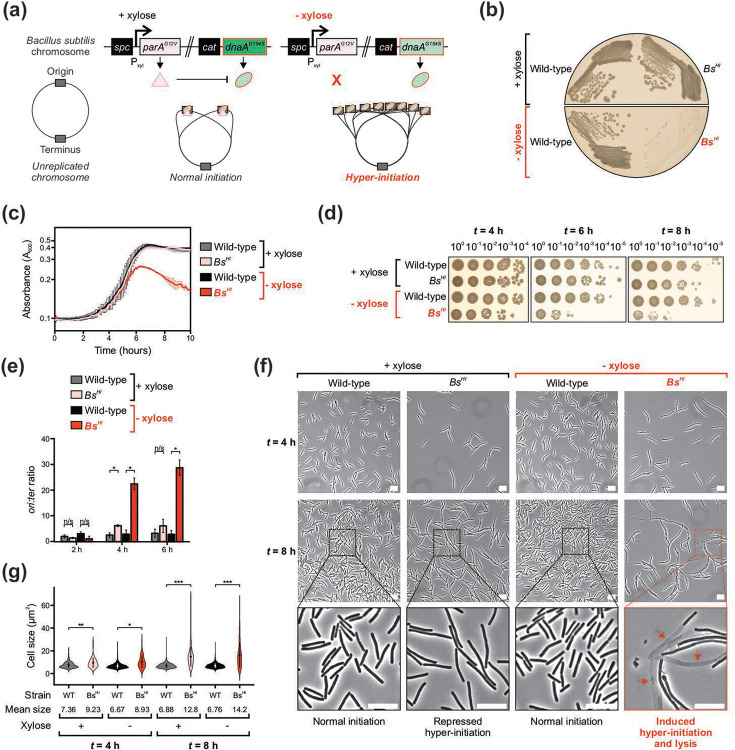 Fig. 1
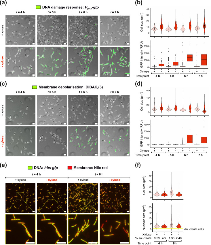 Fig. 2
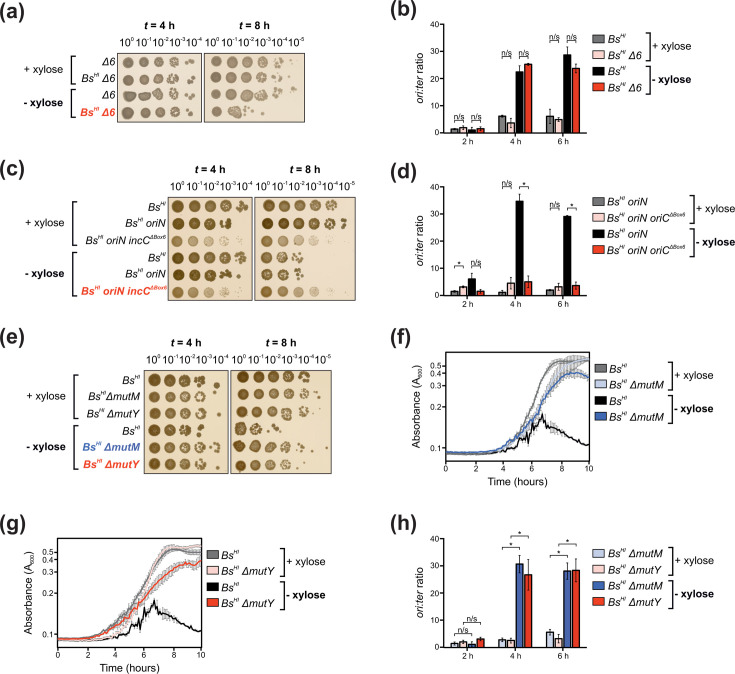 Fig. 3
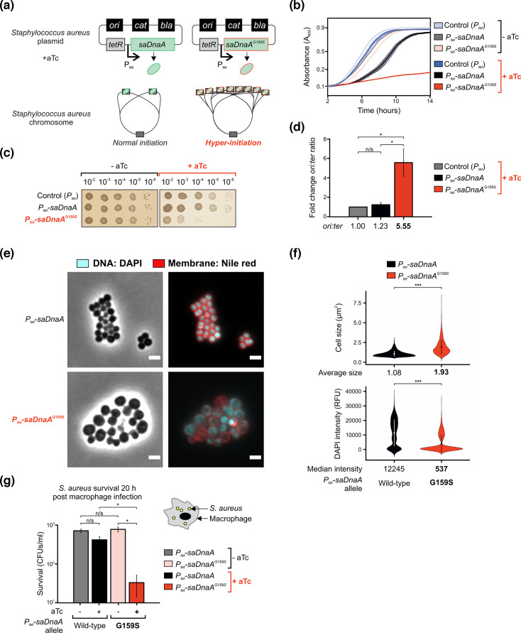 Fig. 4
- —http://dx.doi.org/10.13039/501100000265 Medical Research Council
- —http://dx.doi.org/10.13039/501100000265 Medical Research Council
- —http://dx.doi.org/10.13039/100010269 Wellcome Trust
- —http://dx.doi.org/10.13039/100010269 Wellcome Trust
Peer Reviews
No public reviews on file for this paper yet. If you reviewed it on a platform where reviews are public (OpenReview, ICLR, NeurIPS, ICML), you can paste yours below so the community can read it here.
Videos
No videos yet. Explain this paper in a talk, walkthrough, or lecture? Add one.
Taxonomy
TopicsDNA Repair Mechanisms · Bacterial Genetics and Biotechnology · Cancer Research and Treatments
Data Availability
Supplementary material is available through Figshare at 10.6084/m9.figshare.29988040 [1].
Introduction
A successful cell cycle requires replication and segregation of the genome. In most cells, DNA synthesis begins at specific chromosomal loci called origins and requires accurate coordination with chromosome segregation and cell division to produce viable daughter cells [23]. Importantly, while replication initiation culminates in loading of ring-shaped helicases enabling dsDNA unwinding prior to synthesis, the molecular mechanisms required to achieve helicase loading in prokaryotes and eukaryotes are distinct [4]. Therefore, the unique and essential characteristics of the bacterial DNA replication initiation pathway make it an attractive target for drug development [5].
In bacteria, chromosome replication generally initiates from a single origin (oriC), where the universal prokaryotic initiator protein DnaA binds to sequence-specific elements (DnaA-boxes and DnaA-trios) and unwinds the DNA duplex [68], followed by recruitment and loading of helicases around ssDNA [911]. During this process, DnaA activity is tightly regulated to ensure genome replication occurs only once per cell cycle, while bidirectional DNA synthesis proceeds up to a region called the terminus where converging replication forks collide [1214]. Failure to maintain this homeostasis can lead to dramatic consequences affecting a range of cellular processes, resulting in genomic instability [1516].
Abnormal initiation of genome duplication can result in either replication inhibition or excessive DNA synthesis, both of which can inhibit bacterial proliferation. Intuitively, under-initiation of DNA replication produces cells that lack a copy of the genome [17]. A more complex situation is observed during hyper-initiation of DNA replication. Excessive genome replication has been extensively studied and remains an active field of research in the Gram-negative model organism Escherichia coli, where under aerobic growth conditions, hyper-initiation causes DNA double-strand breaks (DSBs) as a result of the DNA replication machinery (replisome) encountering repair systems acting on the genome [1819]. These detrimental events affect the normal cell cycle and can result in significant cell death via improper completion of DNA synthesis or chromosome segregation defects, yet E. coli may employ several strategies to overcome lethal over-initiation [2023]. However, the lethal phenotype associated with hyper-initiation of DNA replication remains enigmatic, as suppression of cell death did not correlate with a significant decrease in DSBs [18]. Moreover, it is still unclear whether hyper-initiation of DNA replication generally inhibits bacterial proliferation in Gram-positive bacteria, and if so, the specific pathways leading to potential cell death are yet to be determined across species.
In this study, we investigated the consequences of DNA replication hyper-initiation in the Gram-positive model organism Bacillus subtilis and the opportunistic pathogen Staphylococcus aureus. In both cases, hyper-initiation resulted in the loss of cell viability; however, the lethal phenotypes were not identical. The B. subtilis strain displayed cell elongation, DNA damage, membrane depolarization and eventually cell lysis. The S. aureus strain also displayed altered cell morphology with clear defects in chromosome inheritance, but here, loss of cell viability occurred through a lysis-independent mechanism. Interestingly, induction of S. aureus hyper-initiation inhibited bacterial growth in a murine macrophage model, highlighting the potential of stimulating DNA replication initiation as an approach to reduce bacterial load during infection.
Methods
Reagents and growth conditions
Nutrient agar (NA; Oxoid) was used for routine selection and maintenance of E. coli and B. subtilis strains. Tryptic soy agar (TSA; Oxoid) was used for routine selection and maintenance of S. aureus strains. Unless otherwise stated, B. subtilis and S. aureus strains were grown at 30 °C and E. coli was grown at 37 °C. Supplements were added as required: ampicillin (100 µg ml^−1^), erythromycin (1 µg ml^−1^) in conjunction with lincomycin (25 µg ml^−1^), chloramphenicol (5 µg ml^−1^ for B. subtilis and 10 µg ml^−1^ for S. aureus), kanamycin (5 µg ml^−1^), spectinomycin (50 µg ml^−1^), tetracycline (10 µg ml^−1^), xylose (1% w/v) and anydrotetracycline (aTc, 20 ng ml^−1^). All chemicals and reagents were obtained from Sigma-Aldrich. RAW-Blue cells were maintained in Dulbecco’s Modified Eagle’s medium (DMEM, Sigma Cat#D6429) supplemented with 5% FBS at 37 °C and 5% CO_2_.
Biological resources: E. coli, B. subtilis and S. aureus strains
All strains and cell lines used in this study are listed in Table S1 (available in the online Supplementary Material).
B. subtilis strain construction
Transformation of competent B. subtilis cells was performed using an optimized two-step starvation procedure as previously described [24]. Briefly, recipient strains were grown overnight at 30 °C in transformation medium (Spizizen salts supplemented with 1 µg ml^−1^ Fe-NH_4_-citrate, 6 mM MgSO_4_, 0.5% w/v glucose, 0.02 mg ml^−1^ tryptophan and 0.02% w/v casein hydrolysate) supplemented with xylose where required. Overnight cultures were diluted 1 : 20 into fresh transformation medium supplemented with xylose where required and grown at 30 °C for 3 h with continual shaking. An equal volume of prewarmed starvation medium (Spizizen salts supplemented with 6 mM MgSO_4_ and 0.5% w/v glucose) was added, and the culture was incubated at 30 °C for 2 h with continuous shaking. DNA was added to 350 µl cells and the mixture was incubated at 30 °C for 1 h with continual shaking. 20–200 µl of each transformation was plated onto selective media supplemented with xylose where required and incubated at 30 °C for 24–48 h. Note that strains harbouring the dnaA^G154S^/parA^G12V^ hyper-initiation system and/or mutM/mutY knockouts are prone to accumulation of suppressor mutations upon storage or repeated rounds of propagation. For best practice, it is recommended to rebuild these strains using sequence-validated recombinant DNA and to perform whole-genome sequencing on candidate colonies prior to use.
S. aureus strain construction
Transformation of competent S. aureus cells was performed using plasmid electroporation [25]. Briefly, recipient cells were grown overnight in tryptic soy broth (TSB; Oxoid). Overnight cultures were diluted 1 : 100 in fresh TSB and grown to an absorbance at 600 nanometres (A600) of 0.6. Cells were rapidly cooled down in an ice water bath and washed three times using an equal volume of ice-cold sterile deionized water followed by two washes in an equal volume of ice-cold 10% w/v glycerol solution. After the final centrifugation, cells were resuspended in 1 : 100 of the initial culture volume in ice-cold 10% w/v glycerol, snap frozen using liquid nitrogen and stored at −80 °C for up to 6 months. Before electroporation, electrocompetent cells were thawed at room temperature for 5 min, centrifuged at 10,000 g for 1 min and resuspended in 100 µl of electrocompetent buffer (10% w/v glycerol supplemented with 500 mM sucrose). DNA was added to the cell mixture and electroporation was performed at 2.3 kV, 100 Ω, 25 µF (Bio-Rad Gene Pulser II) using a 1 mm gap cuvette (VWR #732-1135). Cells were immediately resuspended in 1 ml of TSB supplemented with 500 mM sucrose, incubated for 1.5 h at the permissive temperature, plated onto selective medium and incubated at permissive temperature for 24–48 h.
E. coli plasmid construction and propagation
E. coli transformation was performed in DH5α via heat shock following the Hanahan method [26]. All plasmids were sequenced and descriptions of plasmid construction, where necessary, are provided below.
pDS150 was generated using Gibson assembly to introduce saDnaA into pRAB11 [27].
pDS157 was created using Quickchange mutagenesis on pDS150 to introduce the saDnaA^G159S^ point mutation.
Spot titre assays
B. subtilis cells were grown in penassay broth (PAB) medium overnight at 30 °C in the presence of xylose. Overnight cultures were diluted 1 : 100 into fresh PAB with or without xylose (time = 0 h). Samples were harvested at time points indicated, tenfold serial dilutions were made into PAB with xylose and 5 μl aliquots were spotted onto NA plates supplemented with xylose. For S. aureus spot titre assays, cells were grown in TSB supplemented with chloramphenicol overnight at 30°C and 5 μl of serial dilutions were spotted onto TSA supplemented with chloramphenicol in the presence or absence of aTc. All plates were incubated at 30 °C for 24 h. Experiments were performed independently at least three times and representative data are shown.
Automated plate reader analyses
Strains were grown overnight at 30 °C in PAB with xylose (B. subtilis) or TSB supplemented with chloramphenicol (S. aureus). Overnight cultures were diluted 1 : 100 into PAB with or without xylose (B. subtilis), or 1 : 1000 into TSB with or without aTc (S. aureus), and automated absorbance measurements were captured over time at 30 °C using a Tecan Sunrise plate reader (high shaking parameters with 2.8 mm shake width at 12.3 Hz and absorbance measurement every 6 min). All experiments were independently performed at least three times and representative data are shown.
Sample preparation for MFA
B. subtilis cells were grown overnight in PAB at 30°C with xylose. Overnight cultures were diluted 1 : 100 into PAB with or without xylose (time = 0 h) and incubated at 30 °C. For S. aureus, cells were grown overnight in TSB in the presence of chloramphenicol, followed by 1 : 100 dilution into TSB with or without aTc for 2 h before harvesting samples. Samples (500 μl) were harvested at times indicated and immediately mixed with sodium azide (1% w/v final concentration) to arrest growth and genome replication. Cultures were collected by centrifugation, the supernatant was discarded and pellets were flash frozen in liquid nitrogen before genomic DNA extraction using the DNeasy blood and tissue kit (Qiagen).
Quantitative PCR
Quantitative PCR (qPCR) was performed using the Luna qPCR mix (NEB) to measure the relative amount of origin DNA compared to the terminus. All PCR reactions were run in a Rotor-Gene Q instrument (Qiagen) using 20 µl reaction volumes in a Rotor-Disc 100 (Qiagen). Standard curves were obtained using the Rotor-Gene Q software version 2.0.2 (Qiagen) to calculate the efficiency of each primer pair, which varied ∼5% between sets. Oligonucleotide primers designed to amplify incC (qSF19/qSF20 for B. subtilis or oDS263/oDS264 for S. aureus) and the terminus (qPCR57/qPCR58 for B. subtilis or oDS269/oDS270 for S. aureus) were typically 20–25 bases in length and amplified a ~100 bp PCR product (Table S2). Individual ori:ter ratios were obtained in three steps: first, every Ct value was converted to 1/2^Ct^ and technical triplicates were averaged to generate a single enrichment value; second, origin enrichment was normalized by corresponding terminus values; third, relative ori:ter values were normalized by the enrichment obtained in control conditions. For B. subtilis, data was normalized to spore DNA as non-replicating control (ori:ter ratio=1). For S. aureus, data was normalized to cells grown with the pRAB11 plasmid (empty vector used for the construction of saDnaA variants) harvested at the same time point in exponential phase. Error bars indicate the standard error of the mean for 2–4 biological replicates.
Statistical analyses
Statistical analyses were performed using Student’s t-tests, and the significance of P values is displayed on individual figure panels and explained in figure legends. The exact value of n is given in the Methods section and represents the number of biological repeats for an experiment. Tests were based on the mean of individual biological replicates. Differences were considered as significant if their associated P value was below 0.05.
Microscopy and image analysis
Microscopy was performed on an inverted epifluorescence microscope (Nikon Ti) fitted with a Plan Apochromat Objective (Nikon DM 100×/1.40 Oil Ph3). Light was transmitted from a CoolLED pE-300 lamp through a liquid light guide (Sutter Instruments), and images were collected using a Prime CMOS camera (Photometrics). Fluorescence filter sets were from Chroma: DAPI (49000, EX350/50, DM400lp, EM460/50), GFP (49002, 537 EX470/40, DM495lpxr, EM525/50) and mCherry (49008, EX560/40, 538 DM585lprx, EM630/75). Digital images were acquired using Metamorph (version 7.7) and NIS-Elements software. Cells were mounted on ~1.2% agar pads and a 0.13–0.17 mm glass coverslip (VWR) was placed on top. Wavelengths used: Brightfield and mCherry (150 ms), GFP/DAPI (50 ms), all at 100% exposure.
For time-lapse microscopy experiments, a GeneFrame was used to create a flat agarose pad using 0.7% agarose dissolved into PAB with or without xylose (B. subtilis) or TSB with or without aTc (S. aureus). An aliquot of cells (1.5 µl) was spotted onto the pad, and a 0.13–0.17 mm glass coverslip (VWR) was placed on top. Digital images were acquired every 15 min for 8–12 h.
For cultures stained with DiBAC_4_(3) (Invitrogen, Thermo Fisher), 0.5 µl dye was added to 100 µl of cells and the mixture was incubated at 30 °C with shaking at 800 r.p.m. for 5 min using a Thermomixer C (Eppendorf) and then spotted onto an agarose slide. DiBAC_4_(3) preferentially binds cellular membranes that are depolarized, emitting a green fluorescence signal to allow for detection (i.e. cells appear fluorescent if the cell envelope is compromised).
For cultures stained with DAPI (Thermo Fisher), the dye was added to 100 µl of cells at a final concentration of 1 µg ml^−1^, and the mixture was incubated at 30 °C with shaking at 800 r.p.m. for 5 min using a Thermomixer C (Eppendorf) and then spotted onto an agarose slide.
Fiji software was used for initial image analysis [28]. Brightness and contrast used for each time point and condition remained consistent and all images are scaled to 0.065 microns. Cell segmentation was performed on phase-contrast images using Omnipose with the bact_phase_omni model, further refined for S. aureus using custom training [29]. Image processing and analysis were conducted in scikit-image [30]. Objects smaller than 0.845 µm² (B. subtilis) or 0.634 µm² (S. aureus) were excluded, and mean background fluorescence was subtracted. Nucleoids were segmented with Ilastik, with labels manually corrected using the membrane channel [31]. Anucleate B. subtilis cells were defined as those with nucleoid area below 5% of individual cell sizes.
Macrophage infection with S. aureus and gentamycin protection assay
RAW-Blue cells were seeded the day before bacterial infection experiments at 0.5×10^6^ cells per ml in DMEM 5% FBS medium in 25 cm angled neck flasks (NUNC). Macrophages were challenged at an m.o.i. of 1 : 5 in duplicate for S. aureus infection with CW1150 or CW1151 bacterial strains. The challenge was carried out in 10 ml of DMEM 5% FBS, and cells were incubated for 1 h at 37 °C and 5% CO_2_ to allow bacterial uptake by the macrophages. Following this incubation, gentamycin was added to all flasks (100 ug ml^−1^ final concentration) to kill extracellular bacteria, and aTc was supplemented to one of each of the duplicates (400 ng ml^−1^ final concentration) to induce SaDnaA variant overexpression. Flasks were then incubated for 20 h at 37 °C and 5% CO_2_. Adherent cells were dislodged with a scrapper, and 1 ml was transferred into an Eppendorf tube and spun down at 13,000 r.p.m. The supernatant was aspirated and the pellet was resuspended by pipetting up and down in 1 ml of Triton X100 (0.5% v/v) to lyse macrophages. Serial dilutions were performed and plated on TSA supplemented with 10 ug ml^−1^ chloramphenicol for selection of S. aureus carrying the inducible plasmid. Bacterial cells were incubated overnight at 37 °C and used to quantify c.f.u. for survival plots. Macrophage experiments were performed as biological duplicates.
Results
DNA replication hyper-initiation causes cell lysis in B. subtilis
A genetic system was employed to control the frequency of DNA replication initiation in B. subtilis. This system exploits a hyperactive variant of DnaA (DnaA^G154S^) that causes cells to over-initiate DNA replication [32]. To control hyper-initiation, a variant of the regulator ParA (ParA^G12V^) was employed to inhibit DnaA activity [33]. The experimental strain encodes dnaA^G154S^ at the endogenous locus (under the control of its native expression system) and parA^G12V^ at an ectopic locus (under the control of a xylose-inducible promoter). Hereafter, we will refer to this strain (dnaA^G154S^ P_xyl_-parA^G12V^) as Bs^HI^ (for B. subtilis hyper-initiation). The Bs^HI^ strain was cultured in the presence of xylose to repress hyper-initiation, followed by growth in the absence of xylose to repress and dilute ParA^G12V^, thereby enabling full activity of the hypermorphic DnaA^G154S^ variant (Fig. 1a).
Hyper-initiation of DNA replication causes lysis and cell death in B. subtilis. (a) Genetic system used to artificially modulate DNA replication hyper-initiation. Xylose induction controls the expression of parAG12V, which enables ParAG12V to downregulate the activity of the hyperactive DnaAG154S variant, resulting in repression of hyper-initiation. In the absence of xylose, parAG12V is no longer expressed and DnaAG154S allows B. subtilis to hyper-initiate. (b) Restreaks showing the phenotype associated with dnaAG154S Pxyl-parAG12V (BsHI) cells in the absence of xylose after 24 h growth. (c) Growth curves showing the arrest of cell growth in BsHI cells after 6-h incubation in the absence of xylose. (d) Spot titre analyses showing the loss of viability observed in BsHI cells after 6-h growth in the absence of xylose. (e) Marker frequency analyses demonstrate that BsHI cells start to hyper-initiate DNA replication after 4-h growth in the absence of xylose. (f) Phase-contrast microscopy showing the lysis phenotype observed in BsHI cells after 8-h growth in the absence of xylose. Scale bar indicates 10 µm. Red arrows highlight lysed cells. (g) Microscopy analyses showing quantification of individual cell sizes from the data shown in (f). Each condition highlights mean cell size (circles within violins) and standard deviation (sd, vertical line crossing the mean). (b–g) Strains: WT (168CA), BsHI (HM946). Non-significant (n/s), P values: <0.05 (), <0.001 (), <0.0001 ().
Using this system, strains were streaked onto solid growth medium with or without xylose and incubated overnight. While both WT and Bs^HI^ strains showed similar growth in the presence of inducer, the Bs^HI^ strain displayed a severe growth defect in the absence of xylose (Fig. 1b). To further characterize this phenotype, cell growth in liquid media was followed over time. The results confirmed that the Bs^HI^ strain had a similar growth rate to WT in the presence of xylose, whereas Bs^HI^ cells could not reach the same density and appeared to lyse after ~6 h in the absence of the inducer (Fig. 1c).
The impact of hyper-initiation on cell viability was quantified by growing cells in liquid medium with or without xylose, followed by aliquoting a serial dilution of each sample onto growth medium containing xylose (i.e. to repress hyper-initiation and allow growth of any viable cells). This spot titre analysis showed a similar number of c.f.u. between WT and Bs^HI^ strains after 4 h of growth. However, by six and 8 h of growth without xylose, the Bs^HI^ strain displayed a >100-fold decrease in c.f.u. compared to both WT and Bs^HI^ cultured in the presence of xylose (Fig. 1d).
Marker frequency analysis (MFA) was used to determine the levels of DNA replication initiation under these experimental conditions. Over a 6-h time course, MFA showed ori:ter ratios ranging from 2.0 to 3.3 for WT B. subtilis (Fig. 1e). During the lag phase, the Bs^HI^ strain showed comparable ori:ter ratios to WT regardless of the presence or the absence of inducer (2-h time point, Fig. 1e). Strikingly, Bs^HI^ cells displayed a significantly increased ori:ter ratio following 4 h of growth without xylose (ori:ter=22 in exponential phase), whereas the same strain grown in the presence of inducer only mildly over-initiated (ori:ter=6 compared to WT ori:ter=2.5, Fig. 1e). The results contrast with a previous report of hyper-initiation in B. subtilis where cells with a milder increase in replication initiation remained viable [34], which indicates that the hyperactive dnaA^G154S^ allele is more penetrant. Taken together, the data suggest that under this growth regime of depleting ParA^G12V^ to allow full activity of DnaA^G154S^, hyper-initiation of DNA replication precedes significant cell death.
Phase-contrast microscopy was used to investigate the fate of single B. subtilis cells experiencing DNA replication hyper-initiation. Regardless of the presence or absence of xylose in conditions preceding significant cell death (e.g. 4-h time point), Bs^HI^ cells showed a ~25% increase in cell size compared to WT (Fig. 1f, g), indicating that the normal cell cycle is perturbed in this strain [35]. We attribute morphological defects observed in the presence of inducer to pleiotropic activities of ParA^G12V^, which is known to affect other cellular processes (summarized in [36]). Consistent with inherent stress under these growth conditions, Bs^HI^ cell size was further increased to approximately twice the size of WT by 8 h of growth (Fig. 1g). However, incubation in the absence of xylose resulted in dramatic changes to the cell envelope (‘bulging’ phenotype) with many cells appearing phase light, a hallmark of cell lysis (Fig. 1f). These phenotypes can be seen in greater detail in time-lapse videos of Bs^HI^ cells grown in the presence or absence of xylose (Videos S1 and S2, respectively). Together, these data indicate that in B. subtilis, DNA replication hyper-initiation causes a loss of viability via cell lysis.
Hyper-initiation activates the DNA damage response and leads to membrane depolarization
In bacteria, it is established that severe DNA damage elicits a stress response (the standard meaning of a distress call (SOS) response), allowing time to repair the damage and segregate replicating chromosomes [353738]. To induce the SOS response, the critical recombination protein RecA binds to ssDNA and stimulates the autocleavage of LexA, the transcriptional repressor of the SOS regulon [39]. Upon cleavage, LexA can no longer bind to DNA to repress transcription, resulting in induction of the SOS response.
One of the SOS-responsive genes most strongly upregulated in B. subtilis encodes the cell division inhibitor YneA [40]. We hypothesized that the cell size increase and lysis observed during hyper-initiation (Fig. 1f) may be caused by induction of the DNA damage response. To test this model, a fluorescent transcriptional reporter under control of the LexA-dependent yneA promoter (P_yneA_-gfp) was transformed into Bs^HI^ (Fig. S1A). As controls, it was confirmed using spot titre assays that the addition of the yneA reporter cassette did not alter the phenotype associated with hyper-initiation (Fig. S1B) and that YneA was not causative of hyper-initiation-induced cell death (Fig. S2A, B). Following growth in liquid media with or without xylose throughout the time window separating the onset of hyper-initiation from accumulation of growth defects leading to cell death (Fig. 1e–g, four to 8 h), the expression of P_yneA_-gfp was observed using fluorescence microscopy. It was validated that the induction of the DNA damage response is a rare event in WT cells in the presence or the absence of xylose (Fig. S3A, B). While a minority of Bs^HI^ cells grown in the presence of xylose appeared to express the GFP reporter, in sharp contrast, the majority of cells displayed a GFP signal gradually increasing following depletion of ParA^G12V^ from 4 h onwards (Figs 2a, b and S3C). Induction of the SOS response suggests that DNA replication hyper-initiation leads to the production of DNA damage intermediates (i.e. ssDNA) recognized by RecA.
Hyper-initiation causes DNA damage followed by membrane depolarization and cell lysis via an apoptotic-like mechanism. (a) Microscopy experiments showing activation of the DNA damage response in BsHI cells in the presence/absence of xylose to control hyper-initiation. Phase-contrast images are overlayed with fluorescence signal corresponding to the expression of the PyneA-gfp SOS regulon reporter. Strain: HG287. (b) Microscopy analyses showing quantification of individual cell sizes (top) and GFP fluorescence (bottom, DNA damage reporter signal) from the data shown in (a). (c) Microscopy experiments showing membrane depolarization in BsHI cells in the presence/absence of xylose to control hyper-initiation. Phase-contrast images are overlayed with fluorescence signal corresponding to DiBAC4(3) entering depolarized cells. Strain: HM946. (d) Microscopy analyses showing quantification of individual cell sizes (top) and GFP fluorescence (bottom, membrane depolarization signal) from the data shown in (c). (e) Microscopy experiments showing the nucleoid in BsHI cells in the presence/absence of xylose to control hyper-initiation. Images are merged between green fluorescence for visualization of DNA content (Hbs-GFP signal) and red fluorescence for membrane stain (Nile red dye). Strain: HM1974. (f) Microscopy analyses showing quantification of individual cell sizes (top) and areas covered by the nucleoid (bottom, highlights the number of anucleate cells) from the data shown in (e). n/a indicates that no anucleate cells were found. (a, c, e) Scale bar indicates 10 µm. (b, d, f) Each condition highlights mean cell size (circles within violins) and sd (vertical line crossing the mean), and boxplots indicate median lines (within boxes) and outliers (circles outside boxes).
It is established that RecA plays a role during hyper-initiation in E. coli, but whether this function is conserved in Gram-positive species had not been fully elucidated [1941]. To test the importance of RecA during hyper-initiation in B. subtilis, we constructed a complementation system by placing an ectopic copy of recA under the control of a constitutive promoter (P_veg_-recA). It was confirmed that introducing this cassette in Bs^HI^ cells yielded a similar number of c.f.u. to control strains in the presence or the absence of xylose (Fig. S4A). Spot titre analyses revealed that the deletion of recA resulted in synthetic lethality in Bs^HI^ cells grown in the absence of xylose, whereas the presence of P_veg_-recA was able to rescue this phenotype (Fig. S4B), indicating that RecA is causative of growth sensitization under these conditions. Consistent with E. coli literature [19], the results suggest that RecA is essential during hyper-initiation in B. subtilis to prevent replication fork collapse.
Under conditions enabling hyper-initiation, time course and time-lapse microscopy indicated that morphological abnormalities and lysis occur after DNA damage is generated (Fig. 1f and Video S2). To further explore the observed compromise to cell envelope integrity, strains were incubated with the fluorescent dye DiBAC_4_(3), which preferentially enters cells with depolarized phospholipid membranes [42]. Under the conditions tested to investigate hyper-initiation in liquid medium, fluorescence microscopy indicated that membrane depolarization is a rare event in WT B. subtilis (Fig. S5A, B). However, a significant number of Bs^HI^ cells displayed fluorescent signals following the depletion of ParA^G12V^ for 6 h (Fig. 2c, d). Note that a size reduction was observed in Bs^HI^ cells grown in the absence of xylose for samples matching the onset time of lysis and onwards (Figs 2d and S5C), which can be attributed to uneven DiBAC_4_(3) staining affecting morphology and phase detection of compromised cells [42]. Using an hbs-gfp cassette to visualize DNA [43], after 8 h of growth in the absence of xylose, the Bs^HI^ strain showed abnormal chromosome content and a significant proportion of anucleate cells (2.4% of the population excluding lysed cells), whereas only a mild over-replication phenotype was observed in the presence of xylose, with fewer cells lacking DNA (Fig. 2e, f). Together with cell growth analyses (Fig. 1c, d), the results indicate that membrane depolarization occurs after DNA damage is detected, at a similar time as the onset of cell lysis.
The lethal phenotype caused by DnaAG154S is independent of prophage induction
The bacterial DNA damage response is known to activate lysogenic bacteriophages that can contribute to cell lysis. To determine whether the lysis phenotype observed in the Bs^HI^ strain was due to prophage activation, the hyper-initiation system was transformed into a B. subtilis strain lacking the six prophage elements harboured in the parental lab strain (Δ6) [4446]. Spot titre analyses showed that after 8 h of growth in the absence of inducer, the Bs^HI^ Δ6 strain displayed a ~100-fold decrease in c.f.u. (Fig. 3a). MFA validated that Bs^HI^ Δ6 cells experienced significant hyper-initiation (Fig. 3b). These results indicate that prophage induction cannot explain the growth defects observed in cells hyper-initiating DNA replication. Nonetheless, to avoid issues of prophage induction, all subsequent experiments were performed using the Δ6 genetic background.
Cell lysis caused by hyper-initiation is not due to prophage induction, is dependent on the replication origin oriC and can be rescued by limiting DNA repair. (a, c, e) Spot titre analyses showing that the hyper-initiation induced lethal phenotype (a) is not due to the activity of major prophages, (c) is dependent on the presence of the functional replication origin oriC recognized by DnaA and (e) can be rescued by a knockout of DNA repair genes mutM or mutY. (b, d, h) Marker frequency analyses showing that (b) a strain lacking major prophages remains capable of hyper-initiating DNA replication, (d) the presence of the ectopic origin oriN does not affect the ability of BsHI cells to hyper-initiate, whereas combination with an inactive endogenous origin (oriN oriCΔBox6) abolishes hyper-initiation, and (h) deletion of mutM or mutY does not affect the ability of dnaAG154S Pxyl-parAG12V cells to hyper-initiate. (f, g) Growth curves showing that the deletion of (f) mutM or (g) mutY is associated with a fitness cost slowing the growth of BsHI cells in the absence of xylose. (a, b) Strains: Δ6 (CW483) and BsHI Δ6 (HM1971). (c, d) Strains: BsHI (HM1970), BsHI oriN (HM2014) and BsHI oriN oriCΔbox6 (HM2015). (e–h) Strains: BsHI (HM1971), BsHI ΔmutM (HM2012) and BsHI ΔmutY (HM2011). Non-significant (n/s), P values: <0.05 ().*
The lethal phenotype caused by DnaAG154S requires DNA replication hyper-initiation from oriC
In addition to being the master initiator of DNA replication in bacteria, DnaA also plays roles in diverse cellular processes such as transcription regulation, chromosome organization, cell division, cell differentiation and metabolism [4748]. To ascertain whether the lethal phenotype observed with DnaA^G154S^ was dependent upon hyper-initiation from the endogenous replication origin oriC, a strain that replicates from a DnaA-independent origin oriN (Bs^HI^ oriN) was constructed, thus allowing mutagenesis of an essential DnaA-box within oriC (Bs^HI^ oriN incC^ΔBox6^) [49]. Grown in the absence of xylose for 8 h, the Bs^HI^ oriN strain displayed a similar drop in c.f.u. to the parental strain, indicating that the presence of oriN does not alleviate the hyper-initiation phenotype (Fig. 3c). In contrast, the Bs^HI^ oriN incC^ΔBox6^ strain showed no decrease in c.f.u. following growth in the absence of xylose (Fig. 3c). MFA confirmed that the Bs^HI^ oriN strain hyper-initiates DNA replication, whereas the addition of the incC^ΔBox6^ mutation alleviated the ability of DnaA^G154S^ to over-activate oriC (Fig. 3d). These results indicate that hyper-initiation of DNA replication from oriC is necessary for DnaA^G154S^ to elicit cell death.
Suppression of lethal DNA replication hyper-initiation by limiting base excision repair
Studies in E. coli have suggested that DNA replication hyper-initiation causes DNA damage when the replisome encounters DNA repair events occurring on the chromosome [1819]. Under aerobic growth conditions, nucleobase oxidation has been proposed to be a source of DNA damage requiring repair [5051]. To initiate repair of damaged DNA, well-studied enzymes including the MutM and MutY glycosylases must first excise damaged bases before replacing them, potentially resulting in single-stranded DNA discontinuities that can promote replisome collapse and emergence of DNA DSBs, which elicit recruitment of further critical repair complexes [5254]. To explore this model, genes encoding base excision repair factors MutM or MutY were deleted in the Bs^HI^ strain, and whole-genome sequencing validated the absence of suppressor mutations in essential genes (Fig. S6A, B, mutM/mutY mutants are known to be mutagenic [55]). Spot titre analyses revealed that removing either of these DNA repair genes partially suppressed the lethal phenotype observed in the parental Bs^HI^ strain (Fig. 3e, note the five- to tenfold reduction in c.f.u. compared to cells grown with xylose), and plate reader analyses identified that these mutations had a fitness cost resulting in slower growth (Fig. 3f, g). Critically, MFA showed that the strains harbouring either mutM or mutY knockouts remained capable of hyper-initiating DNA replication (Fig. 3h). Taken together and consistent with observations made in E. coli [18], the data suggest that excessive replication from oriC can lead to conflicts between DNA replication and base excision repair. Here, in B. subtilis, the results show general elevation of the bacterial stress response by the production of DNA damage substrate of RecA, induction of the SOS response, accumulation of morphological changes and ultimately cell death via lysis.
Hyper-initiation-induced cell death is conserved in S. aureus but independent of lysis
The replication initiator protein DnaA is broadly conserved in prokaryotes, and we wondered whether DNA hyper-initiation could be exploited to inhibit the growth of bacterial pathogens. To test this idea, the consequence of DNA replication hyper-initiation was determined in S. aureus. An ectopic copy of S. aureus WT saDnaA (encoding SaDnaA) or saDnaA^G159S^ (encoding SaDnaA^G159S^, analogous to the hypermorphic B. subtilis DnaA^G154S^ variant) was placed under the control of an anhydrotetracycline-inducible (aTc) promoter on a self-replicating plasmid (Fig. 4a), and the empty inducible plasmid was used as a control to recapitulate conditions where only the native endogenous saDnaA is present [2756].
Hyper-initiation in S. aureus causes cell death and reduces survival in murine macrophages. (a) Genetic system used to artificially modulate DNA replication hyper-initiation in S. aureus. Anhydrotetracycline (aTc) induction controls the expression of saDnaA variants (WT or hyper-initiator allele saDnaAG159S). (b) Plate reader analyses showing the absence of cell growth in cells harbouring the hyper-initiation allele saDnaAG159S in the presence of aTc. (c) Spot titre assay showing that the hyper-initiation allele saDnaAG159S inhibits the growth of S. aureus. (d) Marker frequency analyses showing that saDnaAG159S cells hyper-initiate DNA replication in the presence of aTc. Fold change ori:ter ratios are normalized to the empty vector control (Ptet). (e) Microscopy experiments showing that hyper-initiation (Ptet-saDnaAG159S cells) leads to the accumulation of morphological changes. Fluorescence images are merged between cyan channel for visualization of DNA content (DAPI dye) and red signal for membrane stain (Nile red). Scale bar indicates 2 µm. (f) Microscopy analyses showing quantification of individual cell sizes (top) and UV fluorescence (bottom, nucleoid signal via DAPI) from the data shown in (e). Each condition highlights the mean (circles within violins) and sd (vertical line crossing the mean). (g) Survival plot showing that S. aureus infection is attenuated when inducing the hyper-initiator allele saDnaAG159S. (b–f) Strains: control Ptet (CW1095), Ptet-saDnaA (CW1150) and Ptet-saDnaAG159S (CW1151). Non-significant (n/s), P values: <0.05 (), <0.0001 (**).
To characterize the effect of the SaDnaA^G159S^ variant, strains were grown in liquid media in the absence or presence of aTc (e.g. repression or induction of saDnaA/saDnaA^G159S^). Plate reader analyses showed that S. aureus harbouring the empty vector control was minimally affected by this plasmid system in the absence or the presence of aTc, with comparable growth dynamics to cells encoding the uninduced saDnaA construct (Fig. 4b). The expression of SaDnaA^G159S^ significantly inhibited growth of the culture, while the expression of WT SaDnaA led to a longer lag phase but comparable doubling time during exponential growth (Fig. 4b). These results are consistent with a previous report of cytotoxic SaDnaA overexpression in S. aureus [56].
To quantify the effect of SaDnaA^G159S^ on cell viability, cultures were grown overnight in the absence of aTc, then serially diluted and aliquoted onto solid media with or without inducer. Spot titre analyses showed that in the absence of inducer to express saDnaA alleles, each strain produced a similar number of c.f.u., indicating that these plasmids are not cytotoxic under the conditions tested (Fig. 4c). In the presence of aTc, both the strain with the empty vector and the strain expressing WT SaDnaA showed a small colony phenotype, but nonetheless produced similar c.f.u. compared to cultures grown without an inducer. In contrast, the expression of SaDnaA^G159S^ was associated with a ~100-fold loss in c.f.u. (Fig. 4c, note that this loss of viability is comparable to the growth inhibition observed using the B. subtilis hyper-initiation system in Figs1d3a). Consistent with the literature yet contrasting with the Gram-negative bacterium E. coli, the overexpression of WT saDnaA did not significantly affect replication initiation in S. aureus (Fig. 4d) [1957]. MFA demonstrated that induction of the hyper-active allele saDnaA^G159S^ led to a significant ~5-fold increase in the levels of DNA replication initiation compared to controls, whereas induction of WT saDnaA yielded similar results to the empty vector condition (Fig. 4d). Together, these data indicate that DNA replication hyper-initiation inhibits bacterial growth in S. aureus.
To follow the consequence of hyper-initiation at a single cell level, time-lapse microscopy was used to observe the growth of a strain harbouring the plasmid expressing SaDnaA^G159S^. Cultures were grown to the early exponential phase and then spotted onto agarose pads in the presence or absence of inducer aTc. Over 8 h, uninduced S. aureus cells were able to grow and fill in the field of view without notable morphological abnormalities (Video S3). However, in the presence of aTc, strong growth inhibition and a significant increase in cell size were observed (Video S4). Note that in this context, cell lysis (light phasing) was not detected, likely reflecting inherent differences between the cell envelopes of S. aureus and B. subtilis (i.e. thick staphylococcal cell wall).
To further understand the growth inhibition observed during hyper-initiation in S. aureus, fluorescence microscopy was employed to visualize DNA content and the cell membrane using the dyes DAPI and Nile red, respectively. WT and hyper-initiation SaDnaA proteins were induced with aTc in the early exponential phase, and cells were imaged after 90 min. Under these conditions, cells expressing WT SaDnaA showed uniform DAPI staining (i.e. normal chromosome content, Fig. 4e). In contrast, cells expressing the SaDnaA^G159S^ variant appeared significantly larger (approximately twice the average cell size of the WT control) with heterogeneous DNA content including many cells devoid of DNA, many of which featuring extremely low DAPI intensity (Fig. 4e, f, median fluorescence intensity over 22 times lower than control). Thus, DNA replication hyper-initiation in S. aureus leads to chromosome loss and cell death.
Hyper-initiation decreases S. aureus survival in murine macrophages
S. aureus is an opportunistic pathogen able to survive within immune cells and can cause respiratory, gut, blood and skin infections [58]. During the immune response, macrophages engulf S. aureus and are thought to release reactive oxygen species, potentially creating DNA damage. We hypothesized that DNA replication hyper-initiation in S. aureus might provide synergy with DNA damage generated in macrophages to inhibit bacterial survival during infection.
To test this model, RAW 264.7 murine macrophage-like cells were infected with S. aureus harbouring plasmids encoding either WT or saDnaA^G159S^ alleles. Following a 1-h incubation to enable phagocytosis, extracellular bacteria were killed using gentamycin treatment, and cultures were incubated for 20 h in the presence or absence of aTc. Macrophages were then harvested and lysed to assess the number of surviving intracellular S. aureus cells. In uninduced conditions, strains expressing SaDnaA and SaDnaA^G159S^ yielded similar S. aureus survival (Fig. 4g). In the presence of aTc, macrophages infected with WT saDnaA showed a negligible reduction in bacterial load compared to uninduced cells (1.4× less CFUs). In contrast, the expression of SaDnaA^G159S^ produced a 25-fold reduction in S. aureus survival compared to the control (Fig. 4g). Together, these results suggest that S. aureus experiences oxidative DNA damage within macrophages which is exacerbated by DNA replication hyper-initiation, thereby inhibiting bacterial proliferation.
Discussion
Here, we report that hyper-initiation of DNA replication, achieved via expressing variants of the master initiator DnaA, inhibits the proliferation of both B. subtilis and S. aureus. The data support previous hyper-initiation studies focused on the Gram-negative bacterium E. coli [1819] and add incremental knowledge to a recent investigation that identified pathways able to rescue over-initiation in B. subtilis [20]. Interestingly, we identify that the pathways underlying hyper-initiation-induced cell death across Gram-positive species share some similarities but appear to be distinct.
In B. subtilis, hyper-initiation dysregulates several important cellular processes including broad elevation of the DNA damage response, accumulation of morphological deformations and membrane depolarization, which together lead to cell death via lysis (Figs12). Importantly, these mechanisms cannot be attributed to the activation of prophage and are oriC-dependent, indicating that DNA replication hyper-initiation is causative of the induced lethality (Fig. 3a–d). Consistent with observations made in E. coli and mycobacteria [181959], limiting base excision repair partially suppresses growth inhibition, suggesting that the repair activity of MutM/MutY generates a second-line challenge to oncoming replication forks by promoting fork collapse (Fig. 3e–h). These results imply that a source of stress during hyper-initiation likely results from conflicts between the replication and repair machineries, leading to the emergence of DSBs that elicit critical RecA-mediated homologous repair processes (Fig. S4).
Inspired by these findings, we further found that hyper-initiation can be exploited to limit proliferation of the human pathogen S. aureus (Fig. 4). The ability of S. aureus to survive within immune cells is thought to contribute to bacterial persistence [58]. Therefore, exploring approaches to reduce the intracellular pool of bacteria could favour the clearance of S. aureus associated infections. Under standard laboratory growth conditions, we note that hyper-initiation of DNA replication generates significant morphological defects associated with cell death. During infection, experiments showed that hyper-initiation can also decrease S. aureus survival in macrophages. We speculate that hyper-initiation and associated replication/repair conflicts provide synergy with reactive oxygen species produced by macrophages during infection, thereby resulting in increased DNA damage and strong bacterial growth inhibition.
Our findings open new avenues for the development of alternative antimicrobial strategies. In this context, while several adverse approaches have been proposed to inhibit bacterial DNA replication [60], hyper-activation of DNA replication remains relatively unexplored. Interestingly, clinical isolates of Mycobacterium tuberculosis with mutations in dnaA were found to modulate resistance upon exposure to different classes of antimicrobials [61]. Therefore, the development of novel compounds targeting DnaA to exploit hyper-initiation as an antibiotic adjuvant may be an attractive alternative to potentiate antimicrobials targeting DNA synthesis.
The reference list from the paper itself. Each links out to its DOI / PubMed record.
- 1Gaimster H Stevens D Grimshaw J Hubbard J Mickiewicz K et al Conflicts between the DNA replication and repair machineries promote cell death in gram-positive bacteria Microbiology Society Figshare 202510.6084/m 9.figshare.29988040 PMC 1259141541196727 · doi ↗ · pubmed ↗
- 2Haeusser DP Levin PA The great divide: coordinating cell cycle events during bacterial growth and division Curr Opin Microbiol 200811949910.1016/j.mib.2008.02.00818396093 PMC 2397022 · doi ↗ · pubmed ↗
- 3Cooper G The Cell: A Molecular Approach Sunderland, Ma ASM Press, Sinauer Associates 2000
- 4Bell SP Kaguni JM Helicase Loading at Chromosomal Origins of Replication Cold Spring Harb Perspect Biol 201310.1101/cshperspect.a 010124 PMC 366083223613349 · doi ↗ · pubmed ↗
- 5Yi L LüX New strategy on antimicrobial-resistance: inhibitors of DNA replication enzymes Curr Med Chem 2019261761178710.2174/092986732466617110616032629110590 · doi ↗ · pubmed ↗
- 6Fujikawa N Kurumizaka H Nureki O Terada T Shirouzu M et al Structural basis of replication origin recognition by the Dna A protein Nucleic Acids Res 2003312077208610.1093/nar/gkg 30912682358 PMC 153737 · doi ↗ · pubmed ↗
- 7Richardson TT Harran O Murray H The bacterial Dna A-trio replication origin element specifies single-stranded DNA initiator binding Nature 201653441241610.1038/nature 1796227281207 PMC 4913881 · doi ↗ · pubmed ↗
- 8Pelliciari S Dong M-J Gao F Murray H Evidence for a chromosome origin unwinding system broadly conserved in bacteria Nucleic Acids Res 2021497525753610.1093/nar/gkab 56034197592 PMC 8287927 · doi ↗ · pubmed ↗