Primary Cutaneous Nocardiosis of the Scrotum Following Presurgical Inguinal Shaving
Nathalia Chebli-de-Abreu, Júlia Amélia Ricci, Débora Dumont Cruz Nunes

Abstract
Genes, proteins, chemicals, diseases, species, mutations and cell lines named across the full text — each resolved to its canonical identifier and authoritative record.
Click any figure to enlarge with its caption.
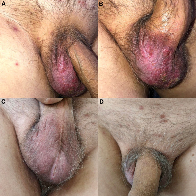 Figure 1
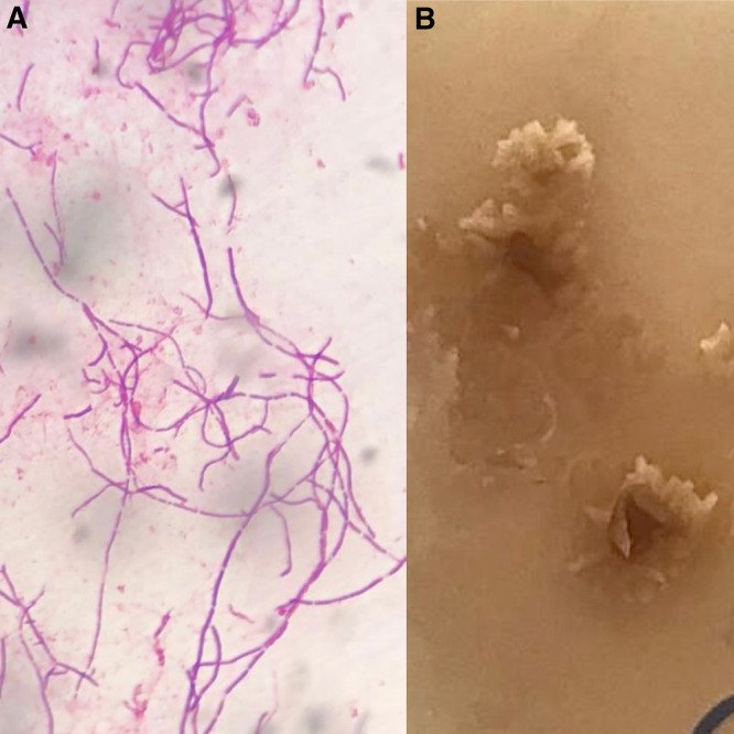 Figure 2
Peer Reviews
No public reviews on file for this paper yet. If you reviewed it on a platform where reviews are public (OpenReview, ICLR, NeurIPS, ICML), you can paste yours below so the community can read it here.
Videos
No videos yet. Explain this paper in a talk, walkthrough, or lecture? Add one.
Taxonomy
TopicsActinomycetales infections and treatment · Diagnosis and treatment of tuberculosis · Diverticular Disease and Complications
A 69-year-old man presented with a 3-week history of pruritic scrotal lesions after presurgical hair removal for a coronary angioplasty. One week before presentation, he had undergone inguinal shaving with a disposable razor in a hospital surgical setting. Physical examination revealed an erythematous plaque with crusts and exulcerations on the right scrotum, accompanied by satellite papules and pustules (Figure 1). A direct smear from a pustule revealed numerous thin, clustered, and branching Gram-positive filaments (Figure 2A), which were partially acid-fast with Kinyoun stain. A culture of skin swabs on Sabouraud dextrose agar at 25°C yielded Nocardia spp., which formed small, chalky-white, heaped, wrinkled, or verrucous colonies with a distinctive odor reminiscent of freshly turned soil (Figure 2B). Lymphatic and systemic involvement were excluded by imaging.
A diagnosis of superficial cutaneous nocardiosis, likely due to direct inoculation, was confirmed. Oral trimethoprim (160 mg) and sulfamethoxazole (800 mg), taken three times daily, were initiated. Significant improvement was noted after 1 month (Figure 1C and D), and therapy was continued for an additional 3 months until complete resolution.
Nocardiosis is an uncommon opportunistic infection caused by aerobic, Gram-positive bacilli of the Nocardia genus. It mimics a variety of dermatoses, often delaying diagnosis despite the availability of straightforward microbiologic methods.1?^–^3 A high index of suspicion, clinical awareness, and timely microbiologic evaluation are critical for accurate diagnosis. In this case, an unusual route of inoculation through presurgical shaving is illustrated, highlighting the importance of infection control practices and the recognition of atypical cutaneous infections.
The reference list from the paper itself. Each links out to its DOI / PubMed record.
- 1Dodiuk-Gad R Cohen E Ziv M Goldstein LH Chazan B Shafer J Sprecher H Elias M Keness Y Rozenman D, 2010. Cutaneous nocardiosis: Report of two cases and review of the literature. Int J Dermatol 49: 1380–1385.21155087 10.1111/j.1365-4632.2010.04554.x · doi ↗ · pubmed ↗
- 2Ramos-e-Silva M Lopes RS Trope BM, 2020. Cutaneous nocardiosis: A great imitator. Clin Dermatol 38: 152–159.32513396 10.1016/j.clindermatol.2019.10.009 · doi ↗ · pubmed ↗
- 3Hu M Bao F Zhang F, 2024. Primary cutaneous nocardiosis. JAMA Dermatol 160: 1237–1238.39356533 10.1001/jamadermatol.2024.3834 · doi ↗ · pubmed ↗