Case Report: Rare invasive aspergillosis with brain abscess in a non–classically immunosuppressed patient, and pooled analysis of individual patient data (2000–2024)
Moksada Regmi, Shikun Liu, Yuwei Dai, Jingyi Ye, Xiaodong Chen, Jun Yang, Chenlong Yang

TL;DR
A rare case of brain abscess caused by Aspergillus in a non-immunosuppressed diabetic patient is reported, along with a pooled analysis showing improved survival rates over time.
Contribution
A case report and pooled analysis of intracranial aspergillosis in non-classically immunosuppressed patients, highlighting the role of molecular diagnostics and azole therapy.
Findings
A 71-year-old diabetic patient with Aspergillus brain abscess showed improvement with azole therapy and multidisciplinary care.
Pooled analysis of 343 cases showed 34.6% overall mortality, with 21.8% in non-classically immunosuppressed patients.
Improved survival is likely due to earlier diagnosis and increased use of azoles, though evidence remains limited.
Abstract
Intracranial aspergillosis is uncommon but often lethal, especially in classically immunocompromised hosts. We report a 71-year-old man with poorly controlled diabetes (a non-classical risk factor) who developed bilateral frontal abscesses due to Aspergillus fumigatus. After an initial craniotomy with negative cultures and galactomannan, recurrent disease was confirmed by stereotactic biopsy with next-generation sequencing (NGS). Targeted azole therapy (voriconazole, isavuconazole) and multidisciplinary care led to marked clinical and radiographic improvement. We also pooled 343 published cases (2000–2024): overall mortality was 34.6%, and 21.8% among patients without classical immunosuppression (including some with non-classical factors such as diabetes). Improved survival in recent decades likely reflects earlier diagnosis and broader azole use, though inference is limited by…
Genes, proteins, chemicals, diseases, species, mutations and cell lines named across the full text — each resolved to its canonical identifier and authoritative record.
Click any figure to enlarge with its caption.
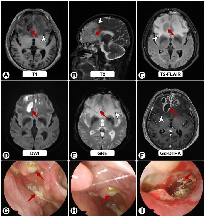 Figure 1
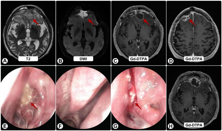 Figure 2
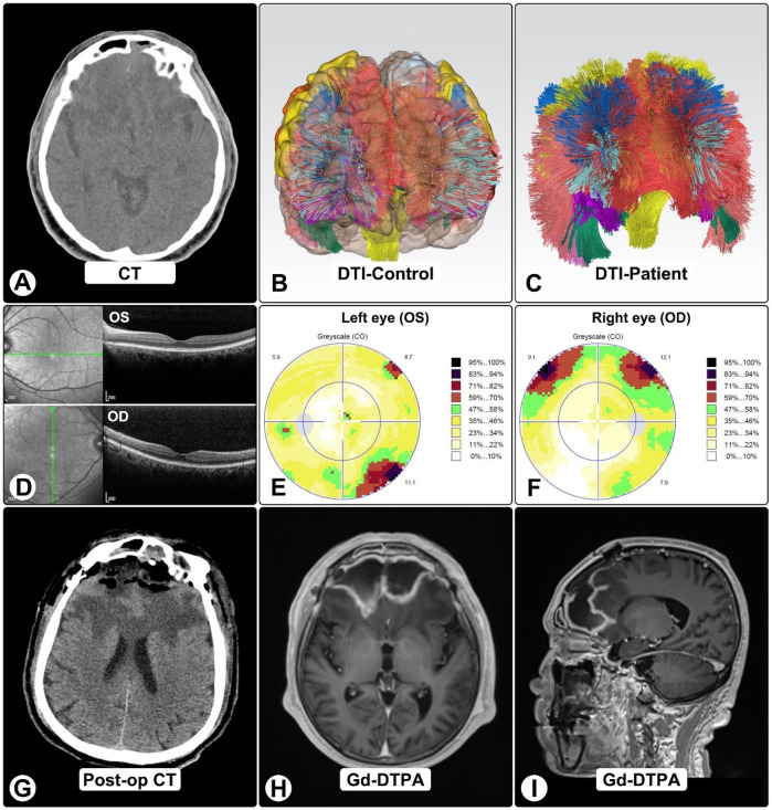 Figure 3
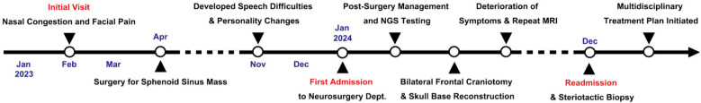 Figure 4
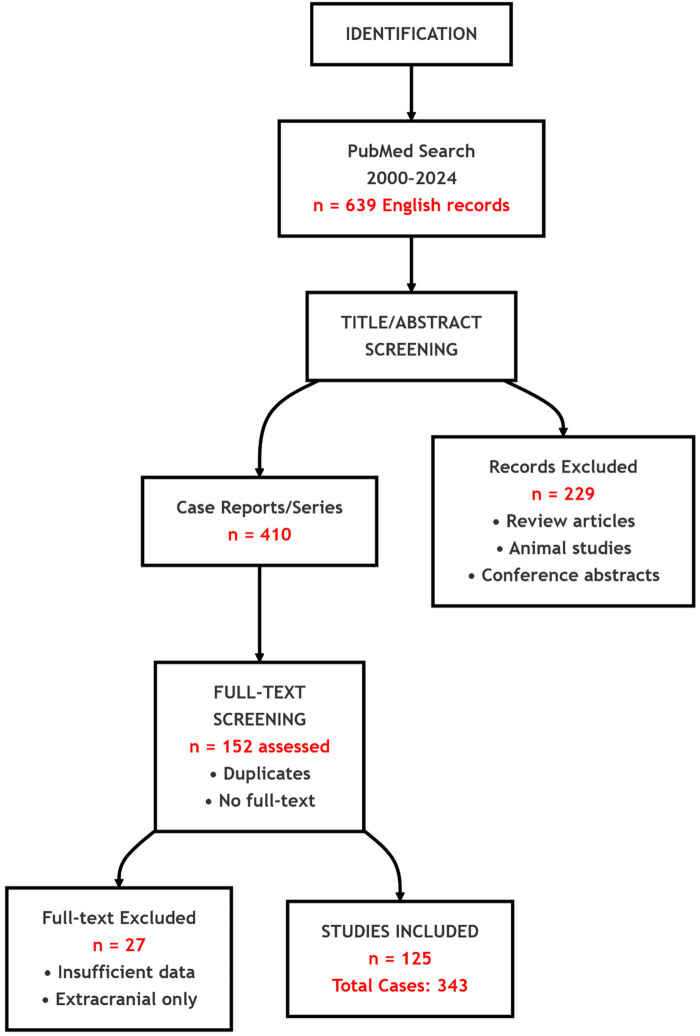 Figure 5
Peer Reviews
No public reviews on file for this paper yet. If you reviewed it on a platform where reviews are public (OpenReview, ICLR, NeurIPS, ICML), you can paste yours below so the community can read it here.
Videos
No videos yet. Explain this paper in a talk, walkthrough, or lecture? Add one.
Taxonomy
TopicsAntifungal resistance and susceptibility · Fungal Infections and Studies · Pneumocystis jirovecii pneumonia detection and treatment
Background
Intracranial Aspergillus infection is a severe complication of invasive aspergillosis, primarily affecting immunocompromised patients (e.g., those with hematologic malignancies, transplant recipients, or on chronic corticosteroids) (1–5). Historically, reported mortality rates for intracranial aspergillosis were as high as 85% to 99%, especially in immunocompromised hosts, and when complicated by brain abscesses, the rate reportedly approaches 100% in such patients (6–14).
Aspergillus typically reaches the brain through hematogenous spread from a primary pulmonary focus or by direct extension from the paranasal sinuses (10, 15). The clinical manifestations of cerebral aspergillosis are often nonspecific, leading to delayed diagnosis (16, 17). Common symptoms include headache, altered mental status, focal neurological deficits, seizures, and visual disturbances. In patients without classical immunosuppression but with non-classical risk factors (e.g., diabetes), the course may be more indolent and may present as meningitis or granulomatous mass (18).
Intracranial aspergillosis is an exceedingly rare condition, even more so among immunocompetent patients. The existing literature primarily comprises single case reports or small retrospective studies, which provide insufficient data for robust prognostic assessments. Over the last 20–25 years, medical diagnostics and treatments have improved significantly. To contextualize our case, we compiled a pooled individual-patient analysis of reports since 2000, explicitly comparing outcomes by immune status. For terminology consistency throughout, we use “non-classically immunosuppressed” to denote patients lacking classical immunosuppressive conditions (e.g., no hematologic malignancy, transplant, HIV/AIDS, active chemotherapy, prolonged high-dose steroids, or primary immunodeficiency) but who carry risk modifiers such as diabetes.
Case presentation
A 71-year-old man with a 17-year history of poorly controlled type 2 diabetes (HbA1c 9.5%) underwent endoscopic resection of a sphenoid sinus mass with sinusotomy for chronic sinusitis (15 months before definitive diagnosis). Histopathology showed inflammation without fungi, and symptoms initially improved. Six months later, he developed aphasia, cognitive slowing, and personality change without fever. MRI at a local hospital suggested bilateral frontal abscesses; mannitol partially improved speech.
Two months thereafter he presented to our center. MRI demonstrated bilateral frontal lesions with extensive edema and ring enhancement (Figures 1A–F); nasal endoscopy confirmed purulence (Figures 1G–I). Pre-operative CT showed anterior skull-base sclerosis and erosion consistent with osteomyelitis (Figure 3A). Laboratory tests revealed leukocytosis with neutrophilia and mildly elevated CRP; HIV screening was negative. Serum galactomannan (GM) was negative.
Brain MRI and nasal endoscopy on admission. (A–F) White arrowheads indicate surrounding edema; red arrows show abscesses. MRI shows bilateral frontal lesions appearing hyperintense on T1 (A) and T2 (B), and isointense on FLAIR (C) Post-contrast images (D–F) reveal ring enhancement of lesions with extensive vasogenic edema and midline shift. (G–I) Nasal endoscopy shows purulent sinus secretions (red arrows).
Given encapsulated abscesses with sinus and skull-base involvement, he underwent bilateral frontal craniotomy for evacuation and anterior skull-base reconstruction. Abundant pus was encountered; bacterial, fungal, and mycobacterial cultures and histopathology were negative.
Postoperatively, he received vancomycin and ceftriaxone plus fluconazole (guided by early nanopore reads suggesting possible Candida). He was discharged to complete IV antibiotics and fluconazole. Two weeks later he worsened neurologically. Follow-up MRI showed progression with persistent ring enhancement and diffusion restriction (Figures 2A–D); endoscopy again revealed purulence (Figures 2E–G).
Follow-up brain MRI and nasal endoscopy. (A) T2-weighted image showing enlargement of frontal lesions (red arrows) with increased edema. (B) DWI shows persistent restricted diffusion. (C,D,H) Gd-DTPA–enhanced images demonstrate continued ring enhancement, with some solid components (H), (E–G) Endoscopy reveals persistent purulent secretions (red arrows) on the nasal mucosa (E), left middle meatus (F), and sphenoid sinus ostium (G), despite antibiotics.
Four months after craniotomy, stereotactic biopsy confirmed A. fumigatus by NGS; serum GM and β-D-glucan were now positive. Sinonasal biopsies demonstrated invasive hyphae. DTI tractography showed marked frontal tract loss (Figures 3B,C). OCT was normal, while visual fields showed dense bilateral defects consistent with compressive optic neuropathy (Figures 3D–F).
DTI tractography, OCT, and visual findings. (A) Pre-op CT shows bilateral frontal lesions with vasogenic edema and mass effect. (B,C) DTI tractography in normal brain (B) and patient (C), showing marked frontal tract loss, including bilateral anterior thalamic radiations, genu of corpus callosum, cingulum bundles, and forceps minor. (D) OCT imaging. (E,F) Octopus visual fields show right superior altitudinal defect (E) and left inferotemporal scotoma (F), consistent with compressive optic neuropathy. (G) Post-op CT showing expected changes after craniotomy and abscess evacuation. (H,I) One-month post-op MRI shows residual ring-enhancing lesions with reduced size and mass effect.
Multidisciplinary management with CNS-penetrant azoles (voriconazole and isavuconazole), blood-pressure control, and supportive care led to steady improvement. One-month MRI showed smaller lesions and reduced mass effect (Figures 3H,I). At six months, he was independent in daily activities with mild residual cognitive impairment and persistent visual-field loss (Figure 4).
Timeline of clinical events and interventions.
Methods for pooled analysis
Search strategy and eligibility
We searched PubMed for English-language reports from January 1, 2000 to May 10, 2024 using (“intracranial aspergillosis” OR “Aspergillus brain abscess” OR “central nervous system Aspergillus” OR “cerebral Aspergillus” OR “brain aspergillosis”). Inclusion required: (1) histopathological, culture, or molecular confirmation of intracranial aspergillosis; (2) radiologic evidence of an intracranial lesion (intraparenchymal or extradural); and (3) documented immune status and outcome. We excluded reviews, animal studies, conference abstracts, studies with insufficient intracranial data, and reports limited to extracranial infection. PRISMA flow: 639 records → 229 excluded at screening; after removing 258 duplicates/inaccessible texts, 152 full texts reviewed; 27 excluded; 125 studies included (343 cases) (Figure 5).
PRISMA flowchart of study selection.
Terminology
“Classically immunocompromised” comprised hematologic malignancy, solid-organ or stem-cell transplantation, HIV/AIDS, active cytotoxic chemotherapy, prolonged high-dose corticosteroids (>20 mg/day prednisone-equivalent for >2 weeks), or primary immunodeficiency. “Non-classically immunosuppressed” included conditions such as diabetes that alter host defenses without meeting classical criteria. For clarity, we report outcomes for patients “without classical immunosuppression,” which may include non-classical factors.
Results
Among 343 patients (201 male, 107 female, 35 unspecified), there were 119 deaths, with 11 additional deaths unrelated to aspergillosis; 213 were recovered or stable at last follow-up. Overall mortality was 34.6%; mortality among patients without classical immunosuppression (including some with non-classical factors) was 21.8%. Detailed per-case data are provided in Supplementary Table S1.
Discussion and conclusion
This case underscores diagnostic pitfalls in intracranial aspergillosis, particularly when early biomarkers are negative. The GM assay has variable sensitivity: at an optical density index of 0.5, pooled sensitivity is ∼82% (≈18% false-negative rate); at 1.0, sensitivity declines to ∼72% (≈28% false-negative rate) (19–21). Thus, negative GM should not preclude biopsy or molecular testing when imaging and clinical evolution are concerning. The eventual detection of Aspergillus DNA via NGS on a biopsy specimen was pivotal, enabling targeted antifungal therapy and likely preventing a fatal outcome.
Surgery remains important for encapsulated collections, mass effect, or diagnostic uncertainty, both to decompress and to obtain tissue for definitive identification (culture, histology, PCR/NGS). In our patient, delayed confirmation by NGS ultimately enabled targeted therapy.
Therapeutically, outcomes have improved with CNS-penetrant triazoles—especially voriconazole—compared with amphotericin B or itraconazole, which historically showed poor CNS efficacy and tolerability (22–33). Isavuconazole may offer additional options, as illustrated by this case, though further CNS-specific data are needed (34, 35). Non-classical risk factors (e.g., diabetes), disruption of anatomic barriers (e.g., sinus surgery), and transient immune dysfunction can permit angioinvasion even without classical immunosuppression. Recognizing these scenarios can prompt earlier imaging, biopsy, and azole initiation (36).
In the future, standardization of molecular diagnostics, therapeutic drug monitoring for azoles, and optimized combination/sequencing strategies may further lower mortality; exploratory immunomodulatory approaches merit study but lie beyond the scope of this report.
Conclusion
Intracranial aspergillosis remains a high-stakes neurosurgical and infectious-disease emergency, yet patients without classical immunosuppression—including those with non-classical risks such as diabetes—can achieve good outcomes when clinicians maintain suspicion despite early false-negative biomarkers, pursue early tissue diagnosis with biopsy/NGS, and combine indicated surgical management with CNS-penetrant azoles (e.g., voriconazole, isavuconazole) under multidisciplinary care.
The reference list from the paper itself. Each links out to its DOI / PubMed record.
- 1Cadena J Thompson GRIII Patterson TF. Aspergillosis: epidemiology, diagnosis, and treatment. Infect Dis Clin North Am. Jun. (2021) 35(2):415–34. 10.1016/j.idc.2021.03.00834016284 · doi ↗ · pubmed ↗
- 2Godoy MCB Ferreira Dalla Pria HR Truong MT Shroff GS Marom EM. Invasive fungal pneumonia in immunocompromised patients. Radiol Clin North Am. (2022) 60(3):497–506. 10.1016/j.rcl.2022.01.00635534133 · doi ↗ · pubmed ↗
- 3Azoulay E Russell L Van de Louw A Metaxa V Bauer P Povoa P Diagnosis of severe respiratory infections in immunocompromised patients. Intensive Care Med. (2020) 46(2):298–314. 10.1007/s 00134-019-05906-532034433 PMC 7080052 · doi ↗ · pubmed ↗
- 4Marr KA Schlamm HT Herbrecht R Rottinghaus ST Bow EJ Cornely OA Combination antifungal therapy for invasive aspergillosis: a randomized trial. Ann Intern Med. (2015) 162(2):81–9. 10.7326/m 13-250825599346 · doi ↗ · pubmed ↗
- 5Warris A Lehrnbecher T Roilides E Castagnola E Brüggemann RJM Groll AH. ESCMID-ECMM guideline: diagnosis and management of invasive aspergillosis in neonates and children. Clin Microbiol Infect. (2019) 25(9):1096–113. 10.1016/j.cmi.2019.05.01931158517 · doi ↗ · pubmed ↗
- 6Kumar D Nepal P Singh S Ramanathan S Khanna M Sheoran R CNS Aspergilloma mimicking tumors: review of CNS aspergillus infection imaging characteristics in the immunocompetent population. J Neuroradiol. (2018) 45(3):169–76. 10.1016/j.neurad.2017.11.00129273531 · doi ↗ · pubmed ↗
- 7Lin SJ Schranz J Teutsch SM. Aspergillosis case-fatality rate: systematic review of the literature. Clin Infect Dis. (2001) 32(3):358–66. 10.1086/31848311170942 · doi ↗ · pubmed ↗
- 8Lamoth F Mercier T AndréP Pagani JL Pantet O Maduri R Isavuconazole brain penetration in cerebral aspergillosis. J Antimicrob Chemother. (2019) 74(6):1751–3. 10.1093/jac/dkz 05030753519 · doi ↗ · pubmed ↗