Differing metabolic responses of guard cells to blue light
Alisdair R. Fernie, Stefan Timm

Abstract
This article is a Commentary on Bahadar et al. (2025), 248: 2347–2360.
Genes, proteins, chemicals, diseases, species, mutations and cell lines named across the full text — each resolved to its canonical identifier and authoritative record.
Click any figure to enlarge with its caption.
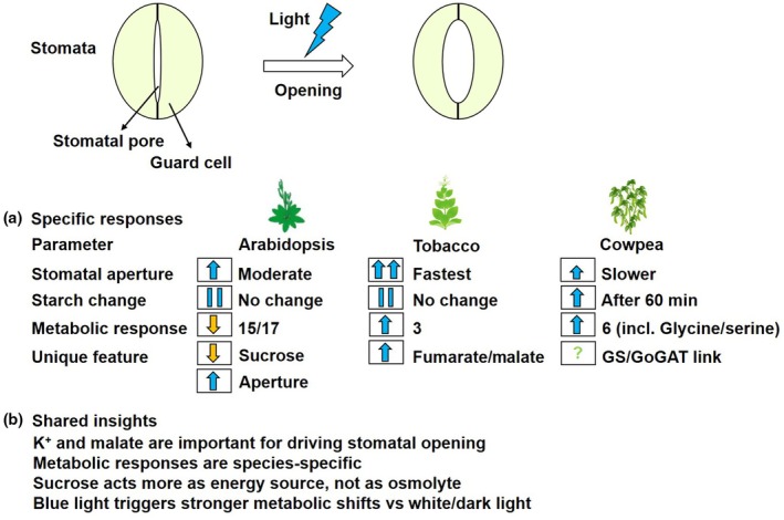 Figure 1
Peer Reviews
No public reviews on file for this paper yet. If you reviewed it on a platform where reviews are public (OpenReview, ICLR, NeurIPS, ICML), you can paste yours below so the community can read it here.
Videos
No videos yet. Explain this paper in a talk, walkthrough, or lecture? Add one.
Taxonomy
TopicsLight effects on plants · Circadian rhythm and melatonin · Photoreceptor and optogenetics research
Plants have adjustable pores, stomata, surrounded by a pair of guard cells. Alterations in the metabolism of these guard cells govern the opening and closure of the stomata in response to both endogenous and environmental signals (Sussmilch et al., 2019). Stomatal opening facilitates the influx of CO_2_ for photosynthesis alongside the transpiratory efflux of water, as they balance optimal growth against dehydration during drought periods. The regulation of stomatal movement is highly complex and subject to a number of triggers, including phytohormones and metabolite levels of both the guard cells themselves and other surrounding cell types (Flütsch et al., 2020). One prominent suggestion in the literature, principally derived from work in Arabidopsis, is that starch degradation is of particular importance during exposure to blue light (Daloso et al., 2017). However, considerable evidence has also been accrued supporting roles for both K^+^ and malate in guard cell osmoregulation (Outlaw & Lowry, 1977; Medeiros et al., 2018). Moreover, while the importance of guard cell starch degradation in stomatal opening is supported by several convincing lines of evidence, several other studies revealed that guard cell starch content was not correlated with aperture under either blue or white light (Heath, 1947; Daloso et al., 2017). In a study published in this issue of New Phytologist, Bahadar et al. (2025; pp. 2347–2360) performed a detailed metabolic and stomatal analysis following the exposure of cowpea and tobacco to blue light, with results suggesting that changes in both starch degradation and downstream aspects of primary metabolism are species‐specific and thus urging caution on making generalized statements concerning guard cell metabolism (Fig. 1). … blue light‐induced stomatal opening does not involve starch remobilization in Arabidopsis, cowpea or tobacco guard cells but rather that the level of starch ranges according to the experimental condition.
As an initial experiment, Bahadar et al. tested the responsiveness of cowpea and tobacco stomata to blue light, finding that stomatal aperture increased linearly for 30 min. Given that contradictory results are presented in the literature for these species, Bahadar et al. carried out a more detailed analysis of the kinetics of stomatal conductance revealing that both species are indeed responsive to blue light but that both the magnitude and the speed of the increase were higher in tobacco than in cowpea (Fig. 1). However, in contrast to previous reports (Flütsch et al., 2020), neither Arabidopsis nor tobacco guard cells exhibited altered starch content in their guard cells, but cowpea displayed elevated starch content after 60 min of blue light treatment, prompting Bahadar et al. to carry out a range of experiments to address the reason behind these discrepancies. In doing so, they were able to discard the possibility that the differences between their results and those previously reported were the consequence of their experimental procedure or guard cell‐enriched epidermal fragments. Leading them to conclude that blue light‐induced stomatal opening does not involve starch remobilization in Arabidopsis, cowpea or tobacco guard cells but rather that the level of starch ranges according to the experimental condition.
In order to better understand the response to blue light, Bahadar et al. next evaluated changes in primary metabolite levels following exposure to blue light using a well‐established gas chromatography mass spectrometry approach. For this purpose, guard cells were extracted predawn, pooled in a hypertonic solution and washed extensively before transfer to blue light. These studies revealed considerable changes in metabolite levels in all three species, with the levels of 15 of the 17 metabolites identified in Arabidopsis decreasing after 60 min of exposure to blue light. By contrast, only three metabolites increased in tobacco during blue light treatment and six metabolites increased in cowpea including the photorespiratory metabolites glycine and serine – a fact that is perhaps pertinent given the recent demonstration of the importance of photorespiratory metabolism in guard cells (Sun et al., 2025). To better gauge which metabolites are associated with the dynamics of blue light‐induced stomatal opening, Bahadar et al. normalized their data in order to carry out a K‐means clustering analysis. For tobacco, no clear patterns between changes in metabolite levels and stomatal aperture were apparent. By contrast, stomatal aperture parameters clustered with aspartate, maltotriose, pyroglutamate, serine and urea in cowpea – with the data also suggesting that sucrose and glutamate are degraded while glucose, maltotriose and pyroglutamate are synthesized during blue light‐induced stomatal opening in cowpea. Moreover, statistical analyses of the data revealed that tobacco exhibited the greatest metabolic response to blue light, followed by cowpea and then tobacco. Collectively, the data suggest that the dynamic of blue light‐induced stomatal opening is associated with diverse metabolic changes in Arabidopsis, tobacco and cowpea. Finally, in order to understand how specific these responses are to blue light, the authors compared their data with that recently obtained for tobacco guard cells exposed to either darkness or white light (Lima et al., 2023). This comparison revealed that the response of metabolism to blue light is quite distinctive, with all metabolites, with the exception of fumarate, displaying higher levels under blue light than white light in tobacco. Notably, both fumarate and malate were higher in both blue light and white light than they were in darkness.
These data collectively provide a more complete picture concerning the stomatal opening properties of blue light, which have now been researched for over 50 years (Zeiger & Hepler, 1977). That said, although blue light responses have been observed in the basal lineages of plants, certain experiments in certain species were not able to identify blue light stomatal responses (Vialet‐Chabrand et al., 2021). The species examined in the study of Bahadar et al. have all been previously documented to exhibit stomatal responses to blue light. In their study, Bahadar et al. also demonstrated responsiveness, but increases in stomatal conductance and speed of response were considerably greater in tobacco than in cowpea (Fig. 1). The kinetic analyses of the responses revealed that they preceded starch degradation under the conditions of the study; thus, while a great deal of evidence has accrued indicating that starch metabolism of guard cells is involved in stomatal closure (Azoulay‐Shemer et al., 2016; Flütsch et al., 2020), starch‐derived metabolites do not appear to be the major contributors to blue light‐induced initial stomatal opening. As the authors correctly underlined, these data do not imply that starch degradation is unimportant in the regulation of blue light‐induced opening, and it is indeed likely important to keep the stomata open following the rapid increase in stomatal opening mediated by the accumulation of K^+^ and its counterions alongside the degradation of storage reserves (McLachlan et al., 2016; Medeiros et al., 2018; Lima et al., 2023). In keeping with such a model, it is important to note that sucrose levels were negatively correlated with stomatal aperture in both Arabidopsis and cowpea, in agreement with the role of sucrose in stomatal movement being purely energetic (Fig. 1). Intriguingly, while the stomatal responses were broadly similar, the changes in metabolite levels were highly diverse across the species studied. That said, a common connection among sugars and tricarboxylic acids was observed, as was a clear alteration in glutamine levels. The latter led to the authors proposing the intriguing hypothesis that the glutamine synthetase/ glutamate synthetase cycle may aid in optimizing stomatal opening. While this hypothesis will need considerable further experimental work to validate the data presented, the study of Bahadar et al. represents a significant milestone in dissecting metabolic aspects of the blue light response of guard cells, which will undoubtedly form a solid foundation for such future studies.
Disclaimer
The New Phytologist Foundation remains neutral with regard to jurisdictional claims in maps and in any institutional affiliations.
The reference list from the paper itself. Each links out to its DOI / PubMed record.
- 1Azoulay‐Shemer T , Bagheri A , Wang C , Palomares A , Stephan AB , Kunz HH , Schroeder JI . 2016. Starch biosynthesis in guard cells but not in mesophyll cells is involved in CO 2‐induced stomatal closing. Plant Physiology 171: 788–798.27208296 10.1104/pp.15.01662 PMC 4902578 · doi ↗ · pubmed ↗
- 2Bahadar H , Morais EG , Freire FBS , Lima VF , Giacomelli ME , dos Anjos L , Antues WG , Daloso DM . 2025. Blue light‐induced stomatal opening is associated with species‐specific changes in primary metabolism but not to starch breakdown in guard cells. New Phytologist 248: 2347–2360.40501107 10.1111/nph.70257 PMC 12589703 · doi ↗ · pubmed ↗
- 3Daloso DM , Medeiros DB , Dos Anjos L , Yoshida T , Araújo WL , Fernie AR . 2017. Metabolism within the specialized guard cells of plants. New Phytologist 216: 1018–1033.28984366 10.1111/nph.14823 · doi ↗ · pubmed ↗
- 4Flütsch S , Wang Y , Takemiya A , Vialet‐Chabrand SRM , Klejchová M , Nigro A , Hills A , Lawson T , Blatt MR , Santelia D . 2020. Guard cell starch degradation yields glucose for rapid stomatal opening in Arabidopsis. Plant Cell 32: 2325–2344.32354788 10.1105/tpc.18.00802 PMC 7346545 · doi ↗ · pubmed ↗
- 5Heath OV . 1947. Role of starch in light‐induced stomatal movement, and a new reagent for staining stomatal starch. Nature 159: 647.20239728 10.1038/159647 b 0 · doi ↗ · pubmed ↗
- 6Lima VF , Freire FBS , Cândido‐Sobrinho SA , Porto NP , Medeiros DB , Erban A , Kopka J , Schwarzländer M , Fernie AR , Daloso DM . 2023. Unveiling the dark side of guard cell metabolism. Plant Physiology and Biochemistry 201: 107862.37413941 10.1016/j.plaphy.2023.107862 · doi ↗ · pubmed ↗
- 7Mc Lachlan DH , Lan J , Geilfus CM , Dodd AN , Larson T , Baker A , Hõrak H , Kollist H , He Z , Graham I et al. 2016. The breakdown of stored triacylglycerols is required during light‐induced stomatal opening. Current Biology 26: 707–712.26898465 10.1016/j.cub.2016.01.019PMC 4791430 · doi ↗ · pubmed ↗
- 8Medeiros DB , Perez Souza L , Antunes WC , Araújo WL , Daloso DM , Fernie AR . 2018. Sucrose breakdown within guard cells provides substrates for glycolysis and glutamine biosynthesis during light‐induced stomatal opening. The Plant Journal 94: 583–594.29543357 10.1111/tpj.13889 · doi ↗ · pubmed ↗