Case Report: Ultrasound-guided fine-needle aspiration for parathyroid cyst
A. Serblin, R. Valcavi

TL;DR
A 55-year-old man had a large parathyroid cyst diagnosed and successfully treated with ultrasound-guided aspiration, showing no recurrence over five years.
Contribution
This case report demonstrates the effectiveness of fine-needle aspiration as a definitive treatment for non-functional parathyroid cysts.
Findings
The cyst was confirmed non-functional with normal PTH, calcium, and vitamin D levels before and after aspiration.
Ultrasound-guided aspiration successfully drained the cyst with no recurrence observed over five years of follow-up.
High PTH levels in the aspirate confirmed the parathyroid origin of the cyst despite normal systemic hormone levels.
Abstract
A 55-year-old man was referred to our Department with a cystic lesion in the lower right lobe of the thyroid, incidentally discovered on ultrasound. The mass measured 52.1 × 55.3 × 66.8 mm, with a volume of 93.2 mL, and caused significant tracheal indentation with contralateral deviation. The patient was asymptomatic and did not have dysphagia, hoarseness or dyspnoea. Ultrasound-guided fine-needle aspiration of the lesion yielded a clear, “rock-water” fluid. Biochemical analysis of the aspirate revealed elevated parathyroid hormone (PTH), leading to a diagnosis of a parathyroid cyst (PCs). This case highlights the importance of considering PCs in the differential diagnosis of large cystic neck masses, particularly when they mimic thyroid nodules. We report on this case and discuss the diagnostic challenges and management strategies for this rare condition. Parathyroid cysts (PCs) are…
Genes, proteins, chemicals, diseases, species, mutations and cell lines named across the full text — each resolved to its canonical identifier and authoritative record.
Click any figure to enlarge with its caption.
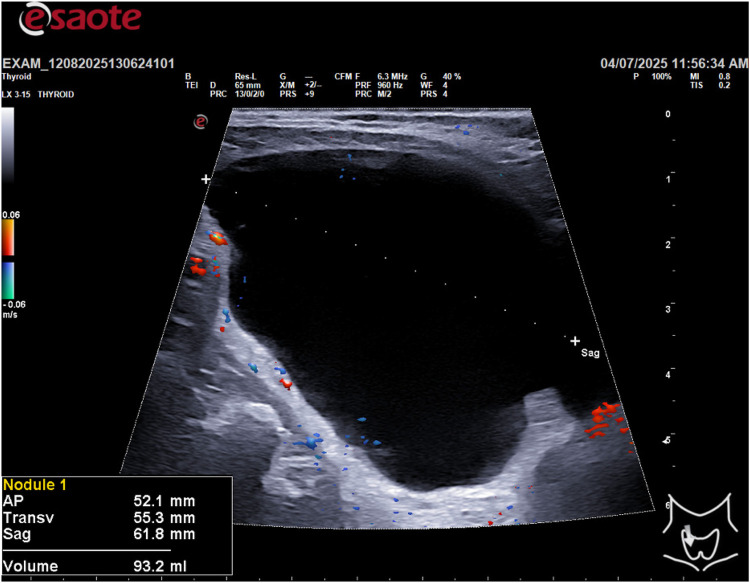 Figure 1
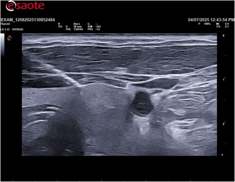 Figure 2
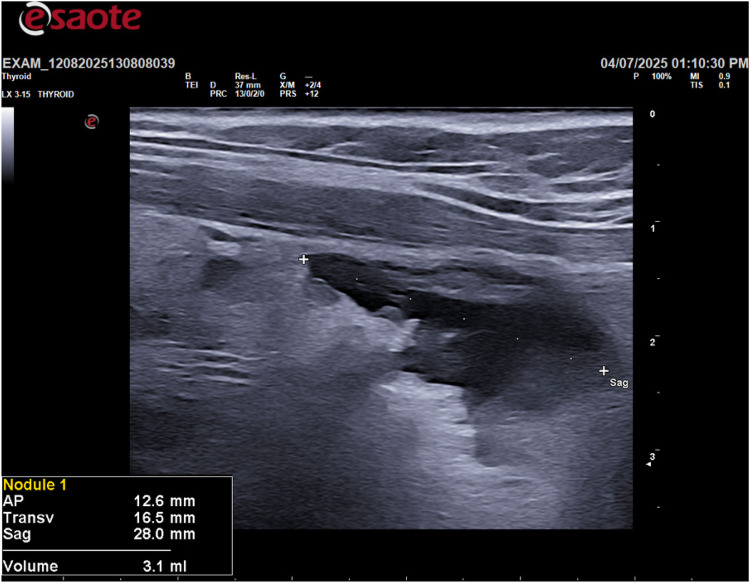 Figure 3
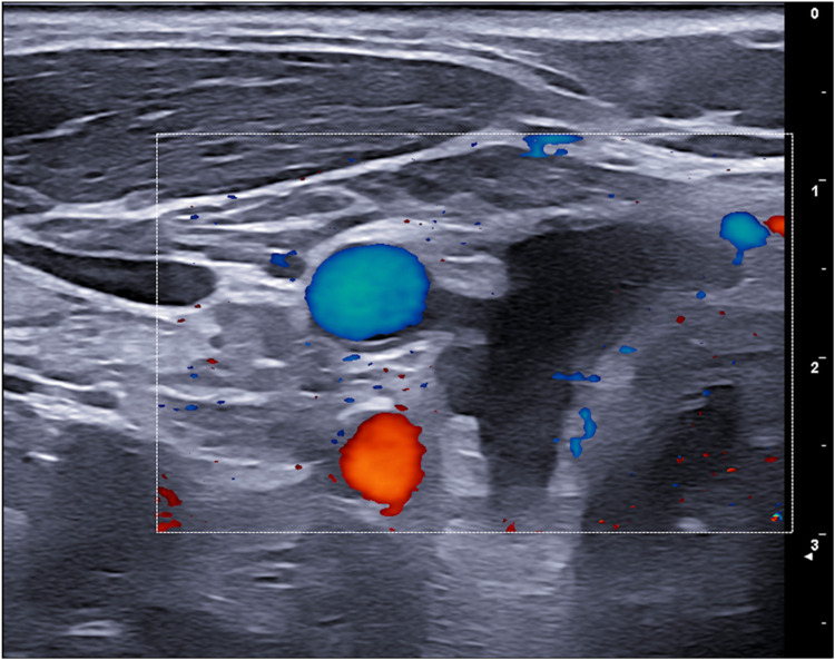 Figure 4
Peer Reviews
No public reviews on file for this paper yet. If you reviewed it on a platform where reviews are public (OpenReview, ICLR, NeurIPS, ICML), you can paste yours below so the community can read it here.
Videos
No videos yet. Explain this paper in a talk, walkthrough, or lecture? Add one.
Taxonomy
TopicsParathyroid Disorders and Treatments · Medical Imaging and Pathology Studies · Pancreatic and Hepatic Oncology Research
Introduction
Parathyroid cysts (PCs) are rare clinical and histological entities, representing a small fraction of parathyroid pathology, typically 1%–3.3% of cases (1, 2). They also account for 1%–5% of all neck masses (2, 3). While they can occur at any age, they are most frequently discovered in women between 40 and 60 years old (1, 4, 5). Based on the provided sources, the embryological derivation of parathyroid cysts is primarily linked to theories concerning the formation of true cysts. Here is an illustration of the potential embryological origins of parathyroid cysts (6):
- 1.The Epithelial Tubule Hypothesis: One theory explaining the origin of cystic parathyroid lesions (CPLs) hypothesizes that varying numbers of epithelial tubules, arising near thymic tissue, develop into canalicular or glandular tissue layers in postnatal life (6).
- 2.Embryological Remnants (True Cysts): Parathyroid cysts are classified into subgroups based on their etiology, including (a) degeneration of an adenoma or hyperplastic gland, and (b) true cysts with an epithelial layer. In clinical practice, microscopic findings can support this origin: the authors reported a case (Patient 2) where the cystic wall was composed of stratified parathyroid cells uniformly lining the CPL. This finding suggested a true parathyroid cyst, potentially derived from an embryological remnant. Ippolito et al. defined “true cysts” exclusively as the non-functioning parathyroid lesions (6).
- 3.Other Etiological Theories (Non-Embryological): It is important to note that while embryological remnants explain the origin of true cysts, another prevalent theory for CPLs involves the degeneration of an adenoma or hyperplastic gland. For instance, in the first case reported by the authors, the pathological finding aligned with cystic degeneration due to intraglandular hemorrhage. In summary, the embryological derivation focuses on the formation of true cysts (often non-functioning) arising from epithelial tubules associated with nearby thymic tissue or other embryological remnants. The distinction between functioning and non-functioning parathyroid cysts (CPLs) primarily rests on hormonal activity, etiology, and required treatment. Functioning Cysts (Hyperfunctioning) cause hyperparathyroidism, evidenced by elevated serum PTH and calcium levels. Diagnosis is confirmed by highly elevated PTH values in the intracystic fluid (e.g., > 5,000 pg/mL). Etiologically, they often arise from the cystic degeneration of a parathyroid adenoma. The definitive treatment for functioning CPLs is surgical excision (6).
Non-functioning Cysts are more common and present with non-specific physical features. They generally cause compressive symptoms (e.g., neck swallowing) rather than systemic hormonal issues. Histologically, non-functioning cysts are often described as pseudocysts lacking cuboidal epithelium. For non-functioning lesions, Fine Needle Aspiration (FNA) can be considered the first-line treatment (6).
Ultrasound is indicative: non-functioning cysts have no intrinsic tissue, while cystic adenomas have observable tissue. Furthermore, true cysts only contain amorphous material in cytology; cystic adenomas, if FNA is directed at observable tissue after emptying, offer a cytological examination with cuboidal cells, similar to thyroid cells, but without background colloid. It is important to empty the cyst before performing FNA for cytology: ultrasound can only detect parenchymal content after the cyst has been emptied, because the parenchyma is compressed by intracystic pressure (6, 7).
The definitive way to distinguish a parathyroid cyst (PC) from other cystic neck masses, such as a thyroid cyst, branchial cleft cyst, or thyroglossal duct cyst (TDC), is through Fine Needle Aspiration (FNA) of the cyst fluid followed by biochemical analysis for specific markers. The key distinguishing feature is the measurement of Parathyroid Hormone (PTH) levels in the aspirated cystic fluid. The measurement of i-PTH in the fluid, which will always be clear water in the case of parathyroid cysts. And with the measurement of thyroglobulin in the fluid—citrine or hemorrhagic—high in thyroid cysts and thyroglossal duct cysts and not in branchial cysts.
Clinical presentation of PCs can be highly variable. Many cases are asymptomatic and discovered incidentally during investigations for other conditions (2, 4, 8). However, PCs frequently manifest as palpable neck masses, which is the most common symptom, occurring in 41.7% of reported patients (2). Due to their location and consistency, they often mimic other cervical pathologies, particularly solitary thyroid nodules, making accurate preoperative diagnosis difficult (2, 9). When PCs reach large dimensions, they can exert a compressive effect on adjacent structures, leading to symptoms such as dysphagia (difficulty swallowing), hoarseness, dyspnoea, and tracheal deviation (2). The pathogenesis of PCs is thought to involve several mechanisms, including congenital acquisition from branchial pouch remnants, coalescence of microcysts, or cystic degeneration/hemorrhage of a parathyroid gland or adenoma (2, 3). The diagnostic workup for PCs is crucial due to their diverse presentations and the need to differentiate them from other neck masses, such as thyroglossal duct cysts, branchial cleft cysts, and thyroid adenomas or carcinomas. Ultrasound (US) is typically the first-line imaging modality, effectively confirming the cystic nature of the mass and assessing its dimensions and relationships with surrounding structures. Computed Tomography (CT) and Magnetic Resonance Imaging (MRI) provide further anatomical detail, confirm the cystic character, and evaluate the extent of compression on vital structures like the trachea and esophagus. 99mTc-sestamibi scintigraphy can be helpful for localizing hyperfunctioning parathyroid tissue, but its sensitivity is limited, and it may yield negative or equivocal results even in functional cysts, depending on the amount of secreting tissue present in the cyst wall (1, 3, 10). Fine-needle aspiration (FNA) of the cyst fluid is a key diagnostic tool. The fluid is often described as clear or “rock-water” fluid, but can also be hemorrhagic or turbid. Biochemical analysis of the aspirate for PTH levels is highly indicative of parathyroid origin; significantly elevated intracystic PTH levels confirm the diagnosis of a PCs, even if serum PTH levels are normal. The histopathological examination of the cyst wall, revealing parathyroid tissue, ultimately confirms the diagnosis. Management strategies depend on the cyst's functionality and clinical presentation (11, 12). For uncomplicated non-functional cysts, initial management may include ultrasound-guided aspiration, although recurrence is common. Sclerotherapy with agents like tetracycline or ethanol has also been used, but carries risks of fibrosis and recurrent laryngeal nerve pals. Surgical excision is a type of treatment for functional PCs, symptomatic non-functional cysts (e.g., due to compression, recurrence), or when diagnostic uncertainty persists. Minimally invasive treatments such as radiofrequency ablation (RFA) and ethanol ablation (EA) have been shown to be effective for various benign parathyroid lesions (13). In particular, EA is effective for symptomatic non-functioning parathyroid cysts (SNPC), leading to significant reductions in size and hormone levels (13). This case report highlights the critical need to include parathyroid cysts in the differential diagnosis of large cystic neck masses, especially given their ability to mimic thyroid nodules and cause compressive symptoms.
Method
This study describes a single case report of a 55-year-old male with a family history of papillary thyroid carcinoma. The patient presented with an asymptomatic thyroid nodule detected on routine ultrasound. All clinical and imaging data were collected from the patient's medical records. The patient provided informed consent for the use of his data for research purposes. A thyroid ultrasound was performed using a high-frequency linear transducer (10–14 MHz). The ultrasound scan revealed a large, cystic lesion in the inferior portion of the thyroid gland with the following dimensions: AP 52.1 mm, Transverse 55.3 mm, and Sagittal 66.8 mm, for a calculated volume of 93.2 ml (Figure 1).
Sagittal diameter and volume of the lesion.
Following the ultrasound, an ultrasound-guided fine-needle aspiration (FNA) was performed on the cystic lesion (Figure 2). Approximately 90 ml of clear, “rock water"-colored fluid was aspirated (Figure 3). The cyst has been completely drained: AP 12.6 mm, Transverse 16.5 mm, and Sagittal 28 mm, for a calculated volume of 3.1 ml (Figure 3). The aspirated cystic fluid was immediately sent for biochemical analysis.
Ultrasound-guided fine-needle aspiration (FNA).
Post fine-needle aspiration (FNA) ultrasound image.
6-month ultrasound follow-up with no recurrence of cysts or cystic degeneration.
Results
Parathyroid hormone (PTH) and thyroglobulin levels were measured in the fluid using standard laboratory assays. The results were as follows:
Aspirate Thyroglobulin: 0.37 μg/L [<55 μg/L]
Aspirate PTH: 1,845.80 ng/L [15.0 ng/L–105.0 ng/L]
Serum PTH, vitamin D, thyroglobulin, and calcium values are as follows:
PTH 49.8 ng/L (6.7–38.8)
Vit D 79.2 mmol/L (<30 deficiency)
Tg 12.2 ng/mL (1.5–25 ng/mL)
Calcium 9.5 mg/dL (8.7–10.4)
Ionized calcium 1.26 mmol/L (1.15–1.35)
Cytological examination of the aspirate showed amorphous acellular material. In addition, serum levels of PTH, calcium, and vitamin D were measured both before and after the FNA procedure to assess for any changes. The patient was monitored by ultrasound and blood tests with a 6-month and yearly follow-up extended to 5 years. No further intervention was required.
Discussion
This case report describes an interesting and rare case of a non-functional, non-PTH-secreting parathyroid cyst, which was successfully treated with a single ultrasound-guided fine-needle aspiration (FNA). The definitive way to distinguish a parathyroid cyst (PC) from other cystic neck masses, such as a thyroid cyst, branchial cleft cyst, or thyroglossal duct cyst (TDC), is through Fine Needle Aspiration (FNA) of the cyst fluid followed by biochemical analysis for specific markers. The key distinguishing feature is the measurement of Parathyroid Hormone (PTH) levels in the aspirated cystic fluid. The measurement of i-PTH in the fluid, which will always be clear water in the case of parathyroid cysts. And with the measurement of thyroglobulin in the fluid—citrine or hemorrhagic—high in thyroid cysts and thyroglossal duct cysts and not in branchial cysts.
At our center, we only perform iPTH and thyroglobulin tests on cysts containing clear water. Thyroglobulin is used to rule out a thyroid cyst. Cytological examination is not sufficient.
In the event of cyst recurrence, radiofrequency thermal ablation is possible as a definitive solution to recurrence (14). If the parathyroid cyst is not treated, possible complications may include: compression symptoms (hoarseness, dysphagia, dyspnea) and hyperparathyroidism in the case of a cystic adenoma.
The six-month follow-up extended to five years showed no recurrence of the disease. This finding highlights the long-term effectiveness of a single FNA procedure in treating such lesions. In conclusion, this case underscores the importance of considering a parathyroid cyst in the differential diagnosis of cystic neck masses, even when serum PTH levels are normal. It also demonstrates the efficacy of ultrasound-guided FNA as a safe, effective, and minimally invasive diagnostic and therapeutic tool for these rare lesions.
The reference list from the paper itself. Each links out to its DOI / PubMed record.
- 1Ahsayen FZ Haddadi Z Aggari HE Benramdane H Aichouni N Nasri S Dysphagia revealing a giant cystic parathyroid adenoma. Radiol Case Rep. (2022) 17(10):3556–8. 10.1016/j.radcr.2022.06.04435923333 PMC 9340078 · doi ↗ · pubmed ↗
- 2Papavramidis TS Chorti A Pliakos I Panidis S Michalopoulos A. Parathyroid cysts: a review of 359 patients reported in the international literature. Medicine (Baltimore). (2018) 97(28):e 11399. 10.1097/MD.000000000001139929995785 PMC 6076162 · doi ↗ · pubmed ↗
- 3El-Housseini YHübner M Boubaker A Bruegger J Matter M Bonny O. Unusual presentations of functional parathyroid cysts: a case series and review of the literature. J Med Case Rep. (2017) 11:333. 10.1186/s 13256-017-1502-129183364 PMC 5706151 · doi ↗ · pubmed ↗
- 4Silva R Cavadas D Vicente C Coutinho J. Parathyroid cyst: differential diagnosis. BMJ Case Rep. (2020) 13:e 232017. 10.1136/bcr-2019-23201733040031 PMC 7552794 · doi ↗ · pubmed ↗
- 5Uehara A Suzuki T Yamamoto Y Hasegawa M Koitabashi K Yazawa M A functional parathyroid cyst from the hemorrhagic degeneration of a parathyroid adenoma. Intern Med. (2020) 59(3):389–94. 10.2169/internalmedicine.3319-1931588082 PMC 7028428 · doi ↗ · pubmed ↗
- 6Princi P Di Fede L Tempera SE Salvi G De Ninno M Veleno M Cystic parathyroid lesion: case report of rare entity. Ann Med Surg (Lond). (2025) 87(1):413–6. 10.1097/MS 9.000000000000286940109624 PMC 11918580 · doi ↗ · pubmed ↗
- 7Mc Coy KL Yim JH Zuckerbraun BS Ogilvie JB Peel RL Carty SE. Cystic parathyroid lesions: functional and nonfunctional parathyroid cysts. Arch Surg. (2009) 144(1):52–6; discussion 56. 10.1001/archsurg.2008.53119153325 · doi ↗ · pubmed ↗
- 8Takenouchi H Anno T Harada A Isobe H Kimura Y Kawasaki F Ectopic PTH-producing parathyroid cyst inside the thymus: a case report. BMC Endocr Disord. (2022) 22(1):327. 10.1186/s 12902-022-01256-436544116 PMC 9769032 · doi ↗ · pubmed ↗