Pediatric Parapharyngeal Space Mass: Successful Outcome Following Transoral Simple Cyst Resection
Christian M. Kabongo, Alexander P. Marston

TL;DR
A 32-month-old girl with a rare parapharyngeal space mass was successfully treated with transoral cyst resection and had no complications.
Contribution
This case adds to the limited literature on pediatric parapharyngeal space masses and their surgical management.
Findings
A transoral resection of a parapharyngeal space mass was successful in a young pediatric patient.
The pathology confirmed the mass was a benign simple cyst.
The patient had no postoperative complications.
Abstract
Parapharyngeal space masses are rare lesions identified in pediatric patients. The present case describes the clinical history and surgical treatment of a parapharyngeal space in a 32-month-old female. The electronic medical record was used for this retrospective case report. A 32-month-old female had a nonsignificant clinical presentation and a negative flexible laryngoscopy. Upon imaging, a mass was seen in the left parapharyngeal space. Resection of the mass was conducted. The final pathology confirmed a benign simple cyst. The patient did not have any postoperative complications. This case illustrates that surgical resection can be an adequate treatment method for a parapharyngeal space mass. It also adds to the literature on types of parapharyngeal space masses that may present in pediatric patients.
Genes, proteins, chemicals, diseases, species, mutations and cell lines named across the full text — each resolved to its canonical identifier and authoritative record.
Click any figure to enlarge with its caption.
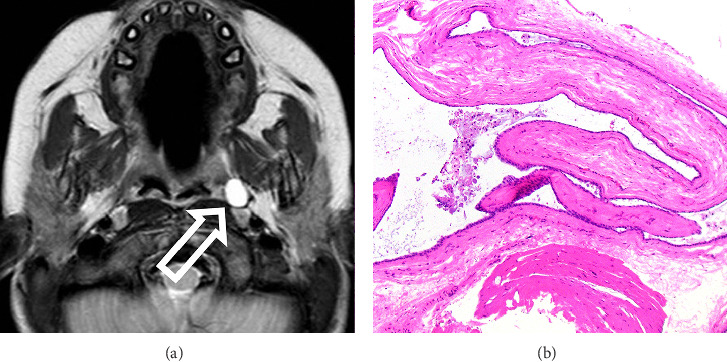 Figure 1
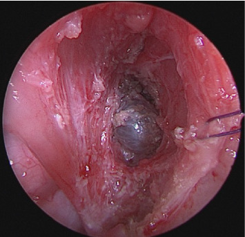 Figure 2
Peer Reviews
No public reviews on file for this paper yet. If you reviewed it on a platform where reviews are public (OpenReview, ICLR, NeurIPS, ICML), you can paste yours below so the community can read it here.
Videos
No videos yet. Explain this paper in a talk, walkthrough, or lecture? Add one.
Taxonomy
TopicsHead and Neck Anomalies · Salivary Gland Tumors Diagnosis and Treatment · Teratomas and Epidermoid Cysts
1. Introduction
Parapharyngeal space masses are rare, typically benign, and range from glioneuronal heterotopias to sarcomas. They are especially uncommon in pediatric patients, with recent studies, such as Starek et al., documenting only 20 cases, while an independent literature review for this report identified 31 [1]. Pediatric presentations vary, including odynophagia or simply incidental findings on imaging [1]. Due to their location, these masses can significantly impact swallowing and respiration, affecting quality of life as the child develops. Given the limited number of reported cases, no standardized approach to characterization or treatment has been established [1]. Given their potential to impair quality of life, identifying the most effective treatment strategies is crucial. This report discusses a case of a benign simple cyst in the left parapharyngeal space of a 32-month-old female, successfully treated with gross resection.
2. Case Presentation
A 32-month-old female was referred to the Pediatric Otolaryngology clinic due to an incidentally identified parapharyngeal space mass on magnetic resonance (MR) imaging. Cervical imaging was previously obtained due to an exam finding of kyphosis, which demonstrated congenital vertebral fusion of L3-L4. The MR also revealed a T2 hyperintense lesion measuring 9 mm in diameter within the left parapharyngeal space (Figure 1). The patient had no symptoms of dysphagia, snoring, or respiratory difficulty. There was a history of congenital vertebral fusion in the patient's maternal grandmother, but the remainder of the family history was unremarkable.
Oropharyngeal examination and flexible fiberoptic nasopharyngoscopy were within normal limits. The cervical exam was negative for masses or lymphadenopathy.
Options of serial MR imaging for surveillance of the parapharyngeal space lesion versus surgical excision were offered to the family. Surgery was selected, and the team proceeded with a unilateral left-sided extracapsular tonsillectomy and transoral parapharyngeal space exploration for complete mass excision.
3. Discussion
The parapharyngeal space is defined by the skull base superiorly, hyoid bone inferiorly, parotid and masticator spaces laterally, and the deep cervical fascia investing the pharyngeal constrictor muscle medially. The space can be further categorized into prestyloid and poststyloid compartments. The prestyloid parapharyngeal space includes fat, minor salivary glands, internal maxillary artery, and mandibular branches of the trigeminal nerve. The poststyloid compartment includes the internal jugular vein, carotid artery, sympathetic chain, and cranial nerves IX through XII. Parapharyngeal space masses are primarily identified in adults and are uncommon in pediatric patients [2]. When identified in pediatric patients, the diagnosis is typically incidental in nature. However, if the mass is large in size, pediatric patients may present with symptoms such as snoring, nasal congestion, and dysphagia [3–6]. Parapharyngeal space masses in adults are reported to be of malignant pathology in approximately 20% of cases, whereas in pediatric patients, malignancy is reported in up to two-thirds of presentations [1, 2]. In the published English-based literature between 1999 and 2025, a total of 31 pediatric parapharyngeal space masses were reported. These were derived from the following pathologies: glioneuronal heterotopia (45.2%), branchial cleft cysts (9.7%), ganglioneuromas (16.1%), fibromas (3.2%), sarcomas (9.7%), adenomas (3.2%), ectomesenchymomas (3.2%), hemangiomas (3.2%), lipoblastomas (3.2%), and schwannomas (3.2%) [1–10]. The mean age at the time of diagnosis for parapharyngeal space masses in pediatric patients was reported to be 3.7 years [1–10].
MR imaging is the ideal imaging modality used in evaluating and characterizing parapharyngeal space masses [6]. The MR findings in the presented case demonstrated a T2 hyperintense, T1 hypointense, and nonenhancing prestyloid parapharyngeal space lesion located just deep to the superior pharyngeal constrictor muscle adjacent to the palatine tonsil (Figure 1). The T2 hyperintense features of the mass in this case correlated with a benign simple cyst, lined by short cuboidal epithelium with abundant extravasated mucin, as confirmed by pathologic analysis (Figure 1). Based on a review of the literature, the presented case is the only description of a simple benign cyst of the parapharyngeal space in a pediatric patient. Since the prestyloid compartment includes minor salivary glands, the origin of this mass may be salivary in nature; however, no definitive salivary architecture was identified in the histopathologic analysis.
Parapharyngeal space masses present a unique set of treatment challenges that necessitate careful consideration of the size, specific location, and relationship of the mass to surrounding structures. Depending on a patient's presentation, exam and imaging characteristics, observation with interval imaging, biopsy, and complete mass excision can all be considered as treatment options. Since up to two-thirds of pediatric parapharyngeal masses can represent a malignant diagnosis, it is important to follow these lesions and have a low threshold for biopsy and/or removal [3]. When surgery is selected, either transoral or transcervical approaches can be utilized to access the parapharyngeal space [2]. In the case of the presented patient, the small size and location in the prestyloid parapharyngeal space, just deep to the pharyngeal constrictor muscle, allowed for complete removal via a transoral approach. Following transoral extracapsular tonsillectomy, the pharyngeal constrictor muscle was carefully transgressed with subsequent access to the prestyloid parapharyngeal space and localization of the mass (Figure 2). The mass was completely excised via this approach with an uncomplicated recovery. Postoperatively, there was no evidence of recurrence at 6 months following the surgical excision.
4. Conclusion
This case uniquely demonstrates an incidentally discovered prestyloid parapharyngeal space mass in a pediatric patient that was completely excised transorally. Although unique tumor pathologies are known to occur in the parapharyngeal space, this report reveals a benign simple cyst and represents a diagnosis that should be considered when encountering T2 hyperintense parapharyngeal space lesions on MR imaging.
The reference list from the paper itself. Each links out to its DOI / PubMed record.
- 1Stárek I. Mihál V. Novák Z. PospísilováD. Vomácka J. Vokurka J. Pediatric Tumors of the Parapharyngeal Space. Three Case Reports and a Literature Review International Journal of Pediatric Otorhinolaryngology 200468560160610.1016/j.ijporl.2003.12.0072-s 2.0-184281567415081237 · doi ↗ · pubmed ↗
- 2Olsen K. D. Mc Caffrey T. V. Tumors and Surgery of the Parapharyngeal Space Otolaryngol Head Neck Surg 1995112510.1016/s 0194-5998(05)80036-12-s 2.0-84970663511 · doi ↗
- 3Fanous A. Couloigner V. Gorphe P. Unusual Presentation of a First Branchial Cleft Cyst Associated With an Abnormal Bony Canal: A Case Report Journal of Otolaryngology-Head & Neck Surgery 2020491 p. 3210.1186/s 40463-020-00426-5PMC 726079532471510 · doi ↗ · pubmed ↗
- 4Suryanarayana K. V. Balakrishnan R. Rao L. Rahim T. A. Parapharyngeal Space Mesenchymal Chondrosarcoma in Childhood International Journal of Pediatric Otorhinolaryngology 1999501697210.1016/s 0165-5876(99)00209-82-s 2.0-003283239710596890 · doi ↗ · pubmed ↗
- 5Xie S. Liu W. Xiang Y. Dai Y. Ren J. A Huge Nodular Fasciitis in Parapharygneal Space in a 7-Year-Old Girl: A Case Report and Review of Literature International Journal of Clinical and Experimental Pathology 20147129023902725674281 PMC 4313988 · pubmed ↗
- 6Zheng Z. Jordan A. C. Hackett A. M. Chai R. L. Pediatric Desmoid Fibromatosis of the Parapharyngeal Space: A Case Report and Review of Literature American Journal of Otolaryngology 201637437237510.1016/j.amjoto.2016.02.0032-s 2.0-8496207451527040413 · doi ↗ · pubmed ↗
- 7Alahmad M. Alkheder A. Alassaf A. Isolated Horner Syndrome as the Sole Presentation of Pediatric Parapharyngeal Rhabdomyosarcoma Ear, Nose & Throat Journal 202510.1177/0145561325135273240563248 · doi ↗ · pubmed ↗
- 8Chen D. Dedhia K. Ozolek J. Mehta D. Case Series of Congenital Heterotopic Neuroglial Tissue in the Parapharyngeal Space International Journal of Pediatric Otorhinolaryngology 201686778110.1016/j.ijporl.2016.04.0262-s 2.0-8496633966327260585 · doi ↗ · pubmed ↗