Bronchoscopic ICG-guided thoracoscopic segmentectomy for removal of an aspirated dental prosthesis in an elderly patient
Eitetsu Koh, Yasuo Sekine, Fumihiro Ishibashi

TL;DR
A novel bronchoscopic ICG-guided technique was used to remove a dental prosthesis from an elderly patient's lung, preserving lung function.
Contribution
First reported use of bronchial ICG-guided segmentectomy for foreign body removal in an elderly patient.
Findings
ICG-guided thoracoscopic segmentectomy successfully removed the foreign body from the right lower lobe.
The technique allowed accurate resection with maximal preservation of lung function.
The patient was discharged without complications on postoperative day 7.
Abstract
An 82-year-old male presented with persistent cough and recurrent pneumonia. Computed tomography revealed a high-density foreign body in the right lower lobe posterior basal segment (S10), later identified as a dental prosthesis. Bronchoscopic removal failed due to severe granulation and impaction. Because of low pulmonary function (FEV1: 920 ml), a lung-preserving procedure was selected. We performed thoracoscopic segmentectomy of S10, guided by bronchoscopic injection of indocyanine green (ICG) into B10a and B10b+c. Intraoperative near-infrared imaging clearly delineated the intersegmental plane. The foreign body was successfully removed, and the patient was discharged on postoperative day 7 without complications. This is the first reported case of bronchial ICG-guided anatomical segmentectomy for foreign body removal in an elderly patient. The technique allowed accurate resection…
Genes, proteins, chemicals, diseases, species, mutations and cell lines named across the full text — each resolved to its canonical identifier and authoritative record.
Click any figure to enlarge with its caption.
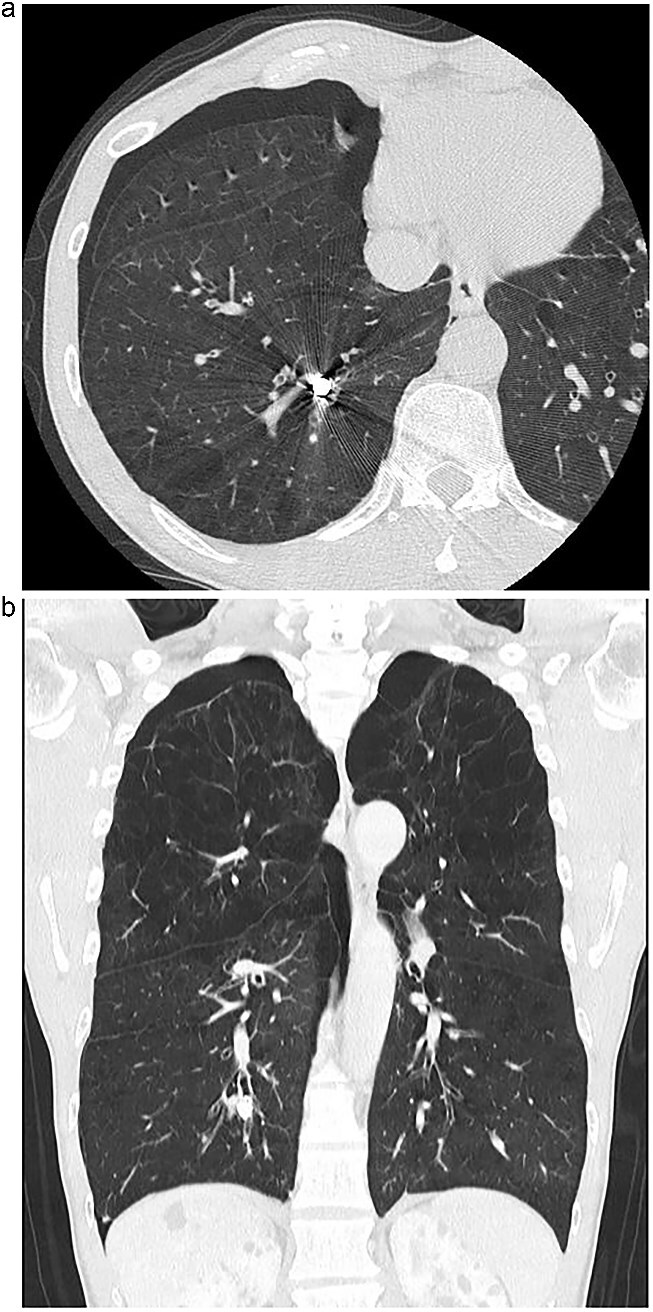 Figure 1
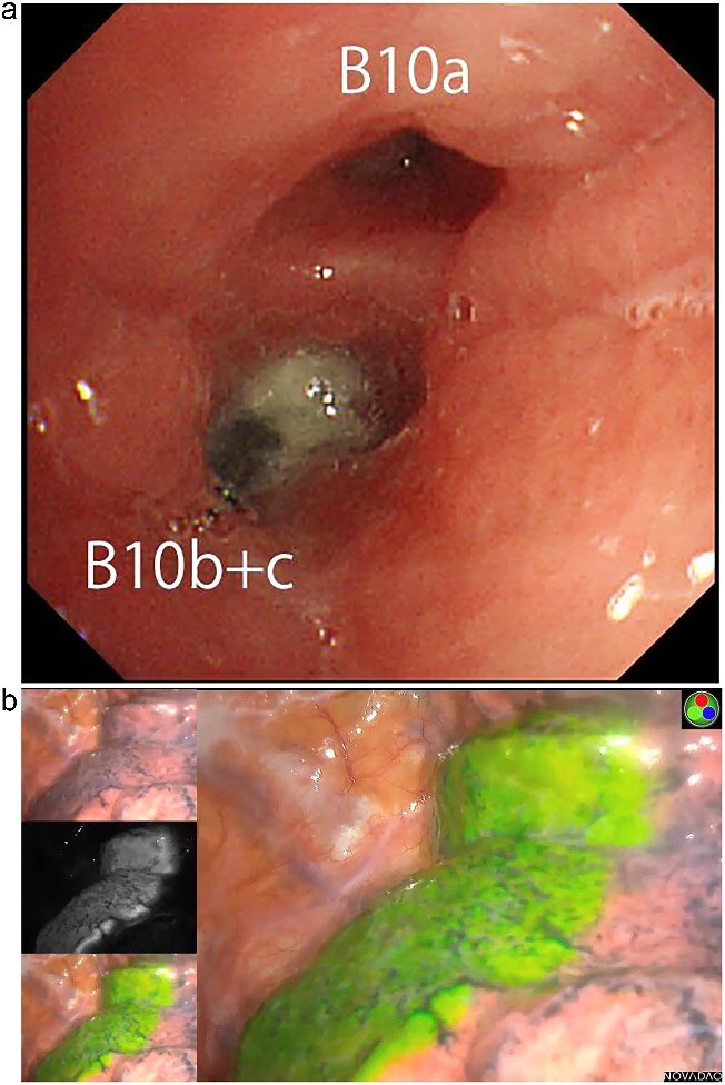 Figure 2
Peer Reviews
No public reviews on file for this paper yet. If you reviewed it on a platform where reviews are public (OpenReview, ICLR, NeurIPS, ICML), you can paste yours below so the community can read it here.
Videos
No videos yet. Explain this paper in a talk, walkthrough, or lecture? Add one.
Taxonomy
TopicsForeign Body Medical Cases · Trauma Management and Diagnosis · Tracheal and airway disorders
Introduction
Foreign body aspiration in the elderly is an uncommon but potentially serious clinical condition. While most cases are managed via bronchoscopy, certain impacted, or chronic foreign bodies may require surgical intervention. In such patients, particularly those with compromised pulmonary function, lung-preserving surgical techniques are preferred. Recent advances in fluorescence-guided surgery using indocyanine green (ICG) have enabled precise intersegmental demarcation during anatomical segmentectomy. Although commonly used in pulmonary oncology, its application for benign conditions, such as foreign body removal, is rarely reported.
Case report
An 82-year-old man presented with chronic cough and a history of repeated right-sided pneumonia. Chest computed tomography (CT) showed a high-density object located in the posterior basal segment (S10) of the right lower lobe (Fig. 1a and b). Bronchoscopy revealed a dental prosthesis impacted in the B10b+c bronchus with surrounding granulation tissue (Fig. 2a). Removal via flexible bronchoscopy was unsuccessful due to firm impaction.
(a) Axial chest CT showing a high-density foreign body in the right S10 bronchus. (b) Coronal CT confirming the position of the dental prosthesis in the posterior basal segment.
(a) Bronchoscopic image showing impacted dental prosthesis in the B10b+c bronchus. (b) Intraoperative near-infrared fluorescence image visualizing the intersegmental plane after ICG bronchial injection.
Pulmonary function tests showed reduced lung capacity (FEV1: 920 ml), making lobectomy undesirable. We therefore planned a thoracoscopic posterior basal segmentectomy (S10) to remove the prosthesis while preserving lung volume. To achieve accurate segmental resection, we adopted the bronchial ICG injection method, as previously described by Sekine et al. [1, 2]. ICG (25 mg diluted in 20 ml saline) was selectively injected into B10a and B10b+c via bronchoscopy. Near-infrared thoracoscopy was used to visualize the intersegmental plane clearly (Fig. 2b), and anatomical S10 segmentectomy was performed via thoracoscopy. The impacted dental prosthesis was successfully retrieved.
The patient’s postoperative course was uneventful. He was discharged on postoperative day 7. No complications occurred during the 2-year follow-up period, and no recurrence of pneumonia or respiratory symptoms was observed.
Discussion
Foreign body aspiration in the elderly may go unnoticed for extended periods due to non-specific symptoms. While bronchoscopy is the first-line approach, chronic impaction and mucosal overgrowth can necessitate surgical retrieval. In this case, the foreign body could not be removed bronchoscopically due to granulation and impaction.
Given the patient’s limited pulmonary reserve, a minimally invasive and lung-preserving approach was critical. Segmentectomy allows for targeted resection with maximal preservation of lung parenchyma. The bronchial ICG injection technique offers a simple and reliable method for intersegmental plane identification. While intravenous ICG has been widely used, selective bronchial ICG instillation provides superior precision, particularly in patients with abnormal perfusion or prior inflammation.
This case demonstrates that bronchial ICG-guided segmentectomy is feasible and effective for non-oncologic indications, such as foreign body removal. It expands the clinical utility of this technique and supports its application in benign thoracic conditions, particularly in elderly or high-risk patients.
Conclusion
Bronchial ICG-guided thoracoscopic segmentectomy enabled safe and effective removal of an aspirated dental prosthesis in a high-risk elderly patient. This technique may represent a valuable surgical option in similar cases, where bronchoscopy fails and parenchymal preservation is essential.
The reference list from the paper itself. Each links out to its DOI / PubMed record.
- 1Sekine Y, Ko E, Oishi H., et al. A novel method of visualizing the intersegmental plane using bronchoscopic instillation of indocyanine green. J Thorac Cardiovasc Surg 2022;163:e 25–7. 10.1016/j.jtcvs.2021.06.036 · doi ↗
- 2Foltran F, Ballali S, Rodriguez H., et al. Foreign bodies in the airways: a meta-analysis of published papers. Int J Pediatr Otorhinolaryngol 2012;76:S 12–9. 10.1016/j.ijporl.2012.02.00422333317 · doi ↗ · pubmed ↗