Imaging Anatomy of the Ankle in Normal and Pathological States: A Clinically Focused Pictorial Review
Omar González-Gutiérrez, Ernesto Roldan-Valadez, Mauricio Molina-Gonzalez, Martin-Romo Garcia-de-Bustamante, Luis-Octavio López-Montoya, Andrea-Paola Gonzalez-Trejo, Jesica Alvarez-Hernández, Samaria Perez-Galindo

TL;DR
This review explains ankle anatomy and imaging techniques for normal and injured ankles to improve diagnosis and treatment.
Contribution
The paper provides a clinically focused, imaging-based review of ankle anatomy and pathology with practical diagnostic correlations.
Findings
Radiographs, ultrasound, CT, and MRI each have specific roles in assessing ankle anatomy and pathology.
Emerging imaging techniques like weight-bearing CT and high-resolution MRI improve detection of subtle injuries.
Combining imaging anatomy with clinical patterns enhances diagnostic accuracy and treatment decisions.
Abstract
This narrative pictorial review presents the imaging anatomy of the ankle in normal and pathologic states and emphasizes practical, clinically oriented correlations. The ankle is a compound synovial joint complex in which osseous architecture, ligamentous systems (lateral collateral, deltoid, and syndesmotic), and tendon compartments interact within a compact space to provide stability and motion during gait and sport. This anatomical complexity underlies a broad spectrum of traumatic and degenerative conditions, including lateral ankle sprains, syndesmotic injury, osteochondral lesions of the talus, tendinopathy/rupture, and post-traumatic osteoarthritis, that are highly prevalent in clinical practice. Normal imaging anatomy provides the baseline for detecting disease: radiographs remain indispensable for initial assessment, ultrasound offers dynamic tendon-ligament evaluation, CT…
Genes, proteins, chemicals, diseases, species, mutations and cell lines named across the full text — each resolved to its canonical identifier and authoritative record.
Click any figure to enlarge with its caption.
 Figure 1
 Figure 2
 Figure 3
 Figure 4
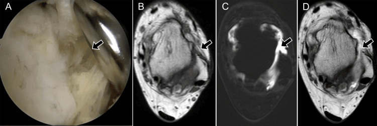 Figure 5
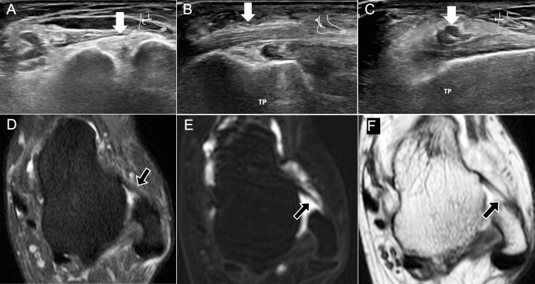 Figure 6
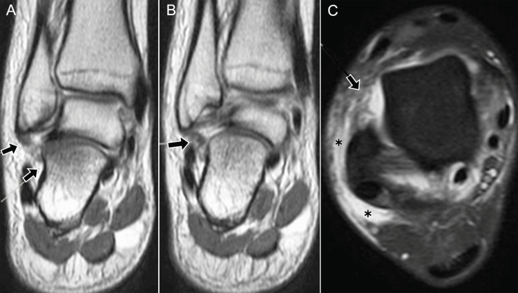 Figure 7
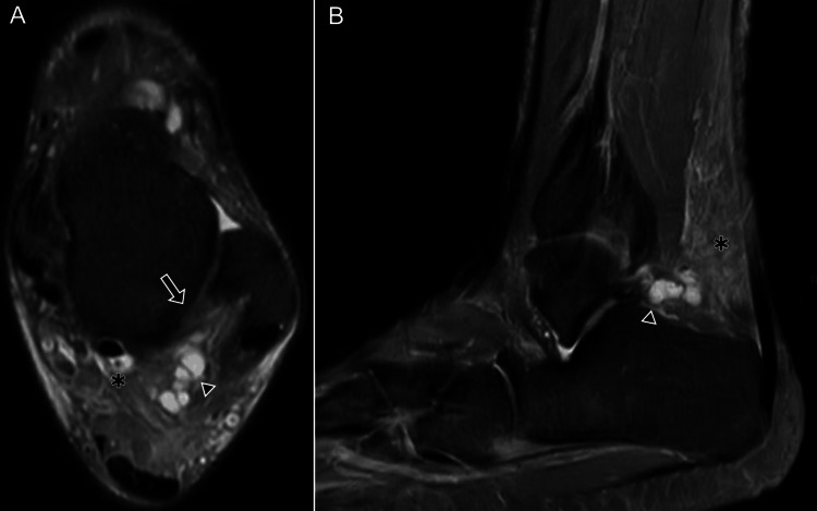 Figure 8
 Figure 9
 Figure 10
 Figure 11
 Figure 12
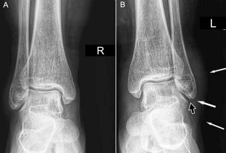 Figure 13
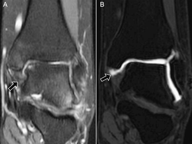 Figure 14
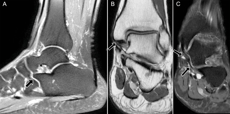 Figure 15
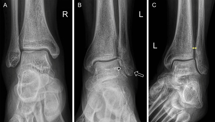 Figure 16
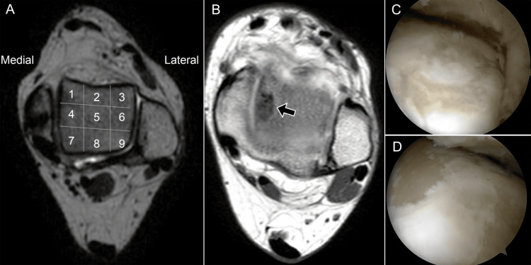 Figure 17
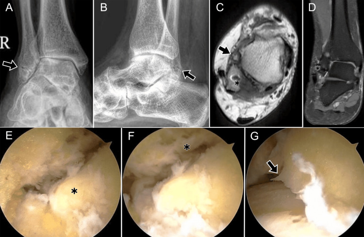 Figure 18
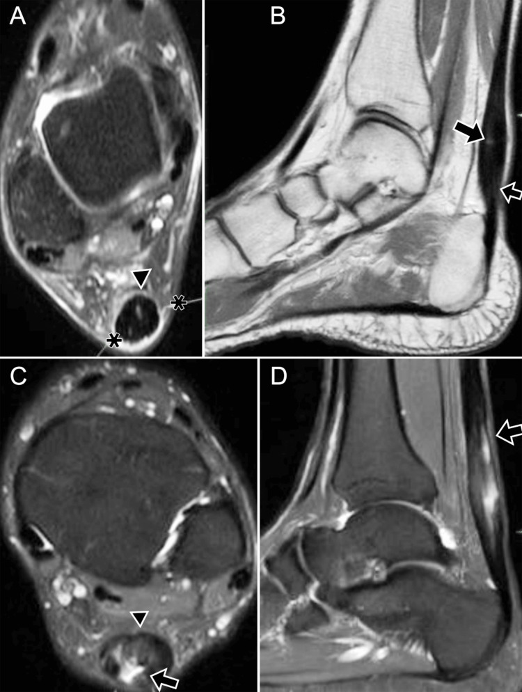 Figure 19
Peer Reviews
No public reviews on file for this paper yet. If you reviewed it on a platform where reviews are public (OpenReview, ICLR, NeurIPS, ICML), you can paste yours below so the community can read it here.
Videos
No videos yet. Explain this paper in a talk, walkthrough, or lecture? Add one.
Taxonomy
TopicsFoot and Ankle Surgery · Musculoskeletal synovial abnormalities and treatments · Tendon Structure and Treatment