Photophysical image analysis for sCMOS cameras: Noise modelling and estimation of background parameters in fluorescence-microscopy images
Dibyajyoti Mohanta, Radhika Nambannor Kunnath, Erik Clarkson, Albertas Dvirnas, Fredrik Westerlund, Tobias Ambjörnsson, Hafiz Muhammad Umer Farooqi, Hafiz Muhammad Umer Farooqi, Hafiz Muhammad Umer Farooqi

TL;DR
This paper introduces a new method to estimate background photon levels in fluorescence microscopy images using sCMOS cameras, improving image analysis accuracy.
Contribution
A novel probabilistic noise modeling approach for sCMOS cameras to estimate the Poisson parameter λbg directly from images.
Findings
The method estimates λbg using a chi-square test and truncated fit technique with strong agreement between sCMOS and EMCCD cameras for low to moderate exposure images.
The approach incorporates Poisson-distributed photon shot noise and Tukey-Lambda read noise modeling for accurate background estimation.
Publicly available software enables photophysical image analysis for sCMOS systems.
Abstract
Fluorescence microscopy is an effective tool for imaging biological samples, yet captured images often contain noises, including photon shot noise and camera read noise. To analyze biological samples accurately, separating background pixels from signal pixels is crucial. This would ideally be guided by the knowledge of a parameter called the Poisson parameter, λbg, representing the mean number of photons collected in a background pixel (for the case when quantum efficiency = 1 and the dark current is negligible). This study introduces a method for estimating λbg, from an image which contains both background and signal pixels, using probabilistic noise modeling for an sCMOS camera. The approach incorporates Poisson-distributed photon shot noise and sCMOS camera read noise modelled with a Tukey-Lambda distribution. We apply a chi-square test and a truncated fit technique to estimate λbg…
Genes, proteins, chemicals, diseases, species, mutations and cell lines named across the full text — each resolved to its canonical identifier and authoritative record.
Click any figure to enlarge with its caption.
 Figure 1
 Figure 2
 Figure 3
 Figure 4
 Figure 5
 Figure 6
 Figure 7
 Figure 8
 Figure 9
 Figure 10
 Figure 11
 Figure 12
 Figure 13
 Figure 14
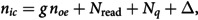 Figure 15
 Figure 16
 Figure 17
 Figure 18
 Figure 19
 Figure 20
 Figure 21
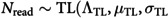 Figure 22
 Figure 23
 Figure 24
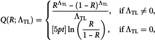 Figure 25
 Figure 26
 Figure 27
 Figure 28
 Figure 29
 Figure 30
 Figure 31
 Figure 32
 Figure 33
 Figure 34
 Figure 35
 Figure 36
 Figure 37
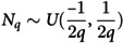 Figure 38
 Figure 39
 Figure 40
 Figure 41
 Figure 42
 Figure 43
 Figure 44
 Figure 45
 Figure 46
 Figure 47
 Figure 48
 Figure 49
 Figure 50
Peer Reviews
No public reviews on file for this paper yet. If you reviewed it on a platform where reviews are public (OpenReview, ICLR, NeurIPS, ICML), you can paste yours below so the community can read it here.
Videos
No videos yet. Explain this paper in a talk, walkthrough, or lecture? Add one.
Taxonomy
TopicsAdvanced Fluorescence Microscopy Techniques · CCD and CMOS Imaging Sensors · Cell Image Analysis Techniques