Sudden deaths of two dogs in a breeding colony associated with enteropathogenic Escherichia coli and necrotoxigenic Escherichia coli
Maho Okumura, Nathan Helgert, Katharine Tuohy, Donna J. Kelly, Stephen D. Cole

TL;DR
Two young dogs suddenly died from a co-infection involving specific strains of Escherichia coli, revealed through whole-genome sequencing.
Contribution
The use of whole-genome sequencing identified a novel co-infection involving EPEC and ExP-NTEC in sudden canine deaths.
Findings
Whole-genome sequencing identified ST5683 and ST127 E. coli strains with distinct virulence factors in deceased pups.
Post-mortem findings included bronchopneumonia, hepatic lipidosis, and gastrointestinal inflammation linked to the E. coli co-infection.
The cases highlight the importance of advanced diagnostics like WGS in understanding complex E. coli infections in neonatal dogs.
Abstract
This case report describes the peracute deaths of two healthy pups in a breeding facility associated with enteropathogenic Escherichia coli (EPEC) and a distinct strain of necrotoxigenic E. coli (ExP-NTEC) co-infection. When E. coli is recovered from post-mortem samples, the role of the organism can be challenging to elucidate. In this case, the use of whole-genome sequencing (WGS) uncovered the virulence factors associated with isolates from both cases and a molecular epidemiological connection between the two deaths. A 7-week-old male and an 8-week-old female intact mixed-breed dogs were found deceased, shortly after routine examination with no preceding clinical signs. Post-mortem examination revealed multiple organ abnormalities, including bronchopneumonia with alveolar collapse, hepatic lipidosis, gastrointestinal inflammation, and bacterial colonization in the lungs and…
Genes, proteins, chemicals, diseases, species, mutations and cell lines named across the full text — each resolved to its canonical identifier and authoritative record.
Click any figure to enlarge with its caption.
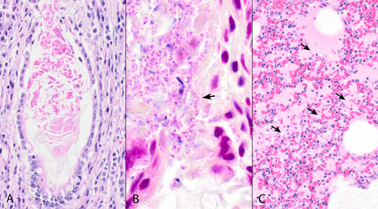 Fig 1
Fig 1| Patient | Source | Other organisms identified | ||||
|---|---|---|---|---|---|---|
| Male pup | Intestine | Beta hemolytic, irregular, dull, spreading, and gray colonies (1+) | Lactose fermenter | ST5683 | O115:H25 | |
| Round, raised, glistening, and gray colonies (1+) | Non-lactose fermenter | ST127 | O6:H31 | |||
| Female pup | Lung | Beta hemolytic, irregular, dull, spreading, and gray colonies | Lactose fermenter | ST5683 | O115:H25 | |
| Round, raised, glistening, and gray colonies | Non-lactose fermenter | ST127 | O6:H31 | |||
| Intestine | Beta hemolytic, irregular, dull, spreading, and gray colonies | Lactose fermenter | ST5683 | O115:H25 | ||
| Round, raised, glistening, and gray colonies | Non-lactose fermenter | ST127 | O6:H31 |
| Virulence factor | % Identity | Function |
|---|---|---|
| ST127 | ||
| | 100 | Cytotoxic necrotizing factor 1 |
| | 100 | Type 1 fimbriae |
| | 100 | Polysialic acid transport protein, group 2 capsule |
| | 100 | Outer membrane protein complement resistance |
| | 99.72 | Tellurium ion resistance protein |
| | 95.21–97.68 | YHD fimbrial cluster |
| ST5683 | ||
| | 100 | Bundle-forming protein subunit |
| | 100 | Type I fimbriae |
| | 99.89 | Intimin gene variant |
| | 99.86 | Outer membrane protein complement resistance |
| | 98.97 | Heat-stable enterotoxin EAST-1 |
Peer Reviews
No public reviews on file for this paper yet. If you reviewed it on a platform where reviews are public (OpenReview, ICLR, NeurIPS, ICML), you can paste yours below so the community can read it here.
Videos
No videos yet. Explain this paper in a talk, walkthrough, or lecture? Add one.
Taxonomy
TopicsEscherichia coli research studies · Clostridium difficile and Clostridium perfringens research · Antibiotic Resistance in Bacteria
INTRODUCTION
Escherichia coli is an important gram-negative bacterium that is a normal inhabitant of many species’ gastrointestinal systems. Although many E. coli strains are not considered pathogenic, some lineages are associated with a wide variety of intestinal and extraintestinal diseases in humans and animals. Intestinal pathogenic E. coli species have been grouped into several major pathotypes, including enteropathogenic E. coli (EPEC), enterotoxigenic E. coli, enteroinvasive E. coli, and Shiga-toxin-producing E. coli/enterohemorrhagic E. coli (1). These are distinct from extraintestinal pathogenic E. coli (ExPEC), which tend to cause infections outside of the gastrointestinal tracts, including uropathogenic, neonatal meningitis, and necrotoxigenic E. coli (NTEC). Knowledge of specific virulence factors produced by bacterial isolates may enhance our understanding of the potential role isolates play in the clinical manifestation of disease. Whole-genome sequencing (WGS) has emerged as a powerful tool to aid in these investigations. This case report describes the investigation into the sudden deaths of two apparently healthy pups in a breeding facility.
CASE PRESENTATION
Patient information
The animals were one 7-week-old intact male and one 8-week-old intact female mixed-breed dogs from different litters and parents, born 11 weeks apart, and were deemed apparently healthy after a routine veterinary examination. They were bred as models to study congenital disease. Both pups received a combination vaccine for canine distemper virus, canine adenovirus-2, canine parvovirus, and canine parainfluenza virus, as well as routine fenbendazole (50 mg/kg) for deworming at 6 weeks of age.
Clinical findings
The pups were found deceased without premonitory signs, 3 months apart, and without prior symptoms, despite normal health checks hours earlier. All other littermates and dogs in the same facility were apparently healthy. The bitches were also apparently healthy. The pups had ideal body condition scores (5/9) with no other clinical issues reported other than mild loose stool in the female pup. Both pups were negative for canine parvovirus on fecal point-of-care antigen testing (SNAP Parvo Test, IDEXX Veterinary Diagnostics, Westbrooke, ME).
Diagnostic assessment
Gross post-mortem examination
At the time of postmortem examination, the male pup had diffusely white mucous membranes, and the right superficial cervical lymph node was mildly enlarged. The left lung lobes were light pink with subtle rib impressions. The right caudoventral lung was dark red and firm, consistent with regional bronchopneumonia. A representative section of the small intestines was saved in a freezer at −20°C as part of the routine sampling for further investigation. The female pup’s stomach contained green-brown mucoid material, and the small intestines contained a mild amount of opaque white fluid. The lungs were diffusely soft and light red. A representative section of the lung and small intestines was separately saved in a freezer at −20°C. Both dogs had evidence of hepatic lipidosis, while other organs appeared grossly normal.
Histopathology
Samples of major organs were fixed in 10% neutral buffered formalin, routinely embedded in paraffin, sectioned, and stained with hematoxylin and eosin or McDonald Gram stain.
Within the small and large intestines of the male pup were mild multifocal crypt ectasia, lymphoid depletion, and abundant gram-negative short bacilli within the brush border (Fig. 1B). In the lungs, alveoli were collapsed (atelectasis) or contained light eosinophilic fluid, mixed morphology bacteria, squamous epithelial cells, and mild numbers of neutrophils and macrophages (Fig. 1C). In the female pup, the duodenum was infiltrated by mild numbers of lymphocytes and plasma cells, and occasional crypts were ectatic and filled with amorphous eosinophilic material (Fig. 1A), and within the brush border of the jejunum and ileum were numerous gram-negative short bacilli. Alveoli contained neutrophils, mixed morphology bacteria, and protein. Apart from confirmation of hepatic lipidosis, other tissues examined were free from histological abnormalities.
Histopathological findings. (A) Jejunum, female puppy. Occasional crypts were moderately to markedly ectatic and filled with cellular debris (crypt ectasia). Original magnification 400×. Hematoxylin and eosin. (B) Small intestine, male puppy. Within the brush border of enterocytes are numerous gram-negative coccobacilli and short bacilli (arrow). Original magnification 1,000×. McDonald's modified Gram stain. (C) Lung, male puppy. Alveoli contain bacteria (arrows), homogeneous, lightly eosinophilic material, and scant inflammatory cells. Original magnification 400×. Hematoxylin and eosin.
Bacteriology
Swabs of thawed lung and intestines from the female pup and intestines from the male pup were collected after the suspicion of an epidemiological link between the cases was established and submitted for aerobic culture. The swabs were streaked on Tryptic Soy with 5% sheep blood, MacConkey, and Columbia Colistin-Nalidixic Acid agar plates (Remel, Lenexa, KS) and incubated for 24–48 hours at 35°C–37°C in 7% carbon dioxide. The sample from the lung was also streaked on chocolate agar (Remel, Lenexa, KS). All bacterial colonies were identified using matrix-assisted laser desorption ionization time of flight (Sirius 1 Biotyper, Bruker, Billerica, MA). E. coli with two different morphologies was isolated from each swab (for a total of six E. coli isolates) along with other bacteria detailed in Table 1.
Whole-genome sequencing
Six E. coli isolates were sequenced: two intestinal and two pulmonary isolates from the female pup and two intestinal isolates from the male pup. Short-read sequencing was performed on the NovaSeq X Plus sequencer (Illumina). Libraries were prepared using the Illumina DNA Prep Kit and custom IDT 10 bp unique dual indices (280 bp). Demultiplexing, quality control, and adapter trimming were performed using a proprietary Illumina software. Draft genomes were assembled using the default parameters of the Bacterial and Viral Bioinformatics Resource Center (BV-BRC) Comprehensive Genome Analysis pipeline. Assemblies were then analyzed using in silico MLST, SeroTypeFinder, and VirulenceFinder applications (1–11). Raw sequence reads (NCBI Bioproject PRJNA1213789) were processed via the Pathogen Genome Annotation Pipeline (12–14), which includes phylogenetic analysis to identify database isolates within 50 single-nucleotide polymorphisms (SNPs) (15, 16).
The six isolates represented a total of two strains of E. coli: ST127 with serotype O6:H31 and ST5683 with serotype O115:H25. One of each detected strain was found in each specimen. The isolates from all sources were found to be very closely related, with the three ST127 isolates from the two dogs being 1 to 2 SNPs apart and the three ST5683 isolates 0 to 1 SNPs apart. The virulence factors that were detected are summarized in Table 2.
Therapeutic intervention
No clinical concerns were observed in the other animals in the colony; therefore, prophylactic treatment was not administered.
Follow-up and outcomes
There were no additional deaths in the colony following these two cases.
DISCUSSION
This case highlights the sudden deaths of two otherwise healthy pups in a breeding colony associated with EPEC and NTEC infection. In veterinary clinical microbiology, we are often asked to contribute to “herd health” investigations, which often rely on post-mortem examination to characterize deaths; however, specimens collected post-mortem are often non-ideal for bacteriology and may yield difficult-to-interpret results. For example, in this investigation, the two cases were separated by approximately 3 months, which led to the submission of limited frozen specimens. While we acknowledge these specimen limitations and the need for cautious interpretation, E. coli infection was determined to be a likely diagnosis based on clinical history, bacteriology, and pathology results. We added additional evidence by further characterizing the E. coli isolates with WGS to determine relatedness and pathogenic potential of isolates.
We believe it is reasonable to conclude that both puppies had gastrointestinal disease that led to vomiting or regurgitation and subsequent aspiration pneumonia. In both cases, bacterial colonization of the small intestinal brush border was suggestive of EPEC infection. EPEC pathogenesis is characterized by the destruction of the microvilli from intimate adherence to the intestinal epithelium, pedestal formation, and aggregation of polarized actin and other cytoskeleton elements (17). In these cases, EPEC infection was confirmed by WGS identification of EPEC-associated virulence factors (bfpA, eae-e01-epsilon, and tir) in the ST5683 isolates. Our EPEC isolates were serotype O115, which is not one of the 12 serotypes described by the WHO, but E. coli O115 has been previously described as an atypical EPEC (17–21).
The aspiration pneumonia was most likely polymicrobial in nature based on histopathology and bacteriology results, which included isolation of both E. coli and Streptococcus canis, which are commonly associated with aspiration bacterial pneumonia in dogs (21). Specifically, in both dogs, we detected E. coli ST127, which was classified as both ExPEC and NTEC by WGS. ST127 has been previously detected in a young dog presenting with vomiting, lethargy, and labored breathing due to a hemothorax, severe hemorrhagic pneumonia, and hemorrhagic enteritis (22, 23). E. coli ST127 has previously been cultured from the lung and small intestines of dogs, minks, and humans who have succumbed to hemorrhagic and non-hemorrhagic pneumonia and sepsis (24–26). Virulence factors found in the sequenced isolates included fimH, sitA, kpsMII, and traT, consistent with ExPEC and cnf1, consistent with NTEC (27, 28). Cnf1 activates the regulatory Rho, Rac, and Cdc42 GTPases in eukaryotic cells, leading to actin cytoskeletal rearrangement and cellular damage (29).
Given the polymicrobial nature of the cultures, it is not feasible to fully characterize to what degree the E. coli relative to other bacteria contributed to disease pathogenesis. Additionally, it is known that in some mammals, EPEC and ExPEC can asymptomatically colonize the gastrointestinal tract, so we cannot fully exclude the possibility of these bacteria being incidental findings in our investigation when isolated from the gastrointestinal tract (30, 31).
The isolates from both dogs were found to be closely related to each other by SNP analysis, suggesting a close epidemiological link. In animal colony settings, biosecurity measures including quarantine and isolation of new or sick animals, use of personal protective equipment by staff, and strict cleaning and disinfection must be used to prevent the spread of pathogens. The isolated E. coli may be part of the normal gastrointestinal flora of an adult dog, which may pose unique challenges for animal care staff when compared to overt pathogens to combat. As these cases were separated by time, further biosecurity interventions were not pursued; however, if future cases were to occur, this investigation has provided valuable information on the potential role of E. coli in these deaths.
The reference list from the paper itself. Each links out to its DOI / PubMed record.
- 1Drolet R, Fairbrother JM, Harel J, Hélie P. 1994. Attaching and effacing and enterotoxigenic Escherichia coli associated with enteric colibacillosis in the dog. Can J Vet Res 58:87–92.8004546 PMC 1263671 · pubmed ↗
- 2Larsen MV, Cosentino S, Rasmussen S, Friis C, Hasman H, Marvig RL, Jelsbak L, Sicheritz-Pontén T, Ussery DW, Aarestrup FM, Lund O. 2012. Multilocus sequence typing of total-genome-sequenced bacteria. J Clin Microbiol 50:1355–1361. doi:10.1128/JCM.06094-1122238442 PMC 3318499 · doi ↗ · pubmed ↗
- 3Clausen PTLC, Aarestrup FM, Lund O. 2018. Rapid and precise alignment of raw reads against redundant databases with KMA. BMC Bioinformatics 19:307. doi:10.1186/s 12859-018-2336-630157759 PMC 6116485 · doi ↗ · pubmed ↗
- 4Bartual SG, Seifert H, Hippler C, Luzon MAD, Wisplinghoff H, Rodríguez-Valera F. 2005. Development of a multilocus sequence typing scheme for characterization of clinical isolates of Acinetobacter baumannii. J Clin Microbiol 43:4382–4390. doi:10.1128/JCM.43.9.4382-4390.200516145081 PMC 1234098 · doi ↗ · pubmed ↗
- 5Griffiths D, Fawley W, Kachrimanidou M, Bowden R, Crook DW, Fung R, Golubchik T, Harding RM, Jeffery KJM, Jolley KA, Kirton R, Peto TE, Rees G, Stoesser N, Vaughan A, Walker AS, Young BC, Wilcox M, Dingle KE. 2010. Multilocus sequence typing of Clostridium difficile. J Clin Microbiol 48:770–778. doi:10.1128/JCM.01796-0920042623 PMC 2832416 · doi ↗ · pubmed ↗
- 6Lemee L, Dhalluin A, Pestel-Caron M, Lemeland JF, Pons JL. 2004. Multilocus sequence typing analysis of human and animal Clostridium difficile isolates of various toxigenic types. J Clin Microbiol 42:2609–2617. doi:10.1128/JCM.42.6.2609-2617.200415184441 PMC 427854 · doi ↗ · pubmed ↗
- 7Wirth T, Falush D, Lan R, Colles F, Mensa P, Wieler LH, Karch H, Reeves PR, Maiden MCJ, Ochman H, Achtman M. 2006. Sex and virulence in Escherichia coli: an evolutionary perspective. Mol Microbiol 60:1136–1151. doi:10.1111/j.1365-2958.2006.05172.x 16689791 PMC 1557465 · doi ↗ · pubmed ↗
- 8Jaureguy F, Landraud L, Passet V, Diancourt L, Frapy E, Guigon G, Carbonnelle E, Lortholary O, Clermont O, Denamur E, Picard B, Nassif X, Brisse S. 2008. Phylogenetic and genomic diversity of human bacteremic Escherichia coli strains. BMC Genomics 9:560. doi:10.1186/1471-2164-9-56019036134 PMC 2639426 · doi ↗ · pubmed ↗