Background parenchymal enhancement of the contralateral breast on preoperative contrast-enhanced breast MRI as a potential predictive factor for disease-free survival in triple-negative breast cancer patients
Xiao-Ting Li, Xing Wang, Hai-Tao Zhu, Nan Sun, Hai-Bin Zhu, Liang You, Xiao-Lei Gu, Yao Luo, Zhao-Qing Fan, Ying-Shi Sun

TL;DR
This study suggests that changes in background breast tissue enhancement on MRI after chemotherapy may predict recurrence risk in triple-negative breast cancer patients.
Contribution
The study identifies contralateral BPE changes as a novel predictive factor for disease-free survival in TNBC patients.
Findings
Post-NAC BPE was significantly associated with disease-free survival in a multivariate Cox model.
Patients with BPE changing from high to low had a lower recurrence rate than those changing from low to high.
BPE levels were linked to ADC values and menopausal status after neoadjuvant therapy.
Abstract
Background parenchymal enhancement (BPE) observed on dynamic contrast-enhanced (DCE) MRI of the contralateral breast is considered to be associated with survival outcomes. However, the prognostic significance of BPE in triple-negative breast cancer (TNBC) is unclear. Between March 2017 and June 2019, 76 TNBC patients undergoing neoadjuvant therapy and subsequent surgery were included in the study. All patients underwent DCE MRI before and after neoadjuvant therapy. Radiologists graded BPE as minimum, mild, moderate, and marked. The BPE level was analyzed according to clinicopathological characteristics and MRI findings. Survival analysis was conducted for clinicopathological characteristics and MRI findings according to disease-free survival (DFS). The mean age was 51.29 ± 9.53 years; 46 (60.5%) patients achieved pathological complete response (pCR), and 13 (17.1%) patients developed…
Genes, proteins, chemicals, diseases, species, mutations and cell lines named across the full text — each resolved to its canonical identifier and authoritative record.
Click any figure to enlarge with its caption.
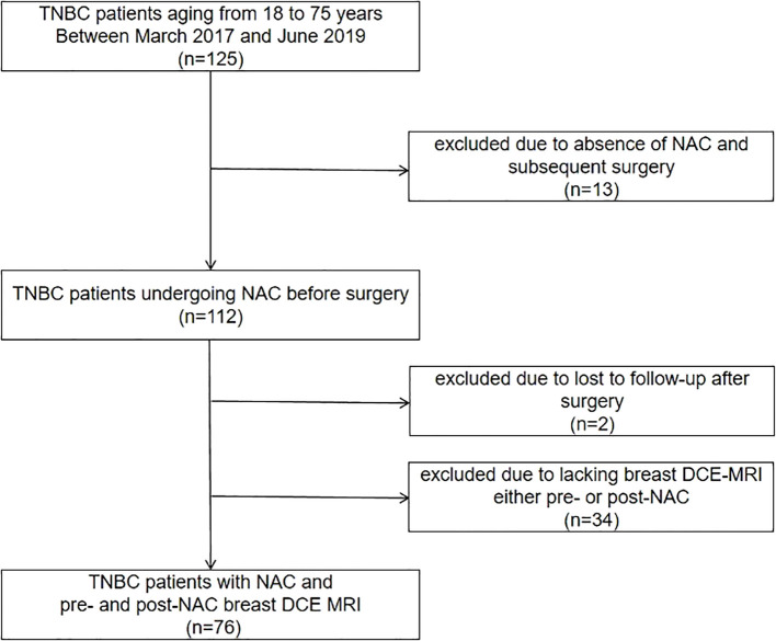 Figure 1
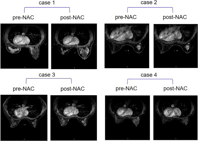 Figure 2
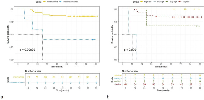 Figure 3
Peer Reviews
No public reviews on file for this paper yet. If you reviewed it on a platform where reviews are public (OpenReview, ICLR, NeurIPS, ICML), you can paste yours below so the community can read it here.
Videos
No videos yet. Explain this paper in a talk, walkthrough, or lecture? Add one.
Taxonomy
TopicsMRI in cancer diagnosis · Breast Cancer Treatment Studies · Medical Imaging Techniques and Applications