Investigation of accessory navicular in local pre-adolescence children: A longitudinal study with 2-year follow-up—KID Locomo Study
Takahide Sasaki, Masatoshi Teraguchi, Kanae Mure, Yoshiki Asai, Yusuke Kido, Makiko Onishi, Kazuyoshi Minamino, Takashi Shimoe, Nobuyuki Miyai, Yukihiro Nakagawa, Hiroshi Hashizume, Hiroshi Yamada

TL;DR
This study tracks the development of accessory navicular in children over two years, revealing how often it fuses and how symptoms resolve.
Contribution
The first longitudinal study to report the natural course of painful accessory navicular in children.
Findings
34.9% of children with accessory navicular showed fusion with the navicular bone after two years.
71.4% of children with painful accessory navicular at baseline experienced resolution of pain after two years.
Female sex and higher BMI were significant risk factors for non-fusion of the accessory navicular.
Abstract
The accessory navicular (AN) is an accessory ossicle located on the medial side of the navicular bone and is often associated with sports-related overuse injuries during adolescence. However, little is known about the natural history of AN. This study aimed to clarify the natural course of AN, including symptomatic cases, in children through a longitudinal epidemiological investigation. Data from the KID Locomo Study, a prospective cohort study aimed at elucidating musculoskeletal disorders in childhood, were used. Of the 834 children recruited in the 2022 baseline survey, 66 children (109 feet) aged 11–13 years with AN were enrolled in this 2-year longitudinal analysis. The presence of AN was assessed using ultrasonography. Data on sex, age, height, weight, presence of pain at the AN site, laterality, flatfoot (based on footprint analysis), and participation in athletic clubs were…
Genes, proteins, chemicals, diseases, species, mutations and cell lines named across the full text — each resolved to its canonical identifier and authoritative record.
Click any figure to enlarge with its caption.
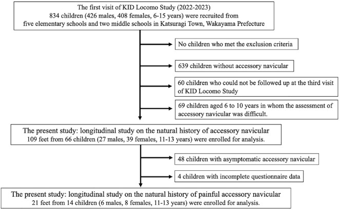 Figure 1
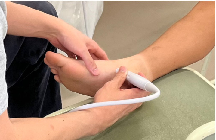 Figure 2
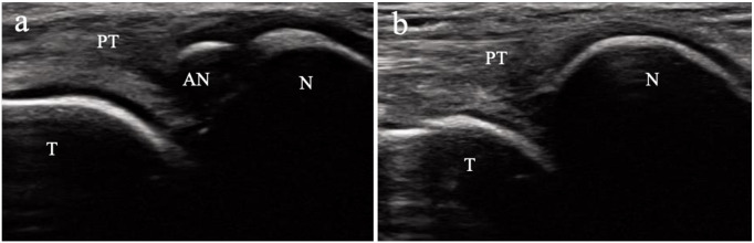 Figure 3
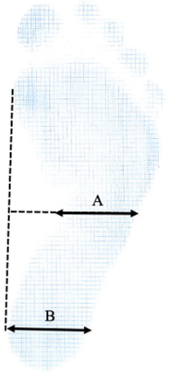 Figure 4
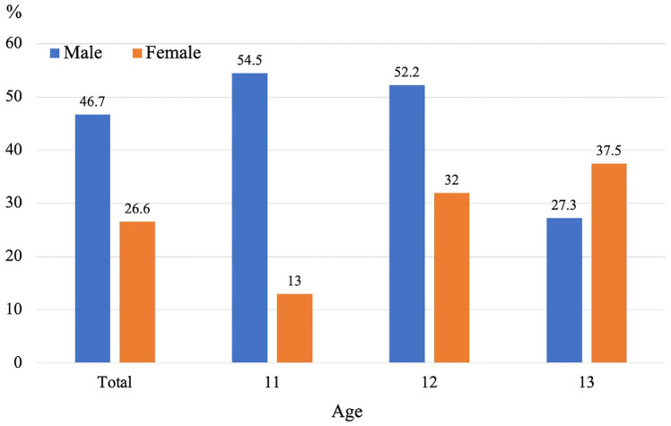 Figure 5
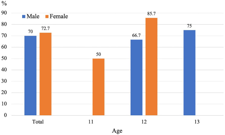 Figure 6
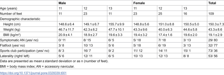 Figure 7
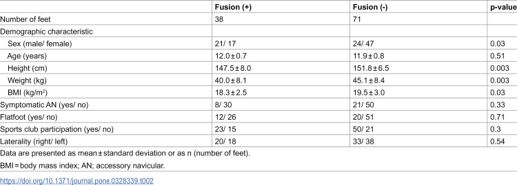 Figure 8
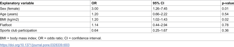 Figure 9
Peer Reviews
No public reviews on file for this paper yet. If you reviewed it on a platform where reviews are public (OpenReview, ICLR, NeurIPS, ICML), you can paste yours below so the community can read it here.
Videos
No videos yet. Explain this paper in a talk, walkthrough, or lecture? Add one.
Taxonomy
TopicsPediatric Pain Management Techniques · Dental Anxiety and Anesthesia Techniques · Ocular Surface and Contact Lens