Bilateral ampiginous choroiditis after COVID-19: a report of two cases
Doğukan Cömerter, Feyza Rümeysa Öz, Eyüp Düzgün

TL;DR
Two patients developed bilateral ampiginous choroiditis after recovering from COVID-19, suggesting a possible link between the virus and eye inflammation.
Contribution
This paper reports two new cases linking post-COVID-19 recovery to ampiginous choroiditis, suggesting a potential immunogenic trigger.
Findings
Two patients with recent COVID-19 infections developed bilateral ampiginous choroiditis.
Multimodal imaging confirmed the diagnosis in both patients.
Treatment with oral steroids and azathioprine was initiated in both cases.
Abstract
Ampiginous choroiditis is a disease that includes features of both acute posterior multifocal placoid pigment epitheliopathy and serpiginous choroiditis. This article presents two patients who were diagnosed with ampiginous choroiditis in our clinic. A 28-year-old female patient presented to our clinic with blurred vision that had persisted for 2 weeks. Her best corrected visual acuity (BCVA) was 20/20. Her medical history included a COVID-19 infection about 1 month before. Fundoscopic examination revealed multiple grayish-yellow lesions with irregular borders and pseudopodial extensions, involving the posterior poles of both eyes diffusely. Fundus findings and multimodal imaging, including optical coherence tomography, fundus autofluorescence imaging, and fundus fluorescein angiography, indicated ampiginous choroiditis. The patient was treated with oral steroids and azathioprine. The…
Genes, proteins, chemicals, diseases, species, mutations and cell lines named across the full text — each resolved to its canonical identifier and authoritative record.
Click any figure to enlarge with its caption.
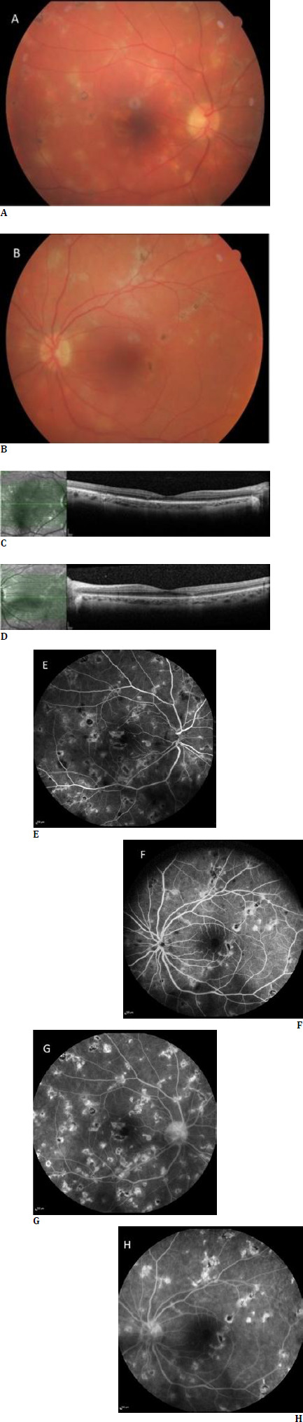 Figure 1
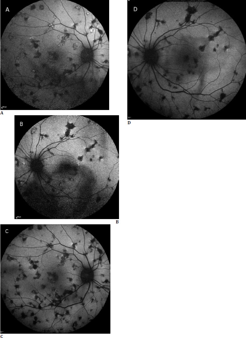 Figure 2
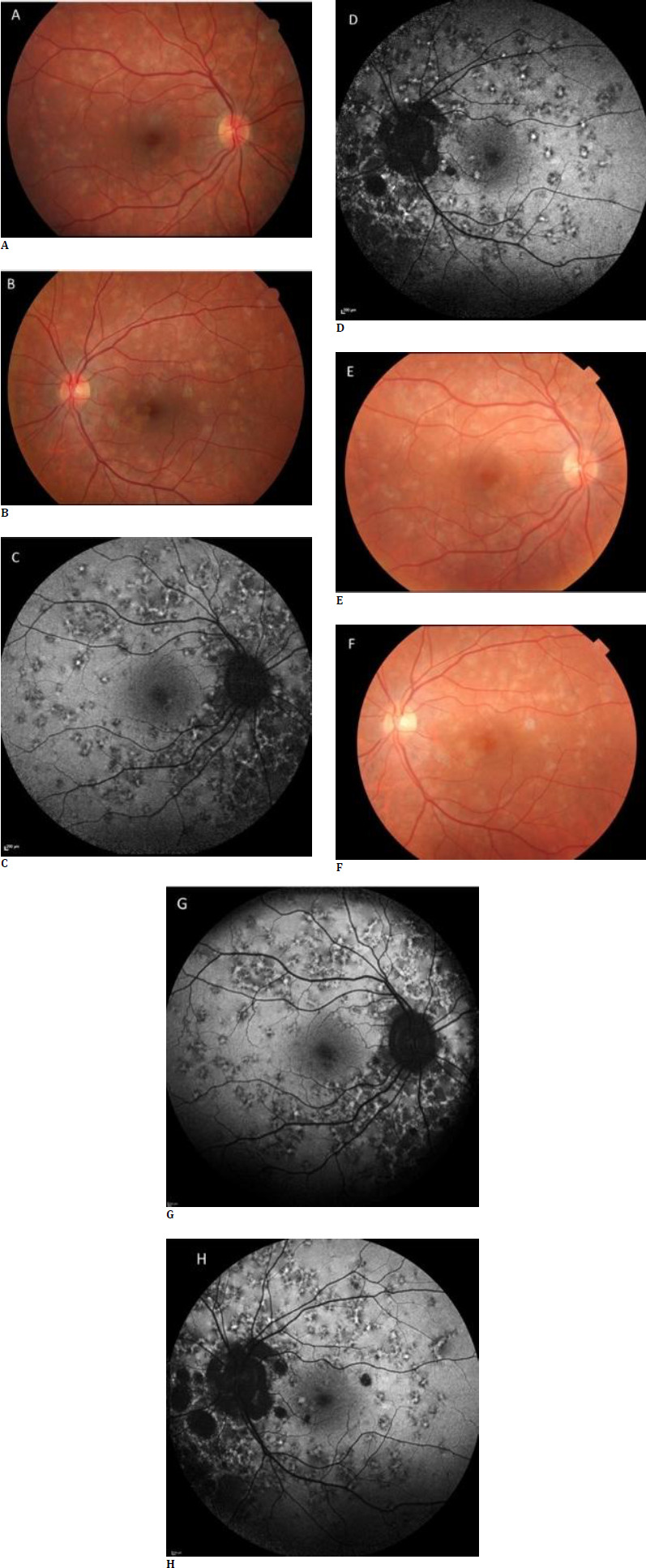 Figure 3
 Figure 4
Peer Reviews
No public reviews on file for this paper yet. If you reviewed it on a platform where reviews are public (OpenReview, ICLR, NeurIPS, ICML), you can paste yours below so the community can read it here.
Videos
No videos yet. Explain this paper in a talk, walkthrough, or lecture? Add one.
Taxonomy
TopicsRetinal and Optic Conditions · Ocular Diseases and Behçet’s Syndrome · Retinal Imaging and Analysis