Penile tourniquet: the Wharton tourniquet
S Nour, GH Lafford, SM Wharton

Abstract
Genes, proteins, chemicals, diseases, species, mutations and cell lines named across the full text — each resolved to its canonical identifier and authoritative record.
Click any figure to enlarge with its caption.
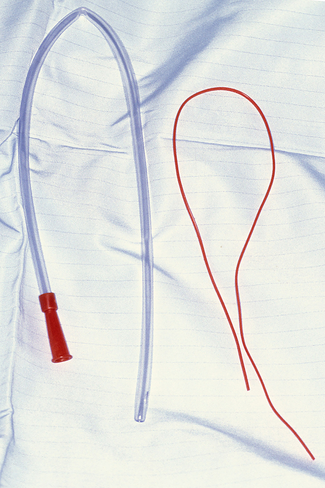 Figure 1
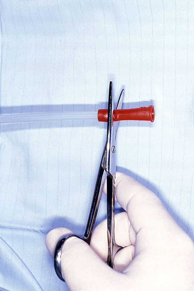 Figure 2
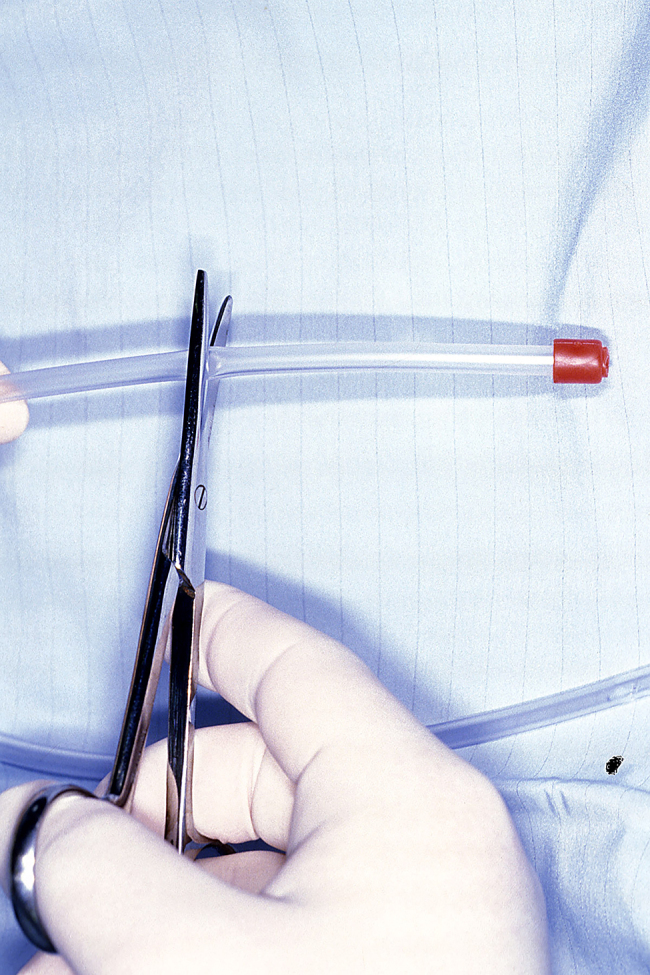 Figure 3
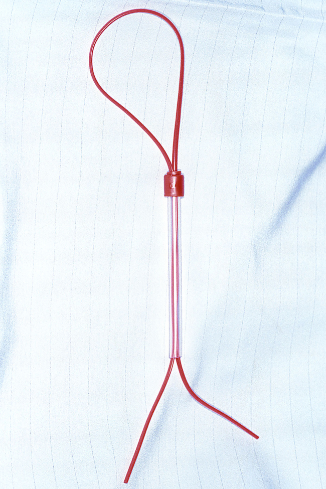 Figure 4
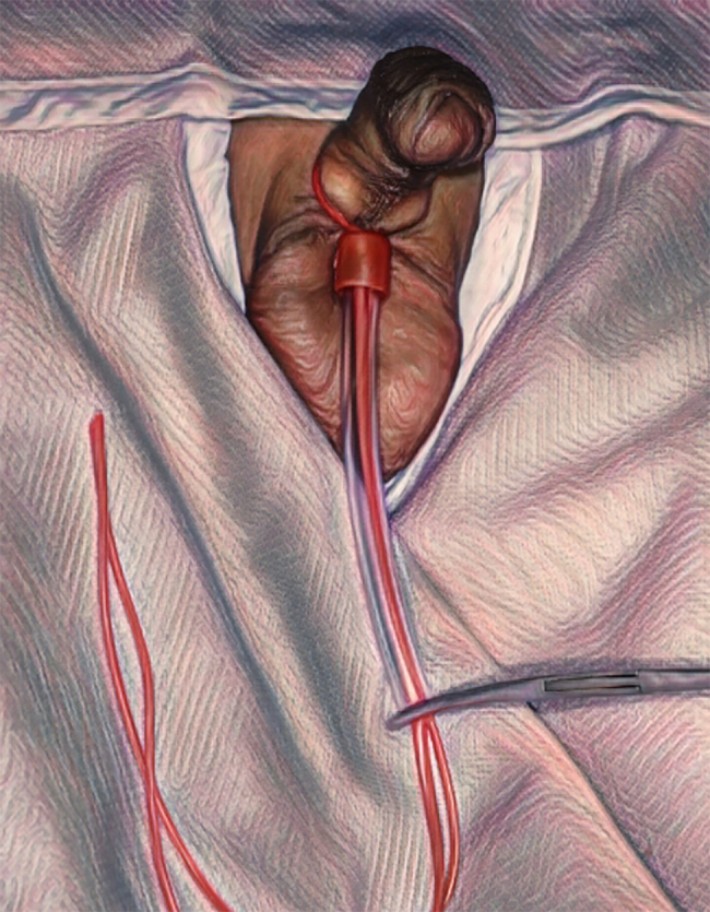 Figure 5
Peer Reviews
No public reviews on file for this paper yet. If you reviewed it on a platform where reviews are public (OpenReview, ICLR, NeurIPS, ICML), you can paste yours below so the community can read it here.
Videos
No videos yet. Explain this paper in a talk, walkthrough, or lecture? Add one.
Taxonomy
TopicsTrauma, Hemostasis, Coagulopathy, Resuscitation · Blood transfusion and management
Introduction
In hypospadias repair surgery, penile tourniquets are used to create a bloodless field, induce an artificial erection, aid in the identification of fistulae and pressure test after closure. Numerous techniques have been described to create the required tourniquet effect. Those include (but are not limited to) the use of red rubber catheters, Penrose tubes, surgical gloves and rubber bands.
Technique
The technique we describe is designed by Mr Simon Wharton. It is used regularly in our Plastic Surgery department and has been adopted in at least two other units. We use a sterile large size 18 Fr suction catheter, a vascular sloop (Figure 1) and an artery clip, all of which are available routinely in the majority of surgical units and trays.
18 Fr suction catheter (left), vascular sloop (right)
The suction tube is cut short at both ends using large Mayo scissors. The end normally attached to suction is shortened to leave a 1cm cuff (Figure 2). The clear end is shortened to about 10cms (Figure 3). The vascular sloop is folded in half to form a loop. A small skin hook is passed down the tube from the red end and catches on to the loop of sloop. The sloop is then drawn through the suction tube to form a loop with two tails at the opposite end (Figure 4).
End usually attached to suction is shortened, leaving a 1cm cuff
Clear end shortened to about 10cms
The vascular sloop is drawn through the suction tube to form a loop with two tails at the opposite end.
The loop is passed over the penis and placed around the base. Both ends of the vascular sloop are stretched to create the required tourniquet effect around the penis and anchored to the shortened end of the suction catheter using an artery clip (Figure 5). This creates a secure clamp. The pressure can be adjusted by increasing or decreasing the tension of the vascular sloop by adjusting where the artery clip sits.
The loop is passed over the penis, stretched at both ends to create a tourniquet effect and secured with an artery clip.
The tourniquet is released by removing the artery clip then suction tube followed by the vascular sloop.
Discussion
Based on our experience this is a safe technique that has been used for well over ten years with no complications or failures.