Longitudinal MRI changes after focal therapy for prostate cancer: cryotherapy vs. microwave tissue coagulation
Nana Kozawa, Kaori Yamada, Bunta Tokuda, Akiko Takahata, Yayoi Iwami, Toshiko Ito-Ihara, Atsuko Fujihara, Takumi Shiraishi, Takashi Ueda, Munehiro Ohashi, Osamu Ukimura, Kei Yamada

TL;DR
This study compares how prostate cancer treatments like cryotherapy and microwave coagulation affect MRI scans over time, showing early differences that eventually converge into similar fibrosis patterns.
Contribution
The study identifies modality-specific MRI patterns and their temporal evolution for cryotherapy and microwave tissue coagulation in prostate cancer treatment.
Findings
Cryotherapy lesions showed T1 hyperintensity, while MTC lesions showed slight hyperintensity on early post-treatment MRI.
Late-stage MRI findings for both treatments converged toward fibrosis, characterized by hypointensity across sequences.
Early rim enhancement was common after both treatments but resolved at different times (23 months for cryotherapy and 41 months for MTC).
Abstract
This study compared the longitudinal changes in multiparametric magnetic resonance imaging (mpMRI) findings following lesion-targeted focal cryotherapy with those after microwave tissue coagulation (MTC) therapy for localized prostate cancer with the aim of determining their modality-specific imaging characteristics and evolution over time. The study included 16 patients (17 procedures) who underwent cryotherapy and 33 patients (34 procedures) who received MTC therapy between March 2017 and February 2024. Serial mpMRI scans were retrospectively reviewed for treatment-induced signal changes on T1-weighted imaging, T2-weighted imaging, diffusion-weighted imaging, and dynamic contrast-enhanced magnetic resonance imaging (MRI). Three radiologists independently reviewed the images, and interobserver agreement was evaluated. Early post-treatment MRI findings indicated distinct…
Genes, proteins, chemicals, diseases, species, mutations and cell lines named across the full text — each resolved to its canonical identifier and authoritative record.
Click any figure to enlarge with its caption.
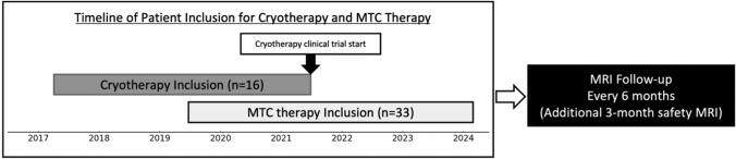 Figure 1
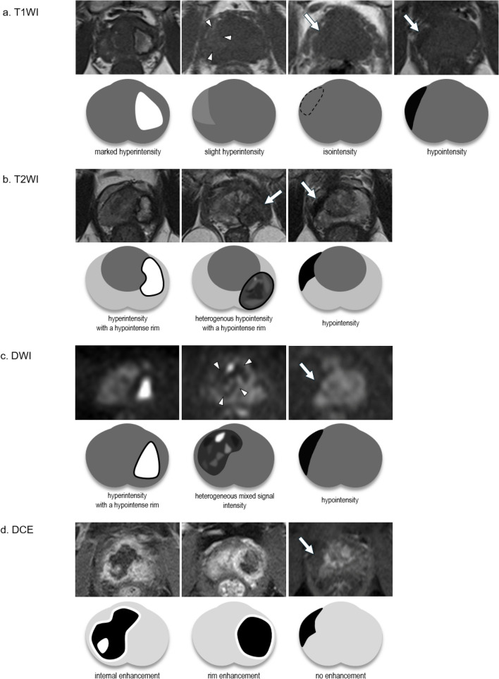 Figure 2
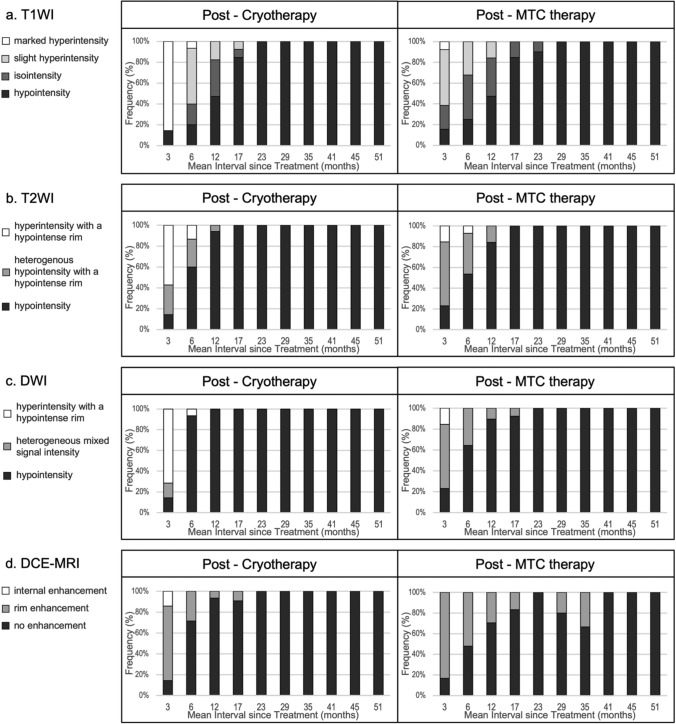 Figure 3
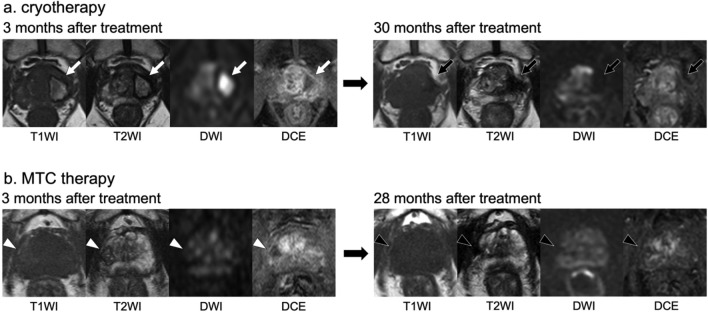 Figure 4
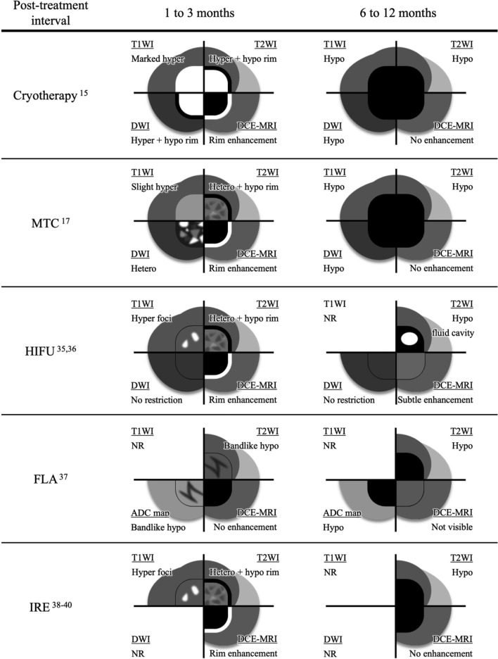 Figure 5
Peer Reviews
No public reviews on file for this paper yet. If you reviewed it on a platform where reviews are public (OpenReview, ICLR, NeurIPS, ICML), you can paste yours below so the community can read it here.
Videos
No videos yet. Explain this paper in a talk, walkthrough, or lecture? Add one.
Taxonomy
TopicsProstate Cancer Diagnosis and Treatment · MRI in cancer diagnosis · Prostate Cancer Treatment and Research