Do eye trackers estimate eyeball rotation? The relationship between tracked eye image feature and estimated saccadic waveform
Marcus Nyström, Diederick C. Niehorster, Roy S. Hessels, Richard Andersson, Marta K. Skrok, Robert Konklewski, Patrycjusz Stremplewski, Maciej Nowakowski, Jakub Lipiński, Szymon Tamborski, Anna Szkulmowska, Maciej Szkulmowski, Ignace T. C. Hooge

TL;DR
This paper investigates how different eye structures affect saccade measurements, showing that signals from the retina differ significantly from those from the pupil or cornea.
Contribution
The study provides a systematic comparison of saccadic waveforms from multiple eye structures, revealing novel differences in saccade parameters.
Findings
Retinal saccades had earlier onsets and smaller amplitudes compared to pupil and P4 signals.
The retinal signal reached peak velocity earlier and stopped faster than other signals.
Saccade parameters vary systematically depending on the tracked eye structure.
Abstract
The eyeball is not rigid and deforms during saccades. As a consequence, the saccade waveform recorded by an eye tracker may depend on which structure of the eye is used to estimate eyeball rotation. Here, we systematically describe and compare signals co-recorded from the retina, the cornea (corneal reflection, CR), the pupil, and the lens (fourth Purkinje reflection, P4) during saccades. We found that several commonly used parameters for saccade characterization differ systematically across the signals. For instance, saccades in the retinal signal had earlier onsets compared to saccades in the pupil and the P4 signals. The retinal signal had the smallest saccade amplitude and reached the peak saccade velocity earlier compared to the other signals. At the end of saccades, the retinal signal came to a stop faster than the other signals. We discuss possible explanations that may account…
Genes, proteins, chemicals, diseases, species, mutations and cell lines named across the full text — each resolved to its canonical identifier and authoritative record.
Click any figure to enlarge with its caption.
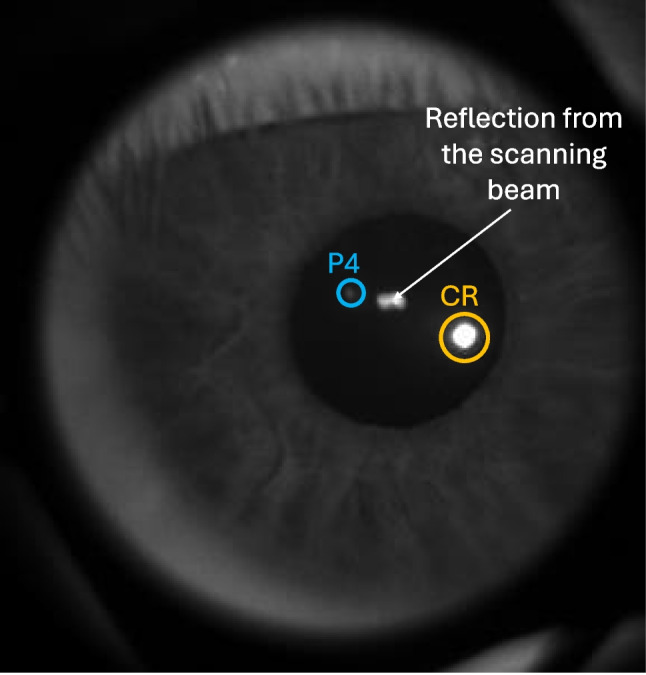 Figure 1
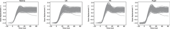 Figure 2
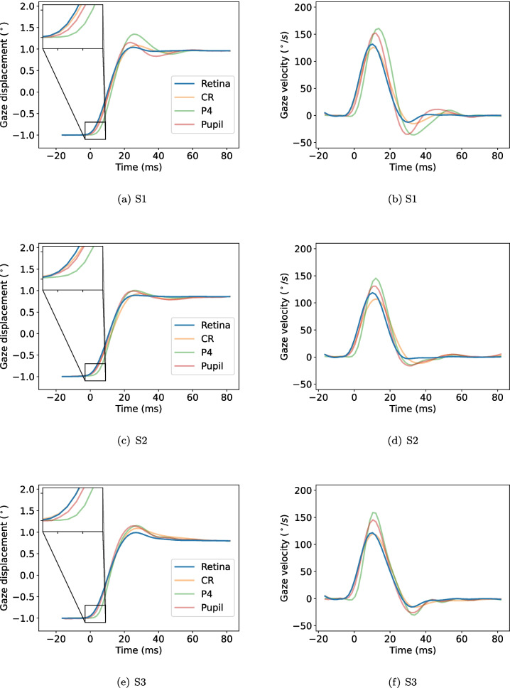 Figure 3
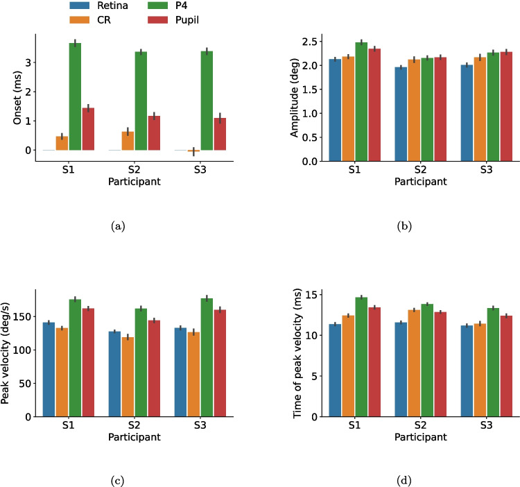 Figure 4
Peer Reviews
No public reviews on file for this paper yet. If you reviewed it on a platform where reviews are public (OpenReview, ICLR, NeurIPS, ICML), you can paste yours below so the community can read it here.
Videos
No videos yet. Explain this paper in a talk, walkthrough, or lecture? Add one.
Taxonomy
TopicsGaze Tracking and Assistive Technology · Ophthalmology and Visual Impairment Studies · Glaucoma and retinal disorders