Diagnostic Value of the Split-Bolus CT Protocol in Blunt Abdominal Trauma: Comparison With Standard Portal Venous Phase Imaging
Arpit Taneja, Narvir S Chauhan, Pankaj Saini, Dinesh Sood, Aman Taneja

TL;DR
This study compares two CT scan protocols for diagnosing abdominal injuries, finding that the split-bolus method offers better image quality without higher radiation risk.
Contribution
The study demonstrates that the split-bolus CT protocol improves vascular and organ contrast in trauma imaging without increasing radiation exposure.
Findings
The split-bolus protocol showed significantly higher contrast enhancement in the spleen, pancreas, and renal cortex compared to the standard portal venous protocol.
Vascular enhancement was consistently better with the split-bolus protocol in key vessels like the portal vein and aorta.
The split-bolus protocol reduced the need for delayed scans by enabling simultaneous arterial and venous injury evaluation.
Abstract
Background Blunt abdominal trauma is a significant cause of morbidity and mortality, particularly in young adults, and accurate imaging is essential for timely diagnosis and management. Computed tomography (CT) is the preferred modality in hemodynamically stable patients, with the routine portal venous (PV) phase protocol commonly employed. Recently, the split-bolus (SB) protocol has been proposed as an alternative, enabling simultaneous arterial and venous phase visualization in a single acquisition. This study compared the image quality, contrast enhancement, diagnostic accuracy, and radiation exposure of the SB protocol versus the standard PV protocol in evaluating blunt abdominal trauma. Methods A prospective observational study was conducted from September 2021 to August 2022 at Dr. Rajendra Prasad Government Medical College, Kangra. Seventy hemodynamically stable patients with…
Genes, proteins, chemicals, diseases, species, mutations and cell lines named across the full text — each resolved to its canonical identifier and authoritative record.
Click any figure to enlarge with its caption.
 Figure 1
 Figure 2
 Figure 3
 Figure 4
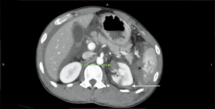 Figure 5
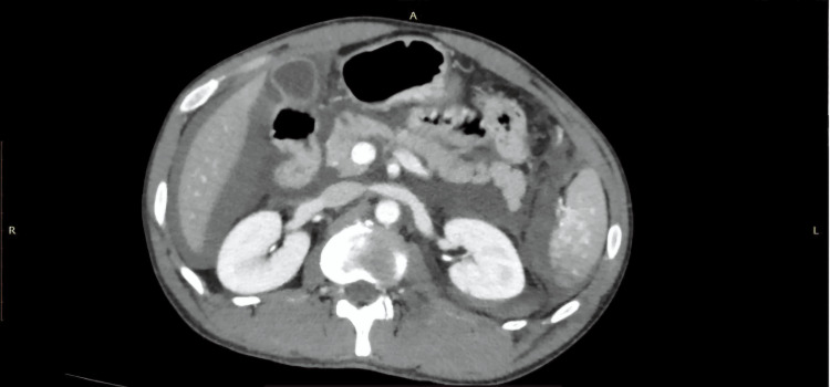 Figure 6
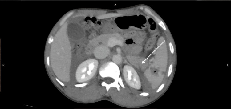 Figure 7
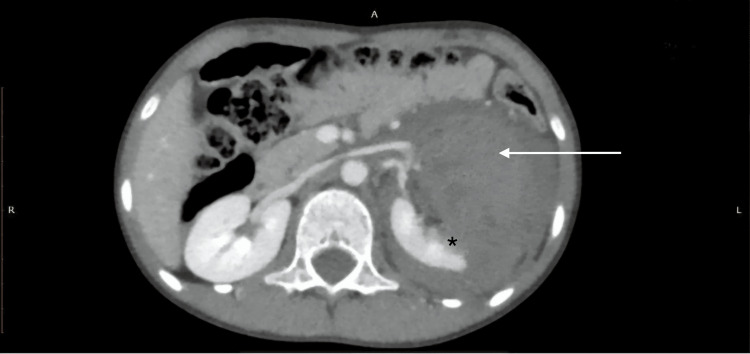 Figure 8
 Figure 9
Peer Reviews
No public reviews on file for this paper yet. If you reviewed it on a platform where reviews are public (OpenReview, ICLR, NeurIPS, ICML), you can paste yours below so the community can read it here.
Videos
No videos yet. Explain this paper in a talk, walkthrough, or lecture? Add one.
Taxonomy
TopicsAbdominal Trauma and Injuries · Pelvic and Acetabular Injuries · Radiation Dose and Imaging