Successful removal of a fractured guidewire from the main pancreatic duct using a novel endoscopic device
Amer Al-Muzaini, Akihisa Ohno, Nao Fujimori, Yosuke Minoda, Keijiro Ueda, Tomohiko Moriyama, Yoshihiro Ogawa

Abstract
Genes, proteins, chemicals, diseases, species, mutations and cell lines named across the full text — each resolved to its canonical identifier and authoritative record.
Click any figure to enlarge with its caption.
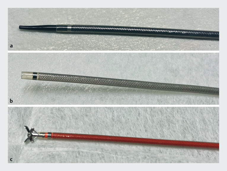 Fig. 1
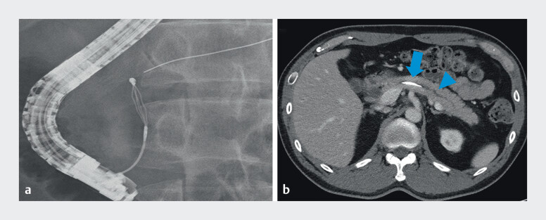 Fig. 2
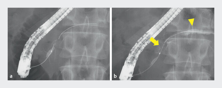 Fig. 3
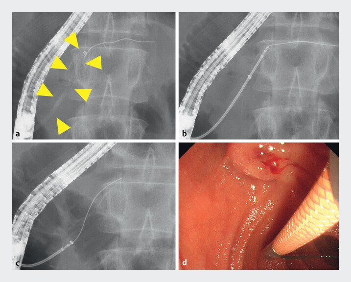 Fig. 4
Peer Reviews
No public reviews on file for this paper yet. If you reviewed it on a platform where reviews are public (OpenReview, ICLR, NeurIPS, ICML), you can paste yours below so the community can read it here.
Videos
No videos yet. Explain this paper in a talk, walkthrough, or lecture? Add one.
Taxonomy
TopicsPancreatitis Pathology and Treatment · Gallbladder and Bile Duct Disorders · Esophageal and GI Pathology
A guidewire fracture in the main pancreatic duct (MPD) is uncommon during endoscopic retrograde cholangiopancreatography (ERCP). The number of reported cases of fractured guidewires in the MPD is limited, and most retrieval attempts present significant technical challenges 1 2 . We used a novel endoscopic sheath (EndoSheather; Piolax Medical Devices, Kanagawa, Japan) with a tapered inner sheath to facilitate passage through strictures as a rescue technique. Instruments with lengths of up to 1.9 mm could be easily inserted after removal of the inner sheath. Several reports have described the use of this device to retrieve migrated stents 3 4 5 , but no previous publication has documented the retrieval of a fractured guidewire in the MPD ( Fig. 1 ).
The novel endoscopic sheath system. a The sheath with both inner and outer components assembled. b The outer sheath remaining after the removal of the inner sheath. c Biopsy forceps inserted through the outer sheath following the removal of the inner sheath.
A 46-year-old man with a history of jaundice and abdominal pain initially underwent ERCP at a different institution for the removal of common bile duct stones. The catheter and guidewire were inadvertently advanced into the MPD during stone extraction, resulting in a guidewire tip fracture inside the MPD ( Fig. 2 a ). The fragment remained lodged in the MPD after multiple retrieval attempts, leading to acute pancreatitis ( Fig. 2 b ). A rescue ERCP was subsequently performed at our institution. Pancreatography revealed a focal stricture extending from the pancreatic head to the body, with mild upstream dilation in the tail ( Fig. 3 ). A novel device (EndoSheater) was inserted proximal to the retained guidewire. Biopsy forceps (Radial Jaw 4 Standard Capacity, Boston Scientific, Marlborough, MA, USA) were inserted through the outer sheath after the inner sheath was removed to grasp the guidewire. The fractured guidewire was successfully removed through the device without any complications ( Fig. 4 and Video 1 ). This case demonstrates the safe and effective retrieval of a fractured guidewire from the MPD using the novel endoscopic sheath.
Fractured guidewire retained in the main pancreatic duct. a Fluoroscopic view revealed the fractured guidewire in the main pancreatic duct (MPD). b Contrast-enhanced computed tomography scan showing impacted guidewire (blue arrow) in the MPD with mild upstream dilation in the pancreatic tail (blue arrowhead).
Fluoroscopic findings of main pancreatic duct (MPD) stricture and guidewire fracture. a Fluoroscopic view showing the initial assessment of the MPD and the location of the fractured guidewire. b Fluoroscopic view showing a focal stricture from the head to body of the MPD (yellow arrow) and upstream dilation in the pancreatic tail (yellow arrowhead).
Retrieval of the fractured guidewire using EndoSheather. a Fluoroscopic view showing the EndoSheather having been passed through the stricture between head and body; image obtained after removal of the inner sheath (yellow arrowhead). b Fluoroscopic view showing the insertion of biopsy forceps through the EndoSheather to grasp the proximal end of the fractured guidewire. Fluoroscopic c and endoscopic d views showing successful and safe retrieval of the fractured guidewire.
Successful retrieval of a fractured pancreatic duct guidewire using a novel endoscopic sheath system.Video 1
Endoscopy_UCTN_Code_CPL_1AK_2AD
The reference list from the paper itself. Each links out to its DOI / PubMed record.
- 1Kitagawa S Successful endoscopic retrieval of a retained fractured guidewire from the main pancreatic duct GE Port J Gastroenterol 20222936436510.1159/00051694836159199 PMC 9485961 · doi ↗ · pubmed ↗
- 2Kim S So H Jung SW Guidewire impaction in the main pancreatic duct in a patient with chronic pancreatitis: A case report Korean J Gastroenterol 202381363936695065 10.4166/kjg.2022.121PMC 12285445 · doi ↗ · pubmed ↗
- 3Fukuda R Takahara N Nakai Y Successful endoscopic removal of a retained guidewire in the intrahepatic bile duct using a novel tapered sheath dilator Video GIE 2024937337510.1016/j.vgie.2024.04.00739233836 PMC 11368704 · doi ↗ · pubmed ↗
- 4Mori Y Kurita A Successful retrieval of a fractured biliary guidewire using a newly developed endoscopic tapered sheath Endoscopy 202355 E 706E 70710.1055/a-2073-514737164336 PMC 10172000 · doi ↗ · pubmed ↗
- 5Higashimori A Maruyama H Maeda N Successful retrieval of a fractured migrated pancreatic stent using an endoscopic tapered sheath for severe pancreatic duct stenosis Endoscopy 202355 E 747E 74810.1055/a-2086-194637236262 PMC 10219759 · doi ↗ · pubmed ↗